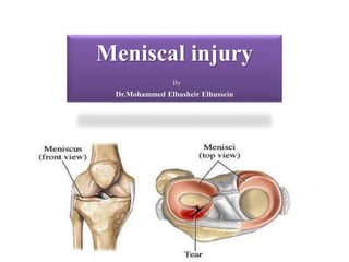
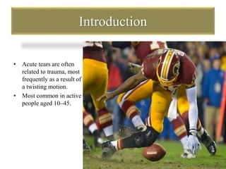
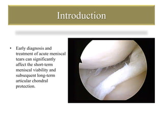
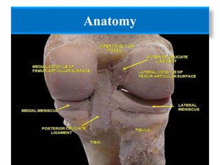
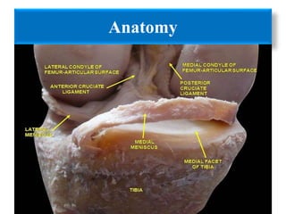
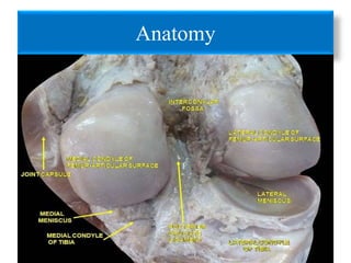
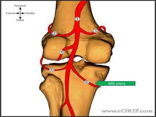
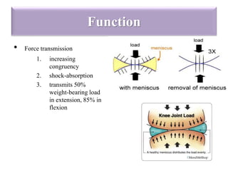
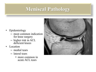
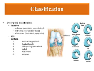
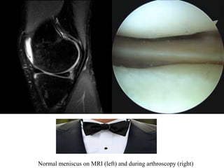
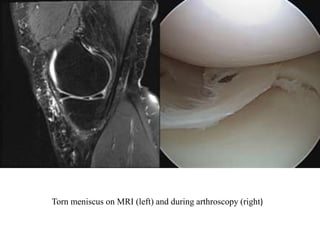
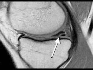
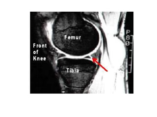
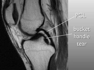
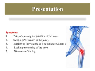
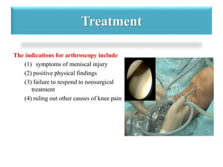
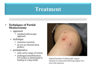
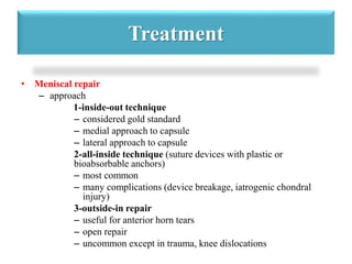
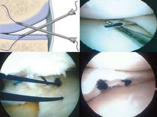
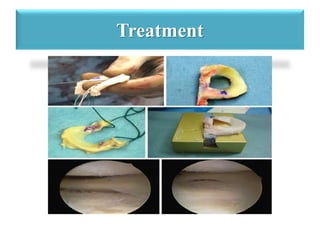
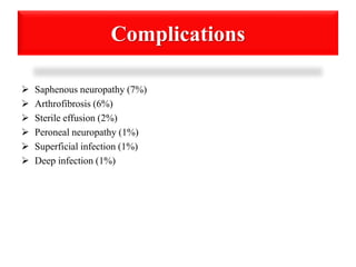
Meniscal injury 
By 
Dr.Mohammed Elbasheir Elhussein
Introduction 
• The menisci are fibrocartilaginous structures that are semilunar in shape 
and wedge-shaped in cross-section. 
• Two menisci(medial and lateral) exist between the femoral and tibial 
articulation.The femoral articulating meniscal surface is concave,whereas 
the tibial articulating surface is convex.These surfaces conform to the 
convex and concave opposing chondral surfaces, respectively. 
• The conforming articulation provides perfect congruency between 
the femoral condyle, meniscus,and tibial plateau, which establishes 
the foundation for the biomechanical function of the menisci.
Introduction 
Meniscal tears can be either 
– Traumatic 
or 
– degenerative.
Introduction 
• Degenerative tears have 
been closely associated 
with osteoarthritis.
Introduction 
• Acute tears are often 
related to trauma, most 
frequently as a result of 
a twisting motion. 
• Most common in active 
people aged 10–45.
Introduction 
• Early diagnosis and 
treatment of acute meniscal 
tears can significantly 
affect the short-term 
meniscal viability and 
subsequent long-term 
articular chondral 
protection.
Anatomy
Anatomy
Anatomy
Anatomy 
• Blood supply 
– medial inferior genicular artery 
– lateral inferior genicular artery
AnatomyPopliteal artery
Anatomy 
ILG artery
Anatomy 
IMG artery
Anatomy 
• Innervation 
– peripheral two-thirds innervated by Type I and II nerve endings 
– posterior horns have highest concentration of mechanoreceptors
Composition 
 Made of 
1. fibroelastic cartilage 
2. Collagen 
3. Fibers
Stability 
• medial meniscus 
– posterior horn of medial 
meniscus is the 
main secondary 
stabilizer to anterior 
translation 
• lateral meniscus 
– is less stabilizing and 
has 2X the excursion of 
the medial meniscus
Function 
• Force transmission 
1. increasing 
congruency 
2. shock-absorption 
3. transmits 50% 
weight-bearing load 
in extension, 85% in 
flexion
Meniscal Pathology 
• Epidemiology 
– most common indication 
for knee surgery 
– higher risk in ACL 
deficient knees 
• Location 
– medial tears 
– lateral tears 
• more common in 
acute ACL tears
Injury & Healing potential 
• Tears in peripheral 25% red zone 
– can heal via fibrocartilage scar formation 
• Tears of central 75% 
– have limited or no intrinsic healing ability
Classification 
• Descriptive classification 
– location 
• red zone (outer third, vascularized) 
• red-white zone (middle third) 
• white zone (inner third, avascular) 
– size 
– pattern 
1. vertical/longitudinal 
2. bucket handle 
3. oblique/flap/parrot beak 
4. radial 
5. horizontal 
6. complex
• The repairability of a meniscus depends on a number of factors these 
include: 
1. Age/strength 
2. Activity level 
3. Tear pattern 
4. Chronicity of the tear 
5. Associated injuries (anterior cruciate ligament injury) 
6. Healing potential
Normal meniscus on MRI (left) and during arthroscopy (right)
Torn meniscus on MRI (left) and during arthroscopy (right)
Presentation 
Symptoms 
1. Pain, often along the joint line of the knee . 
2. Swelling (“effusion” in the joint). 
3. Inability to fully extend or flex the knee without discomfort . 
4. Locking or catching of the knee. 
5. Weakness of the leg.
Presentation 
 Signs 
• Joint line tenderness 
• Effusion 
• Positive McMurray's test
Imaging 
• X-ray: 
– Images (normally during weightbearing) 
to rule out other conditions .
Imaging 
• MRI 
– Indications 
• MRI is most sensitive diagnostic test, but also has a high false 
positive rate
Treatment 
 Non-operative 
Rest, NSAIDS, rehabilitation 
• indications 
– indicated as first line of treatment for degenerative tears
Treatment 
 Operative 
– The definitive treatment of meniscal tears involves either repair or 
excision of the pathologic tissue. 
– Surgery.
Treatment 
The indications for arthroscopy include 
(1) symptoms of meniscal injury 
(2) positive physical findings 
(3) failure to respond to nonsurgical 
treatment 
(4) ruling out other causes of knee pain
Treatment 
– Partial meniscectomy 
• indications 
– tears not amenable to repair (complex, degenerative, radial tear 
patterns) 
• outcomes 
– >80% satisfactory function at minimum follow-up 
– 50% radiographic changes (osteophytes, flattening, joint space 
narrowing)
Treatment 
– Meniscal repair 
• indications 
– best candidate for repair is a tear with the following 
characteristics 
» peripheral in the red zone (vascularized region) 
» rim width correlates with the ability of a meniscal repair to 
heal (lower rim width has better blood supply) 
» vertical and longitudinal tear 
» 1-4 cm in length 
» acute repair combined with ACL reconstruction
Treatment 
• outcomes 
– 70-95% successful 
– highest success when done with concomitant ACL 
reconstruction 
– poor results with untreated ACL-deficiency (30%)
Treatment 
– Total meniscectomy 
– of historical interest only 
• outcomes 
– 20% have significant arthritic lesions and 70% have 
radiographic changes three years after surgery 
– 100% have arthrosis at 20 years 
– severity of degenerative changes is proportional to % of the 
meniscus that was removed
Treatment 
• Techniques of Partial 
Meniscectomy 
– approach 
• standard arthroscopic 
approach 
– technique 
• minimize resection 
• do not use thermal (heat 
probes) 
– postoperative 
• early active range of motion 
• prolonged immobilization 
(10 weeks) is detrimental to 
healing in a dog model 
Typical locations of arthroscopic surgery 
incisions in a knee joint following surgery for a 
tear in the meniscus
Treatment 
• Meniscal repair 
– approach 
1-inside-out technique 
– considered gold standard 
– medial approach to capsule 
– lateral approach to capsule 
2-all-inside technique (suture devices with plastic or 
bioabsorbable anchors) 
– most common 
– many complications (device breakage, iatrogenic chondral 
injury) 
3-outside-in repair 
– useful for anterior horn tears 
– open repair 
– uncommon except in trauma, knee dislocations
Treatment
Treatment 
• Side effects of meniscectomy include: 
1. The knee loses its ability to transmit and distribute load and absorb 
mechanical shock. 
2. Persistent and significant swelling and stiffness in the knee. 
3. The knee may be not fully mobile, there may be the sensation of 
knee locking or buckling in the knee. 
4. The full knee may be in full motion after tear of meniscus
Treatment 
• Meniscal Transplantation 
– technique 
• bone to bone healing 
with plugs at each horn 
or a bridge between 
horns 
• peripheral vertical 
mattress sutures 
• correct sizing of the 
allograft is 
essential (commonly 
based on radiographs, 
within 5-10% error 
tolerated)
Prevention 
 There are three major ways of 
preventing a meniscus tear. 
1. wearing the correct footwear. 
2. Strengthening and stretching the 
major leg muscles. 
3. learning proper technique for the 
movement. 
Proper parallel squat form to improve knee 
stability
Complications 
 Saphenous neuropathy (7%) 
 Arthrofibrosis (6%) 
 Sterile effusion (2%) 
 Peroneal neuropathy (1%) 
 Superficial infection (1%) 
 Deep infection (1%)
Meniscal injury

Meniscal injury

  • 1.
    Meniscal injury By Dr.Mohammed Elbasheir Elhussein
  • 2.
    Introduction • Themenisci are fibrocartilaginous structures that are semilunar in shape and wedge-shaped in cross-section. • Two menisci(medial and lateral) exist between the femoral and tibial articulation.The femoral articulating meniscal surface is concave,whereas the tibial articulating surface is convex.These surfaces conform to the convex and concave opposing chondral surfaces, respectively. • The conforming articulation provides perfect congruency between the femoral condyle, meniscus,and tibial plateau, which establishes the foundation for the biomechanical function of the menisci.
  • 3.
    Introduction Meniscal tearscan be either – Traumatic or – degenerative.
  • 4.
    Introduction • Degenerativetears have been closely associated with osteoarthritis.
  • 5.
    Introduction • Acutetears are often related to trauma, most frequently as a result of a twisting motion. • Most common in active people aged 10–45.
  • 6.
    Introduction • Earlydiagnosis and treatment of acute meniscal tears can significantly affect the short-term meniscal viability and subsequent long-term articular chondral protection.
  • 7.
  • 8.
  • 9.
  • 10.
    Anatomy • Bloodsupply – medial inferior genicular artery – lateral inferior genicular artery
  • 11.
  • 12.
  • 13.
  • 14.
    Anatomy • Innervation – peripheral two-thirds innervated by Type I and II nerve endings – posterior horns have highest concentration of mechanoreceptors
  • 15.
    Composition  Madeof 1. fibroelastic cartilage 2. Collagen 3. Fibers
  • 16.
    Stability • medialmeniscus – posterior horn of medial meniscus is the main secondary stabilizer to anterior translation • lateral meniscus – is less stabilizing and has 2X the excursion of the medial meniscus
  • 17.
    Function • Forcetransmission 1. increasing congruency 2. shock-absorption 3. transmits 50% weight-bearing load in extension, 85% in flexion
  • 18.
    Meniscal Pathology •Epidemiology – most common indication for knee surgery – higher risk in ACL deficient knees • Location – medial tears – lateral tears • more common in acute ACL tears
  • 19.
    Injury & Healingpotential • Tears in peripheral 25% red zone – can heal via fibrocartilage scar formation • Tears of central 75% – have limited or no intrinsic healing ability
  • 20.
    Classification • Descriptiveclassification – location • red zone (outer third, vascularized) • red-white zone (middle third) • white zone (inner third, avascular) – size – pattern 1. vertical/longitudinal 2. bucket handle 3. oblique/flap/parrot beak 4. radial 5. horizontal 6. complex
  • 21.
    • The repairabilityof a meniscus depends on a number of factors these include: 1. Age/strength 2. Activity level 3. Tear pattern 4. Chronicity of the tear 5. Associated injuries (anterior cruciate ligament injury) 6. Healing potential
  • 22.
    Normal meniscus onMRI (left) and during arthroscopy (right)
  • 23.
    Torn meniscus onMRI (left) and during arthroscopy (right)
  • 27.
    Presentation Symptoms 1.Pain, often along the joint line of the knee . 2. Swelling (“effusion” in the joint). 3. Inability to fully extend or flex the knee without discomfort . 4. Locking or catching of the knee. 5. Weakness of the leg.
  • 28.
    Presentation  Signs • Joint line tenderness • Effusion • Positive McMurray's test
  • 29.
    Imaging • X-ray: – Images (normally during weightbearing) to rule out other conditions .
  • 30.
    Imaging • MRI – Indications • MRI is most sensitive diagnostic test, but also has a high false positive rate
  • 31.
    Treatment  Non-operative Rest, NSAIDS, rehabilitation • indications – indicated as first line of treatment for degenerative tears
  • 32.
    Treatment  Operative – The definitive treatment of meniscal tears involves either repair or excision of the pathologic tissue. – Surgery.
  • 33.
    Treatment The indicationsfor arthroscopy include (1) symptoms of meniscal injury (2) positive physical findings (3) failure to respond to nonsurgical treatment (4) ruling out other causes of knee pain
  • 34.
    Treatment – Partialmeniscectomy • indications – tears not amenable to repair (complex, degenerative, radial tear patterns) • outcomes – >80% satisfactory function at minimum follow-up – 50% radiographic changes (osteophytes, flattening, joint space narrowing)
  • 35.
    Treatment – Meniscalrepair • indications – best candidate for repair is a tear with the following characteristics » peripheral in the red zone (vascularized region) » rim width correlates with the ability of a meniscal repair to heal (lower rim width has better blood supply) » vertical and longitudinal tear » 1-4 cm in length » acute repair combined with ACL reconstruction
  • 36.
    Treatment • outcomes – 70-95% successful – highest success when done with concomitant ACL reconstruction – poor results with untreated ACL-deficiency (30%)
  • 37.
    Treatment – Totalmeniscectomy – of historical interest only • outcomes – 20% have significant arthritic lesions and 70% have radiographic changes three years after surgery – 100% have arthrosis at 20 years – severity of degenerative changes is proportional to % of the meniscus that was removed
  • 38.
    Treatment • Techniquesof Partial Meniscectomy – approach • standard arthroscopic approach – technique • minimize resection • do not use thermal (heat probes) – postoperative • early active range of motion • prolonged immobilization (10 weeks) is detrimental to healing in a dog model Typical locations of arthroscopic surgery incisions in a knee joint following surgery for a tear in the meniscus
  • 39.
    Treatment • Meniscalrepair – approach 1-inside-out technique – considered gold standard – medial approach to capsule – lateral approach to capsule 2-all-inside technique (suture devices with plastic or bioabsorbable anchors) – most common – many complications (device breakage, iatrogenic chondral injury) 3-outside-in repair – useful for anterior horn tears – open repair – uncommon except in trauma, knee dislocations
  • 41.
  • 42.
    Treatment • Sideeffects of meniscectomy include: 1. The knee loses its ability to transmit and distribute load and absorb mechanical shock. 2. Persistent and significant swelling and stiffness in the knee. 3. The knee may be not fully mobile, there may be the sensation of knee locking or buckling in the knee. 4. The full knee may be in full motion after tear of meniscus
  • 43.
    Treatment • MeniscalTransplantation – technique • bone to bone healing with plugs at each horn or a bridge between horns • peripheral vertical mattress sutures • correct sizing of the allograft is essential (commonly based on radiographs, within 5-10% error tolerated)
  • 44.
    Prevention  Thereare three major ways of preventing a meniscus tear. 1. wearing the correct footwear. 2. Strengthening and stretching the major leg muscles. 3. learning proper technique for the movement. Proper parallel squat form to improve knee stability
  • 45.
    Complications  Saphenousneuropathy (7%)  Arthrofibrosis (6%)  Sterile effusion (2%)  Peroneal neuropathy (1%)  Superficial infection (1%)  Deep infection (1%)

Editor's Notes

  • #6 These tears are more likely to produce a moveable fragment that can catch in the knee and therefore require surgical treatment
  • #7 This treatment is particularly critical in a younger population
  • #8 The menisci are C-shaped wedges of fibrocartilage located between the tibial plateau and femoral condyles. The menisci contain 70% type I collagen.[3]
  • #9 The larger semilunar medial meniscus is attached more firmly than the loosely fixed, more circular lateral meniscus. The anterior and posterior horns of both menisci are secured to the tibial plateaus.
  • #10  Anteriorly, the transverse ligament connects the 2 menisci; posteriorly, the meniscofemoral ligament helps stabilize the posterior horn of the lateral meniscus to the femoral condyle. The coronary ligaments connect the peripheral meniscal rim loosely to the tibia. Although the lateral collateral ligament (LCL) passes in close proximity, the lateral meniscus has no attachment to this structure Attachment
  • #11 Blood supply 1-medial inferior genicular artery supplies peripheral 20-30% of medial meniscus 2-lateral inferior genicular artery supplies peripheral 10-25% of lateral meniscus central 75% receive nutrition through diffusion-This presents a problem when there is an injury to the meniscus, as the avascular areas tend not to heal without the essential nutrients supplied by blood vessels.
  • #15  Which is a sensory receptors that responds to mechanical pressure or distortion.
  • #16 The menisci are made of 1-fibroelastic cartilage -interlacing network of collagen, proteoglycan, glycoproteins, and cellular elements composed of 65-75% water 2-Collagen- 90 % Type I collagen 3-Fibers -which allow the meniscus to expand under compressive forces and increase contact area of the joint .
  • #17 the meniscus deepens tibial surface and acts as secondary stabilizer
  • #18 The meniscus functions to optimize force transmission across the knee and this will be done by 1-increasing congruency -increases contact area leads to decreased point loading 2-shock-absorption the meniscus is more elastic than articular cartilage, and therefore absorbs shock. 3-transmits 50% weight-bearing load in extension, 85% in flexion
  • #19 Epidemiology most common indication for knee surgery higher risk in ACL deficient knees. Location -medial tears- more common than lateral tears the exception is in the setting of an acute ACL tear where lateral tears are more common degenerative tears in older patients usually occur in the posterior horn medial meniscus -lateral tears -more common in acute ACL tears
  • #20 Fibrochondrocyte are the cells responsible for healing
  • #21 pattern Vertical or longitudinal is more common, especially with ACL tears repair when peripheral bucket handle vertical tear which may displace into the notch. oblique/flap/parrot beak may cause mechanical locking symptoms. radial horizontal more common in older population may be associated with meniscal cysts
  • #22 The functional importance of these classifications, however, is to ultimately determine whether a meniscus is repairable..
  • #23 Normally the medal and lateral menisci appear as low signal bow-tie-shaped structures between the femoral condyles and tibial plateauxThe absent bow tie sign represents the loss of the normal appearance of the menisci on parasagittal MRI images, and is suggestive of meniscal injury.
  • #27 The double PCL sign appears on sagittal MRI images of the knee when there is a bucket-handle tear of the meniscus
  • #28 1-often develops due to inflammation and/or bleeding from the injury 5-particularly the quadriceps muscle. This may be evident when trying to perform a straight leg raise or walk up and down stairs.
  • #29 flex the knee and place a hand on medial side of knee, externally rotate the leg and bring the knee into extension a palpable pop or click is a positive test and can correlate with a medial meniscus tear
  • #30 The menisci themselves cannot be visualised with plain radiographs knee arthroscopy allows quick diagnosis and simultaneous treatment. Recent clinical data shows that MRI and clinical testing are comparable in sensitivity and specificity when looking for a meniscal tear
  • #33 is indicated in patients who have persistent mechanical symptoms and/or pain and have not responded to a course of nonoperative treatment
  • #34 The indications for arthroscopy include (1) symptoms of meniscal injury that affect activities of daily living, work, and/or sports participation, such as instability, locking, effusion, and pain; (2) positive physical findings of joint-line tenderness, joint effusion, limitation of motion, and provocative signs, such as pain with squatting, a positive pinch test, or a positive McMurray test; (3) failure to respond to nonsurgical treatment, including activity modification, medication, and a rehabilitation program; and (4) ruling out other causes of knee pain identified by patient history, physical examination, plain radiographs, or other imaging studies.
  • #35 predictors of success age <40yo normal alignment minimal or no arthritis single tear
  • #36 rim width is the distance from the tear to the peripheral meniscocapsular junction (blood supply).
  • #39 Techniques of Partial Meniscectomy approach standard arthroscopic approach technique minimize resection (DJD proportional to amount removed) do not use thermal (heat probes) postoperative early active range of motion prolonged immobilization (10 weeks) is detrimental to healing in a dog model
  • #40 ???Meniscal repair approach inside-out technique considered gold standard medial approach to capsule expose capsule by incising the sartorius fascia, retracting the pes tendons and semimembranosus posteriorly, and developing the plane between the medial gastrocnemius and capsule. lateral approach to capsule expose capsule by developing plane between the iliotibial band and biceps tendon interval, then retract lateral head of gastrocnemius posteriorly
  • #42 meniscal allografts. Cylindrical bone-plug (C) or keyhole-slot (D) techniques may F, Second-look arthroscopic views of a lateral meniscal allograft.
  • #45 There are three major ways of preventing a meniscus tear The first of which is wearing the correct footwear for the sport and surface that the activity is taking place on The proper footwear is imperative when engaging in physical activity because one off balanced step could mean a meniscus tear. The second way to prevent a meniscus tear is to strengthen and stretch the major leg muscles. The third things learning proper technique for the movement that is taking place For the sports involving quick powerful movements it is important to learn how to cut, turn, land from a jump, and stop correctly