1) Neurons communicate with each other via electrical and chemical signals. Electrical signals allow for faster transmission than just chemical signals alone.
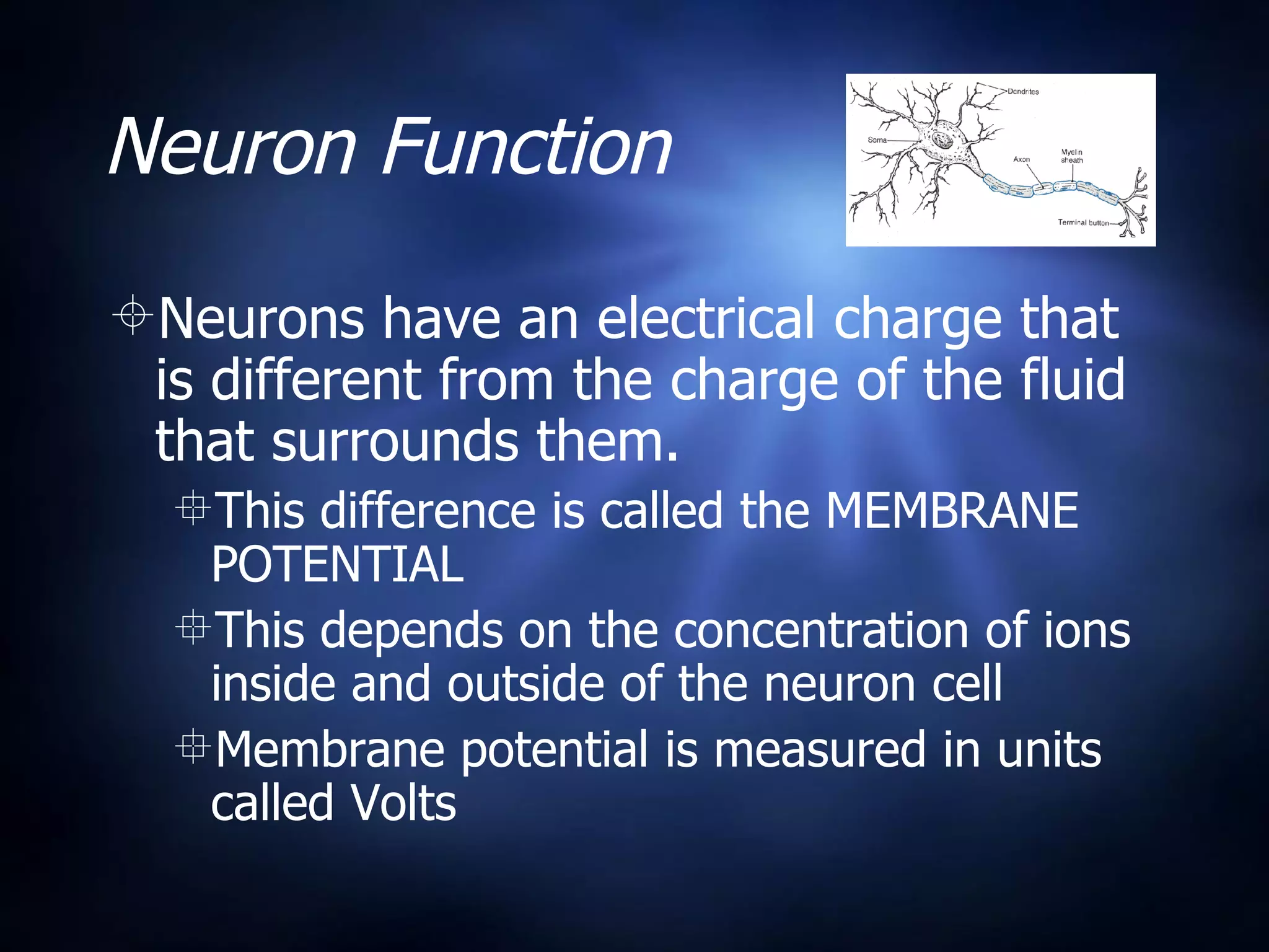
2) The basic parts of a neuron include dendrites that receive signals, an axon that conducts signals, and axon terminals that transmit signals to other cells at synapses. Some neurons have a fatty myelin sheath that insulates the axon and speeds transmission.
3) Neurons maintain a voltage difference across their cell membranes called the membrane potential. An "action potential" is generated when the membrane potential rapidly changes, propagating a nerve impulse down the axon via voltage-gated ion channels.