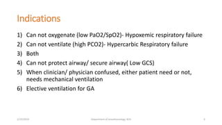
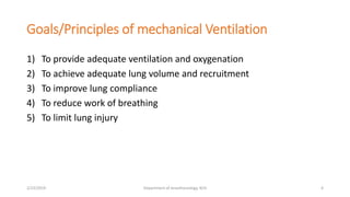
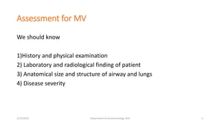
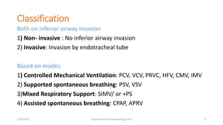
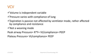
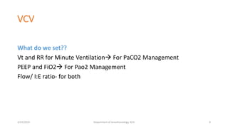
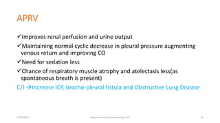
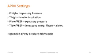
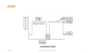
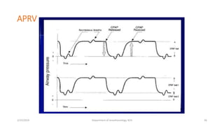
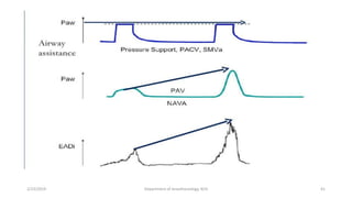
Mechanical Ventilation
Presenter:
Dr. Tirtha Raj Bhandari
2nd year Resident
Department of Pediatric Anesthesiology
KCH,Maharajguj
2/23/2019 Department of Anesthesiology, KCH 1
Objectives
• To discuss indication of mechanical ventilation
• To discuss different modes for mechanical ventilation
2/23/2019 Department of Anesthesiology, KCH 2
Indications
1) Can not oxygenate (low PaO2/SpO2)- Hypoxemic respiratory failure
2) Can not ventilate (high PCO2)- Hypercarbic Respiratory failure
3) Both
4) Can not protect airway/ secure airway( Low GCS)
5) When clinician/ physician confused, either patient need or not,
needs mechanical ventilation
6) Elective ventilation for GA
2/23/2019 Department of Anesthesiology, KCH 3
Goals/Principles of mechanical Ventilation
1) To provide adequate ventilation and oxygenation
2) To achieve adequate lung volume and recruitment
3) To improve lung compliance
4) To reduce work of breathing
5) To limit lung injury
2/23/2019 Department of Anesthesiology, KCH 4
Assessment for MV
We should know
1)History and physical examination
2) Laboratory and radiological finding of patient
3) Anatomical size and structure of airway and lungs
4) Disease severity
2/23/2019 Department of Anesthesiology, KCH 5
Classification
Both on inferior airway invasion
1) Non- invasive : No inferior airway invasion
2) Invasive: Invasion by endotracheal tube
Based on modes
1) Controlled Mechanical Ventilation: PCV, VCV, PRVC, HFV, CMV, IMV
2) Supported spontaneous breathing: PSV, VSV
3)Mixed Respiratory Support: SIMV/ or +PS
4) Assisted spontaneous breathing: CPAP, APRV
2/23/2019 Department of Anesthesiology, KCH 6
VCV
Volume is independent variable
Pressure varies with compliance of lung
Expiration is passive not affected by ventilator mode, rather affected
by compliance and resistance
Not a weaning mode
Peak airway Pressure= R*F+ Vt/compliance+ PEEP
Plateau Pressure= Vt/compliance+ PEEP
2/23/2019 Department of Anesthesiology, KCH 7
VCV
What do we set??
Vt and RR for Minute Ventilation For PaCO2 Management
PEEP and FiO2 For Pao2 Management
Flow/ I:E ratio- for both
2/23/2019 Department of Anesthesiology, KCH 8
PCV
Pressure is independent variable
Volume is dependent variable Depends on: level of pressure, I-time
also it is the function of compliance and airway resistance. That’s why
volume deliver can vary breath to breath.
Ventilator adjust flow to maintain pressure.
Flow decreases throughout the inspiratory cycle.
Good mode to use if patient has large air leak, the ventilator will
increase the flow to compensate it.
Not a weaning mode
2/23/2019 Department of Anesthesiology, KCH 9
PCV
What do we set??
Pressure limit, T-Insp, RR,FIO2,PEEP
P and RR for Minute ventilation PaCO2 management
FiO2 and PEEP for PaO2 management
2/23/2019 Department of Anesthesiology, KCH 10
VCV and PCV
2/23/2019 Department of Anesthesiology, KCH 11
A/C, V/C
2/23/2019 Department of Anesthesiology, KCH 12
PRVC= Pressure Regulated Volume Control
Actually a volume control assist control mode
Target minute ventilation is set
Ventilator will adjust the flow to deliver the volume without
exceeding a target inspiratory pressure
Very useful in patient with high airway pressure.
2/23/2019 Department of Anesthesiology, KCH 13
PRVC
Advantage:
1)No change in MV delivery even if pulmonary condition changes
2) The desired TV will be delivered at lowest PIP, thus minimizes risk of
barotrauma.
Disadvantage:
1)Hard to use on a spontaneously breathing patient or one with air leak
2)Not a weaning mode
2/23/2019 Department of Anesthesiology, KCH 14
2/23/2019 Department of Anesthesiology, KCH 15
Pressure Support=PS
Supports each spontaneous breath with supplemental flow to
achieve preset pressure
All breath are triggered by patient
Preset value  PIP, PEEP, FiO2
Patient determine Rate, Ti, I/E ratio, TV
2/23/2019 Department of Anesthesiology, KCH 16
PS
Needs intact respiratory drive
Helps to overcome airway resistance/ tube resistance, so that
spontaneous breathing will be easier
Can not be use in patient not having spontaneous breathing(i.e.
muscle relaxant)
2/23/2019 Department of Anesthesiology, KCH 17
SIMV= Synchronized intermittent mandatory
ventilation
Preset mechanical breath delivered within interval acc to preset and
wait for spontaneous in between, which it will use as a trigger to
deliver full breath.
If not sensed it will automatically give a breath
Vt on spontaneous breaths depends entirely upon the patient effort
and lung mechanics, can be pressure or volume controlled.
2/23/2019 Department of Anesthesiology, KCH 18
SIMV
Tb(time for breathing) = Tm(mandatory)+Ts(spontaneous)
If patient tries to breath during Tm, ventilator gives a fully assisted
breath
If Patient tries to breath during Ts, the ventilator will allow the
patient to take breath
2/23/2019 Department of Anesthesiology, KCH 19
2/23/2019 Department of Anesthesiology, KCH 20
2/23/2019 Department of Anesthesiology, KCH 21
2/23/2019 Department of Anesthesiology, KCH 22
SIMV+PS
SIMV+PS= provides assistance for spontaneous breath to overcome tube resistance
2/23/2019 Department of Anesthesiology, KCH 23
CPAP
Provides continuous positive pressure throughout the respiratory
cycle
So gives supports to inspiration and resistance to expiration
Can be use both in invasive and non-invasive form.
Very similar to PEEP
2/23/2019 Department of Anesthesiology, KCH 24
PEEP
PEEP is a residual pressure above atmospheric pressure maintained
at the end of expiration.
PEEP can be added to any mode
PEEP helps to recruit alveoli, increase FRC , Redistribute pulmonary
edema, Decrease intrapulmonary shunt, Increase PaO2.(GOOD)
PEEP , decreases venous return/CO, increase ICP/intensify cerebral
ischemia/ risk of barotrauma(Bad)
2/23/2019 Department of Anesthesiology, KCH 25
NASAL CPAP FOR NEONATE/INFANT
Nasal prong/Nasopharyngral tube/ET tube can be use for nasal cpap2/23/2019 Department of Anesthesiology, KCH 26
Indication
Disease condition;
1)Retained lung fluid
2)Post-extubation (if risk of airway collapse)
3)Atelectasis
4)Respiratory distress syndrome
5) For administration of controlled concentration of nitric oxide
2/23/2019 Department of Anesthesiology, KCH 27
CONTD
Physical findings:
1)Increase WOB ,indicated by increase RR by 30-40%
2)Sub-sternal/suprasternal retraction
3)Grunting and nasal flaring
4)Pale or cyanotic skin color
5)Agitation
2/23/2019 Department of Anesthesiology, KCH 28
Contraindication/ Should 'Not Try
C/I:
Choanal atresia
Untreated diaphragmatic hernia
Cleft palate
TEF
Should 'not try:
Cardiovascular instability
Severe ventilatory impairment
Severe hypoxemia
Frequent apnea
High level of sedation
2/23/2019 Department of Anesthesiology, KCH 29
How it Helps??
Reduces grunting and tachypnea
Increases FRC and PaO2
Decreases intrapulmonary shunting
Improves lung compliance
Aids in stabilization of floppy infant chest wall
2/23/2019 Department of Anesthesiology, KCH 30
CONTD
Improves distribution of ventilation
Reduce WOB
Reduces central and obstructive sleep apnea by mechanically
splinting the upper airway.
Better recruitment and oxygenation
Stimulation of infant/neonate for breathing
2/23/2019 Department of Anesthesiology, KCH 31
APRV= Airway Pressure Release Ventilation
It is a form of CPAP with release of pressure in between
It is a inverse ration ventilation
Uses lower PIP to maintain oxygenation and ventilation without
compromising venous return
Improves V/Q matching
2/23/2019 Department of Anesthesiology, KCH 32
APRV
Improves renal perfusion and urine output
Maintaining normal cyclic decrease in pleural pressure augmenting
venous return and improving CO
Need for sedation less
Chance of respiratory muscle atrophy and atelectasis less(as
spontaneous breath is present)
C/I Increase ICP, brocho-pleural fistula and Obstructive Lung Disease
2/23/2019 Department of Anesthesiology, KCH 33
APRV Settings
• P High= Inspiratory Pressure
• T high= time for inspiration
• P low/PEEP= expiratory pressure
• T low/PEEP= time spent in exp. Phase = allows
High mean airway pressure maintained
2/23/2019 Department of Anesthesiology, KCH 34
APRV
2/23/2019 Department of Anesthesiology, KCH 35
APRV
2/23/2019 Department of Anesthesiology, KCH 36
Initial setting for APRV
P high:
20-25 cm H2O- mean airway
pressure
30cmh2o if pplat is >=30cmh2o
Higher setting may be required
for morbid obesity and if thoracic
and abdominal compliance
decrease(ascites)
T high: minimum 2-3 seconds,
progressively increase to 10-15 sec
Helps in oxygenation
P low: Peep set at zero, provides
high Pressure difference for
unimpeded expiratory flow
T low/PEEP: 50-70% of peak
expiratory flow.
0.2 to 0.6 sec
2/23/2019 Department of Anesthesiology, KCH 37
2/23/2019 Department of Anesthesiology, KCH 38
NAVA= Neurally Adjusted Ventilatory Assistance
• It is a spontaneous or supported mode
• Patient initiated synchronized breathing mode
• Breathing support is triggered by the electrical activity of the
diaphragm(EADi)
• NAVA setting(1-4 cm H2o/uv) multiplies measures EADi to provide
pressure, which is proportional and synchronized to patient effort
• It avoids over/under assistance
• Lung protective mode in spontaneous breath
• Avoids asynchrony, so less sedation required and better sleep.
2/23/2019 Department of Anesthesiology, KCH 39
2/23/2019 Department of Anesthesiology, KCH 40
2/23/2019 Department of Anesthesiology, KCH 41
Any Questions???
2/23/2019 Department of Anesthesiology, KCH 42
Summary
We should know
When to start mechanical ventilation
Which mode would be better for particular patient
Setting should be appropriate according to patient to minimize injury
to lung
Steps of changing modes for weaning
2/23/2019 Department of Anesthesiology, KCH 43
References
• Clinical Application of Mechanical Ventilation(Chang)
• Miller Anesthesia- 8th edition
• Barash anesthesia- 8th edition
2/23/2019 Department of Anesthesiology, KCH 44

Mechanical Ventilation -Modes for Pediatrics

  • 1.
    Mechanical Ventilation Presenter: Dr. TirthaRaj Bhandari 2nd year Resident Department of Pediatric Anesthesiology KCH,Maharajguj 2/23/2019 Department of Anesthesiology, KCH 1
  • 2.
    Objectives • To discussindication of mechanical ventilation • To discuss different modes for mechanical ventilation 2/23/2019 Department of Anesthesiology, KCH 2
  • 3.
    Indications 1) Can notoxygenate (low PaO2/SpO2)- Hypoxemic respiratory failure 2) Can not ventilate (high PCO2)- Hypercarbic Respiratory failure 3) Both 4) Can not protect airway/ secure airway( Low GCS) 5) When clinician/ physician confused, either patient need or not, needs mechanical ventilation 6) Elective ventilation for GA 2/23/2019 Department of Anesthesiology, KCH 3
  • 4.
    Goals/Principles of mechanicalVentilation 1) To provide adequate ventilation and oxygenation 2) To achieve adequate lung volume and recruitment 3) To improve lung compliance 4) To reduce work of breathing 5) To limit lung injury 2/23/2019 Department of Anesthesiology, KCH 4
  • 5.
    Assessment for MV Weshould know 1)History and physical examination 2) Laboratory and radiological finding of patient 3) Anatomical size and structure of airway and lungs 4) Disease severity 2/23/2019 Department of Anesthesiology, KCH 5
  • 6.
    Classification Both on inferiorairway invasion 1) Non- invasive : No inferior airway invasion 2) Invasive: Invasion by endotracheal tube Based on modes 1) Controlled Mechanical Ventilation: PCV, VCV, PRVC, HFV, CMV, IMV 2) Supported spontaneous breathing: PSV, VSV 3)Mixed Respiratory Support: SIMV/ or +PS 4) Assisted spontaneous breathing: CPAP, APRV 2/23/2019 Department of Anesthesiology, KCH 6
  • 7.
    VCV Volume is independentvariable Pressure varies with compliance of lung Expiration is passive not affected by ventilator mode, rather affected by compliance and resistance Not a weaning mode Peak airway Pressure= R*F+ Vt/compliance+ PEEP Plateau Pressure= Vt/compliance+ PEEP 2/23/2019 Department of Anesthesiology, KCH 7
  • 8.
    VCV What do weset?? Vt and RR for Minute Ventilation For PaCO2 Management PEEP and FiO2 For Pao2 Management Flow/ I:E ratio- for both 2/23/2019 Department of Anesthesiology, KCH 8
  • 9.
    PCV Pressure is independentvariable Volume is dependent variable Depends on: level of pressure, I-time also it is the function of compliance and airway resistance. That’s why volume deliver can vary breath to breath. Ventilator adjust flow to maintain pressure. Flow decreases throughout the inspiratory cycle. Good mode to use if patient has large air leak, the ventilator will increase the flow to compensate it. Not a weaning mode 2/23/2019 Department of Anesthesiology, KCH 9
  • 10.
    PCV What do weset?? Pressure limit, T-Insp, RR,FIO2,PEEP P and RR for Minute ventilation PaCO2 management FiO2 and PEEP for PaO2 management 2/23/2019 Department of Anesthesiology, KCH 10
  • 11.
    VCV and PCV 2/23/2019Department of Anesthesiology, KCH 11
  • 12.
    A/C, V/C 2/23/2019 Departmentof Anesthesiology, KCH 12
  • 13.
    PRVC= Pressure RegulatedVolume Control Actually a volume control assist control mode Target minute ventilation is set Ventilator will adjust the flow to deliver the volume without exceeding a target inspiratory pressure Very useful in patient with high airway pressure. 2/23/2019 Department of Anesthesiology, KCH 13
  • 14.
    PRVC Advantage: 1)No change inMV delivery even if pulmonary condition changes 2) The desired TV will be delivered at lowest PIP, thus minimizes risk of barotrauma. Disadvantage: 1)Hard to use on a spontaneously breathing patient or one with air leak 2)Not a weaning mode 2/23/2019 Department of Anesthesiology, KCH 14
  • 15.
    2/23/2019 Department ofAnesthesiology, KCH 15
  • 16.
    Pressure Support=PS Supports eachspontaneous breath with supplemental flow to achieve preset pressure All breath are triggered by patient Preset value  PIP, PEEP, FiO2 Patient determine Rate, Ti, I/E ratio, TV 2/23/2019 Department of Anesthesiology, KCH 16
  • 17.
    PS Needs intact respiratorydrive Helps to overcome airway resistance/ tube resistance, so that spontaneous breathing will be easier Can not be use in patient not having spontaneous breathing(i.e. muscle relaxant) 2/23/2019 Department of Anesthesiology, KCH 17
  • 18.
    SIMV= Synchronized intermittentmandatory ventilation Preset mechanical breath delivered within interval acc to preset and wait for spontaneous in between, which it will use as a trigger to deliver full breath. If not sensed it will automatically give a breath Vt on spontaneous breaths depends entirely upon the patient effort and lung mechanics, can be pressure or volume controlled. 2/23/2019 Department of Anesthesiology, KCH 18
  • 19.
    SIMV Tb(time for breathing)= Tm(mandatory)+Ts(spontaneous) If patient tries to breath during Tm, ventilator gives a fully assisted breath If Patient tries to breath during Ts, the ventilator will allow the patient to take breath 2/23/2019 Department of Anesthesiology, KCH 19
  • 20.
    2/23/2019 Department ofAnesthesiology, KCH 20
  • 21.
    2/23/2019 Department ofAnesthesiology, KCH 21
  • 22.
    2/23/2019 Department ofAnesthesiology, KCH 22
  • 23.
    SIMV+PS SIMV+PS= provides assistancefor spontaneous breath to overcome tube resistance 2/23/2019 Department of Anesthesiology, KCH 23
  • 24.
    CPAP Provides continuous positivepressure throughout the respiratory cycle So gives supports to inspiration and resistance to expiration Can be use both in invasive and non-invasive form. Very similar to PEEP 2/23/2019 Department of Anesthesiology, KCH 24
  • 25.
    PEEP PEEP is aresidual pressure above atmospheric pressure maintained at the end of expiration. PEEP can be added to any mode PEEP helps to recruit alveoli, increase FRC , Redistribute pulmonary edema, Decrease intrapulmonary shunt, Increase PaO2.(GOOD) PEEP , decreases venous return/CO, increase ICP/intensify cerebral ischemia/ risk of barotrauma(Bad) 2/23/2019 Department of Anesthesiology, KCH 25
  • 26.
    NASAL CPAP FORNEONATE/INFANT Nasal prong/Nasopharyngral tube/ET tube can be use for nasal cpap2/23/2019 Department of Anesthesiology, KCH 26
  • 27.
    Indication Disease condition; 1)Retained lungfluid 2)Post-extubation (if risk of airway collapse) 3)Atelectasis 4)Respiratory distress syndrome 5) For administration of controlled concentration of nitric oxide 2/23/2019 Department of Anesthesiology, KCH 27
  • 28.
    CONTD Physical findings: 1)Increase WOB,indicated by increase RR by 30-40% 2)Sub-sternal/suprasternal retraction 3)Grunting and nasal flaring 4)Pale or cyanotic skin color 5)Agitation 2/23/2019 Department of Anesthesiology, KCH 28
  • 29.
    Contraindication/ Should 'NotTry C/I: Choanal atresia Untreated diaphragmatic hernia Cleft palate TEF Should 'not try: Cardiovascular instability Severe ventilatory impairment Severe hypoxemia Frequent apnea High level of sedation 2/23/2019 Department of Anesthesiology, KCH 29
  • 30.
    How it Helps?? Reducesgrunting and tachypnea Increases FRC and PaO2 Decreases intrapulmonary shunting Improves lung compliance Aids in stabilization of floppy infant chest wall 2/23/2019 Department of Anesthesiology, KCH 30
  • 31.
    CONTD Improves distribution ofventilation Reduce WOB Reduces central and obstructive sleep apnea by mechanically splinting the upper airway. Better recruitment and oxygenation Stimulation of infant/neonate for breathing 2/23/2019 Department of Anesthesiology, KCH 31
  • 32.
    APRV= Airway PressureRelease Ventilation It is a form of CPAP with release of pressure in between It is a inverse ration ventilation Uses lower PIP to maintain oxygenation and ventilation without compromising venous return Improves V/Q matching 2/23/2019 Department of Anesthesiology, KCH 32
  • 33.
    APRV Improves renal perfusionand urine output Maintaining normal cyclic decrease in pleural pressure augmenting venous return and improving CO Need for sedation less Chance of respiratory muscle atrophy and atelectasis less(as spontaneous breath is present) C/I Increase ICP, brocho-pleural fistula and Obstructive Lung Disease 2/23/2019 Department of Anesthesiology, KCH 33
  • 34.
    APRV Settings • PHigh= Inspiratory Pressure • T high= time for inspiration • P low/PEEP= expiratory pressure • T low/PEEP= time spent in exp. Phase = allows High mean airway pressure maintained 2/23/2019 Department of Anesthesiology, KCH 34
  • 35.
    APRV 2/23/2019 Department ofAnesthesiology, KCH 35
  • 36.
    APRV 2/23/2019 Department ofAnesthesiology, KCH 36
  • 37.
    Initial setting forAPRV P high: 20-25 cm H2O- mean airway pressure 30cmh2o if pplat is >=30cmh2o Higher setting may be required for morbid obesity and if thoracic and abdominal compliance decrease(ascites) T high: minimum 2-3 seconds, progressively increase to 10-15 sec Helps in oxygenation P low: Peep set at zero, provides high Pressure difference for unimpeded expiratory flow T low/PEEP: 50-70% of peak expiratory flow. 0.2 to 0.6 sec 2/23/2019 Department of Anesthesiology, KCH 37
  • 38.
    2/23/2019 Department ofAnesthesiology, KCH 38
  • 39.
    NAVA= Neurally AdjustedVentilatory Assistance • It is a spontaneous or supported mode • Patient initiated synchronized breathing mode • Breathing support is triggered by the electrical activity of the diaphragm(EADi) • NAVA setting(1-4 cm H2o/uv) multiplies measures EADi to provide pressure, which is proportional and synchronized to patient effort • It avoids over/under assistance • Lung protective mode in spontaneous breath • Avoids asynchrony, so less sedation required and better sleep. 2/23/2019 Department of Anesthesiology, KCH 39
  • 40.
    2/23/2019 Department ofAnesthesiology, KCH 40
  • 41.
    2/23/2019 Department ofAnesthesiology, KCH 41
  • 42.
    Any Questions??? 2/23/2019 Departmentof Anesthesiology, KCH 42
  • 43.
    Summary We should know Whento start mechanical ventilation Which mode would be better for particular patient Setting should be appropriate according to patient to minimize injury to lung Steps of changing modes for weaning 2/23/2019 Department of Anesthesiology, KCH 43
  • 44.
    References • Clinical Applicationof Mechanical Ventilation(Chang) • Miller Anesthesia- 8th edition • Barash anesthesia- 8th edition 2/23/2019 Department of Anesthesiology, KCH 44

Editor's Notes

  • #6 Symptom; dyspnea, orthopnea, increased cough/ wheez,somnolence Signs: stridor, tachypnea, use of accessory muscle of respiration, retraction, prolonged expiratory phase, Paradoxical abdominal motion on inspiration, cyanosis Lab: Arterial blood gas measurement, pulse oxymetric study Others; chest radiograph, measurement of pulmonary compliance
  • #28 RDS= TTN, MAS, Pulmonary hemorrhage, paralysis of hemodiaphragm, following repair of diaphragm, congenital cardiac anomalies, RSV, apnea of prematurity, (VLBW baby, premature infant risk for RDS)