Downloaded 128 times

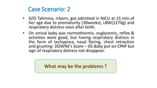


The document describes 3 case scenarios involving newborn infants with respiratory issues. Case 1 involves a term newborn not crying after birth who is not responding to positive pressure ventilation. Case 2 involves a preterm infant admitted to the NICU with respiratory distress. Case 3 involves a preterm infant with symmetrical IUGR who develops repeated apnea while on CPAP support. The document asks what the problem is in each case. It then discusses troubleshooting positive pressure ventilation and various problems that can occur, such as air leaks, obstruction, equipment issues, and abnormal blood gases. Management strategies for different problems are provided.


