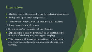
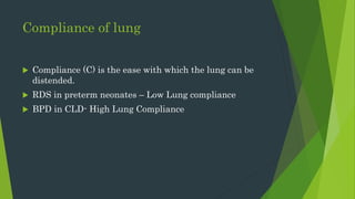
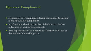
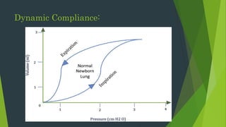
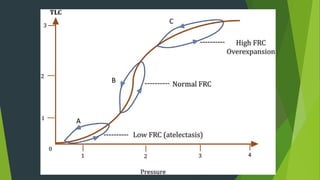
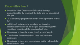
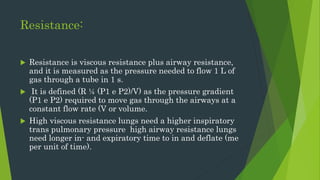
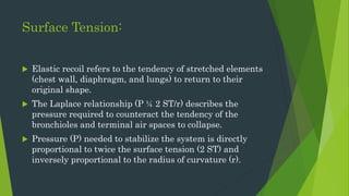
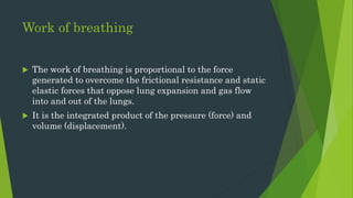
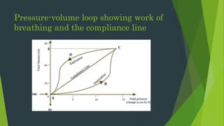
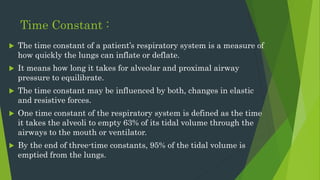
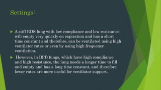
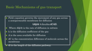
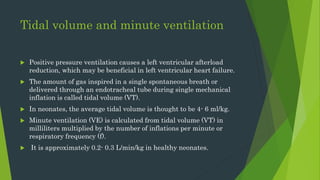
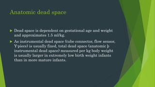
Neonatal Mechanical
Ventilation - Basics
Dr Isha Deshmukh
Assist Prof
Pediatrics
BJGMC, Pune
Why Neonatal Ventilation ?
 The goal of mechanical ventilation is to oxygenate the
baby and to remove carbon dioxide, and while doing so,
attempt to minimize damage to the lungs.
 An attempt to improve lung injury due to intra-
parenchymal lung damage.
 To overcome physiologic dead space
 To improve lung compliance.
I time
 The lung is expanded during inspiration by forces generated
primarily by the diaphragm but also by the intercostal muscles.
 Increasing i-time is one of the possible maneuvers to improve
oxygenation. Inspiration on a ventilator is driven by the flow of air
into the lungs generating pressure to open up the lungs and drive
gas (air or oxygen) into the lungs.
 Normal I –Time for Pre-term infant is 0.30 to 0.35.
 For term infant is – 0.35 to 0.45.
Expiration
 Elastic recoil is the main driving force during expiration.
 It depends upon three components;
a) surface tension produced by an air-liquid interface
b) lung tissue elastic elements
c) the structure/development of the rib cage.
 Expiration is a passive process, but an obstruction to
flow out of the lung may cause gas trapping.
 This is seen with increased secretions, inflammation,
and with tracheo/bronchomalacia as in chronic lung
disease.
Compliance of lung
 Compliance (C) is the ease with which the lung can be
distended.
 RDS in preterm neonates – Low Lung compliance
 BPD in CLD- High Lung Compliance
Static Compliance:
Dynamic Compliance:
 Measurement of compliance during continuous breathing
is called dynamic compliance.
 It reflects the elastic properties of the lung but is also
influenced by resistive components.
 It is dependent on the magnitude of airflow and thus on
the newborn’s breathing rate.
Dynamic Compliance:
Poiseuille’s law :
 Poiseuille’s law (Resistance (R) and is directly
proportional to (L) length of the tube and (n) viscosity of
the gas.
 It is inversely proportional to the fourth power of radius
(r)
 Additional resistance is noted during invasive
mechanical ventilation as the gas passes through the
ventilator circuit and the endotracheal tube.
 Resistance is linearly proportional to tube length.
 The shorter the endotracheal tube, the lower the
resistance.
 Resistance is inversely proportional to the radius of the
endotracheal tube.
Resistance:
 Resistance is viscous resistance plus airway resistance,
and it is measured as the pressure needed to flow 1 L of
gas through a tube in 1 s.
 It is defined (R ¼ (P1 e P2)/V) as the pressure gradient
(P1 e P2) required to move gas through the airways at a
constant flow rate (V or volume.
 High viscous resistance lungs need a higher inspiratory
trans pulmonary pressure high airway resistance lungs
need longer in- and expiratory time to in and deflate (me
per unit of time).
Surface Tension:
 Elastic recoil refers to the tendency of stretched elements
(chest wall, diaphragm, and lungs) to return to their
original shape.
 The Laplace relationship (P ¼ 2 ST/r) describes the
pressure required to counteract the tendency of the
bronchioles and terminal air spaces to collapse.
 Pressure (P) needed to stabilize the system is directly
proportional to twice the surface tension (2 ST) and
inversely proportional to the radius of curvature (r).
Work of breathing
 The work of breathing is proportional to the force
generated to overcome the frictional resistance and static
elastic forces that oppose lung expansion and gas flow
into and out of the lungs.
 It is the integrated product of the pressure (force) and
volume (displacement).
Pressure-volume loop showing work of
breathing and the compliance line
Time Constant :
 The time constant of a patient’s respiratory system is a measure of
how quickly the lungs can inflate or deflate.
 It means how long it takes for alveolar and proximal airway
pressure to equilibrate.
 The time constant may be influenced by both, changes in elastic
and resistive forces.
 One time constant of the respiratory system is defined as the time
it takes the alveoli to empty 63% of its tidal volume through the
airways to the mouth or ventilator.
 By the end of three-time constants, 95% of the tidal volume is
emptied from the lungs.
Settings:
 A stiff RDS lung with low compliance and low resistance
will empty very quickly on expiration and has a short
time constant and therefore, can be ventilated using high
ventilator rates or even by using high frequency
ventilation.
 However, in BPD lungs, which have high compliance
and high resistance, the lung needs a longer time to fill
and empty and has a long time constant, and therefore
lower rates are more useful for ventilator support.
Basic Mechanisms of gas transport
 Fick’s equation governs the movement of any gas across
a semipermeable membrane for diffusion
(dQ/dt ¼ k x A x dC/dl).
 Where dQ/dt is the rate of diffusion in ml/min
 k is the diffusion coefficient of the gas
 A is the area available for diffusion
 dC is the concentration difference of molecule across the
membrane
 dl is the length of the diffusion pathway.
Tidal volume and minute ventilation
 Positive pressure ventilation causes a left ventricular afterload
reduction, which may be beneficial in left ventricular heart failure.
 The amount of gas inspired in a single spontaneous breath or
delivered through an endotracheal tube during single mechanical
inflation is called tidal volume (VT).
 In neonates, the average tidal volume is thought to be 4- 6 ml/kg.
 Minute ventilation (VE) is calculated from tidal volume (VT) in
milliliters multiplied by the number of inflations per minute or
respiratory frequency (f).
 It is approximately 0.2- 0.3 L/min/kg in healthy neonates.
Anatomic dead space
 Dead space is dependent on gestational age and weight
and approximates 1.5 ml/kg.
 As instrumental dead space (tube connector, flow sensor,
Y-piece) is usually fixed, total dead space (anatomic þ
instrumental dead space) measured per kg body weight
is usually larger in extremely low birth weight infants
than in more mature infants.
Alveolar dead space
 A portion of VT may be delivered to un-perfused or under
perfused
alveoli.
 Because gas exchange does not take place in these units,
the volume that they constitute is called alveolar dead
space.
Physiologic dead space and wasted
ventilation
 Together, anatomic dead space and alveolar dead space
make up total or physiologic dead space (VDS).
 The ratio of dead space to VT (VDS/VT) defines wasted
ventilation, the proportion of tidal gas delivered that is
not involved in actual gas exchange.
 As a 23-week, 500 g preterm infant would have a larger
dead space, one would usually start at 5 ml/kg Tidal
Volume when compared to a term baby, where one would
probably aim for 4 ml/kg at the start of volume
ventilation.
Five different ways to increase mean airway
pressure (Adopted from Goldsmith
et al.).
Any maneuver to increase the area under the curve
improves oxygenation in neonates with alveolar lung
disease such as RDS.
 The maneuvers include
1) increase in inspiratory flow rate, producing a more
square-wave inspiratory pattern
2) increase peak inspiratory pressure
3) increasing the I time;
4) increasing PEEP
5) increase the ventilator rate by reducing expiratory time
without changing the inspiratory time.
 Increasing positive end-expiratory pressure (PEEP) is
probably the safest, most effective and most commonly
used way to achieve a higher mean airway pressure(
MAP) , to open lung volume and thus to improve
oxygenation in alveolar lung disease.
Ventilation
 Ventilation is the process of removal of carbon dioxide from the
lungs.
 The retention of CO2 causes respiratory acidosis and deterioration
of the pH
 Carbon dioxide removal in invasive neonatal conventional
ventilation can be achieved by increasing tidal volume (Vt) or
increasing the ventilator rate (which is controlled either directly or
by altering the inspiratory and/or expiratory time).
 Adjustments in tidal volume may be made by measuring the expired
tidal volume and changing the delta P based on that measurement.
Perfusion:
 The two types of pulmonary blood vessels seen in the lungs are
A) alveolar vessels mainly composed of capillaries with its diameter
mainly influenced by the intra-alveolar pressure and the hydrostatic
pressure in capillaries
B) extra-alveolar vessels mainly composed of arteries and veins in
interstitial tissue with its diameter mainly influenced by lung volume.
 A chest X-ray allows to detect hyper expand lungs or lung areas.
 Unexplained increases in PCO2 in an improving RDS following
surfactant could be due to hyper expansion resulting in decreased
ventilation and thus CO2- removal, and decreased blood pressure
due to decreased venous return to the heart.
Summary:
 A good understanding of basic physiology and, in
particular, concepts in neonatal lung physiology is
essential for successful respiratory support.
 This allows a physiology-based approach and application
of these concepts in daily practice for decision making
regarding the use of modes and settings of mechanical
ventilation, with the ultimate aim of providing adequate
gas exchange and minimizing lung injury.
Thank You……!!!!!!!!!

Mechanical ventilation in neonates

  • 1.
    Neonatal Mechanical Ventilation -Basics Dr Isha Deshmukh Assist Prof Pediatrics BJGMC, Pune
  • 2.
    Why Neonatal Ventilation?  The goal of mechanical ventilation is to oxygenate the baby and to remove carbon dioxide, and while doing so, attempt to minimize damage to the lungs.  An attempt to improve lung injury due to intra- parenchymal lung damage.  To overcome physiologic dead space  To improve lung compliance.
  • 3.
    I time  Thelung is expanded during inspiration by forces generated primarily by the diaphragm but also by the intercostal muscles.  Increasing i-time is one of the possible maneuvers to improve oxygenation. Inspiration on a ventilator is driven by the flow of air into the lungs generating pressure to open up the lungs and drive gas (air or oxygen) into the lungs.  Normal I –Time for Pre-term infant is 0.30 to 0.35.  For term infant is – 0.35 to 0.45.
  • 4.
    Expiration  Elastic recoilis the main driving force during expiration.  It depends upon three components; a) surface tension produced by an air-liquid interface b) lung tissue elastic elements c) the structure/development of the rib cage.  Expiration is a passive process, but an obstruction to flow out of the lung may cause gas trapping.  This is seen with increased secretions, inflammation, and with tracheo/bronchomalacia as in chronic lung disease.
  • 5.
    Compliance of lung Compliance (C) is the ease with which the lung can be distended.  RDS in preterm neonates – Low Lung compliance  BPD in CLD- High Lung Compliance
  • 6.
  • 7.
    Dynamic Compliance:  Measurementof compliance during continuous breathing is called dynamic compliance.  It reflects the elastic properties of the lung but is also influenced by resistive components.  It is dependent on the magnitude of airflow and thus on the newborn’s breathing rate.
  • 8.
  • 10.
    Poiseuille’s law : Poiseuille’s law (Resistance (R) and is directly proportional to (L) length of the tube and (n) viscosity of the gas.  It is inversely proportional to the fourth power of radius (r)  Additional resistance is noted during invasive mechanical ventilation as the gas passes through the ventilator circuit and the endotracheal tube.  Resistance is linearly proportional to tube length.  The shorter the endotracheal tube, the lower the resistance.  Resistance is inversely proportional to the radius of the endotracheal tube.
  • 11.
    Resistance:  Resistance isviscous resistance plus airway resistance, and it is measured as the pressure needed to flow 1 L of gas through a tube in 1 s.  It is defined (R ¼ (P1 e P2)/V) as the pressure gradient (P1 e P2) required to move gas through the airways at a constant flow rate (V or volume.  High viscous resistance lungs need a higher inspiratory trans pulmonary pressure high airway resistance lungs need longer in- and expiratory time to in and deflate (me per unit of time).
  • 12.
    Surface Tension:  Elasticrecoil refers to the tendency of stretched elements (chest wall, diaphragm, and lungs) to return to their original shape.  The Laplace relationship (P ¼ 2 ST/r) describes the pressure required to counteract the tendency of the bronchioles and terminal air spaces to collapse.  Pressure (P) needed to stabilize the system is directly proportional to twice the surface tension (2 ST) and inversely proportional to the radius of curvature (r).
  • 13.
    Work of breathing The work of breathing is proportional to the force generated to overcome the frictional resistance and static elastic forces that oppose lung expansion and gas flow into and out of the lungs.  It is the integrated product of the pressure (force) and volume (displacement).
  • 14.
    Pressure-volume loop showingwork of breathing and the compliance line
  • 15.
    Time Constant : The time constant of a patient’s respiratory system is a measure of how quickly the lungs can inflate or deflate.  It means how long it takes for alveolar and proximal airway pressure to equilibrate.  The time constant may be influenced by both, changes in elastic and resistive forces.  One time constant of the respiratory system is defined as the time it takes the alveoli to empty 63% of its tidal volume through the airways to the mouth or ventilator.  By the end of three-time constants, 95% of the tidal volume is emptied from the lungs.
  • 16.
    Settings:  A stiffRDS lung with low compliance and low resistance will empty very quickly on expiration and has a short time constant and therefore, can be ventilated using high ventilator rates or even by using high frequency ventilation.  However, in BPD lungs, which have high compliance and high resistance, the lung needs a longer time to fill and empty and has a long time constant, and therefore lower rates are more useful for ventilator support.
  • 17.
    Basic Mechanisms ofgas transport  Fick’s equation governs the movement of any gas across a semipermeable membrane for diffusion (dQ/dt ¼ k x A x dC/dl).  Where dQ/dt is the rate of diffusion in ml/min  k is the diffusion coefficient of the gas  A is the area available for diffusion  dC is the concentration difference of molecule across the membrane  dl is the length of the diffusion pathway.
  • 18.
    Tidal volume andminute ventilation  Positive pressure ventilation causes a left ventricular afterload reduction, which may be beneficial in left ventricular heart failure.  The amount of gas inspired in a single spontaneous breath or delivered through an endotracheal tube during single mechanical inflation is called tidal volume (VT).  In neonates, the average tidal volume is thought to be 4- 6 ml/kg.  Minute ventilation (VE) is calculated from tidal volume (VT) in milliliters multiplied by the number of inflations per minute or respiratory frequency (f).  It is approximately 0.2- 0.3 L/min/kg in healthy neonates.
  • 19.
    Anatomic dead space Dead space is dependent on gestational age and weight and approximates 1.5 ml/kg.  As instrumental dead space (tube connector, flow sensor, Y-piece) is usually fixed, total dead space (anatomic þ instrumental dead space) measured per kg body weight is usually larger in extremely low birth weight infants than in more mature infants.
  • 20.
    Alveolar dead space A portion of VT may be delivered to un-perfused or under perfused alveoli.  Because gas exchange does not take place in these units, the volume that they constitute is called alveolar dead space.
  • 21.
    Physiologic dead spaceand wasted ventilation  Together, anatomic dead space and alveolar dead space make up total or physiologic dead space (VDS).  The ratio of dead space to VT (VDS/VT) defines wasted ventilation, the proportion of tidal gas delivered that is not involved in actual gas exchange.  As a 23-week, 500 g preterm infant would have a larger dead space, one would usually start at 5 ml/kg Tidal Volume when compared to a term baby, where one would probably aim for 4 ml/kg at the start of volume ventilation.
  • 22.
    Five different waysto increase mean airway pressure (Adopted from Goldsmith et al.).
  • 23.
    Any maneuver toincrease the area under the curve improves oxygenation in neonates with alveolar lung disease such as RDS.  The maneuvers include 1) increase in inspiratory flow rate, producing a more square-wave inspiratory pattern 2) increase peak inspiratory pressure 3) increasing the I time; 4) increasing PEEP 5) increase the ventilator rate by reducing expiratory time without changing the inspiratory time.
  • 24.
     Increasing positiveend-expiratory pressure (PEEP) is probably the safest, most effective and most commonly used way to achieve a higher mean airway pressure( MAP) , to open lung volume and thus to improve oxygenation in alveolar lung disease.
  • 25.
    Ventilation  Ventilation isthe process of removal of carbon dioxide from the lungs.  The retention of CO2 causes respiratory acidosis and deterioration of the pH  Carbon dioxide removal in invasive neonatal conventional ventilation can be achieved by increasing tidal volume (Vt) or increasing the ventilator rate (which is controlled either directly or by altering the inspiratory and/or expiratory time).  Adjustments in tidal volume may be made by measuring the expired tidal volume and changing the delta P based on that measurement.
  • 26.
    Perfusion:  The twotypes of pulmonary blood vessels seen in the lungs are A) alveolar vessels mainly composed of capillaries with its diameter mainly influenced by the intra-alveolar pressure and the hydrostatic pressure in capillaries B) extra-alveolar vessels mainly composed of arteries and veins in interstitial tissue with its diameter mainly influenced by lung volume.  A chest X-ray allows to detect hyper expand lungs or lung areas.  Unexplained increases in PCO2 in an improving RDS following surfactant could be due to hyper expansion resulting in decreased ventilation and thus CO2- removal, and decreased blood pressure due to decreased venous return to the heart.
  • 27.
    Summary:  A goodunderstanding of basic physiology and, in particular, concepts in neonatal lung physiology is essential for successful respiratory support.  This allows a physiology-based approach and application of these concepts in daily practice for decision making regarding the use of modes and settings of mechanical ventilation, with the ultimate aim of providing adequate gas exchange and minimizing lung injury.
  • 28.