Downloaded 1,235 times


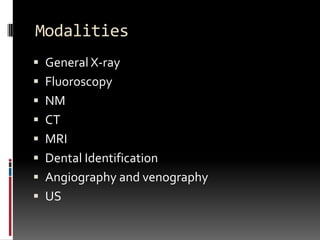

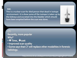


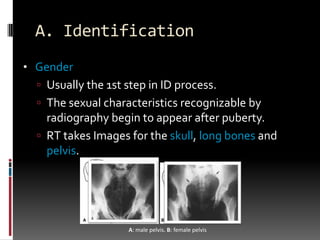


Forensic radiography uses medical imaging techniques like x-rays, CT scans, and MRIs to assist in legal investigations and identify remains. It has been used since the late 19th century. The main applications are identification of individuals by examining bones and teeth for characteristics like age, gender, and injuries or implants/prosthetics. Cause of death can also be determined by identifying foreign objects, injuries, trauma, or disease visible in images. Radiographers play a key role by properly positioning the subject and collecting high quality images that can provide evidence. As technology advances, the use of modalities like CT is increasing in forensic radiology due to benefits like improved scan quality and reduced time and costs.