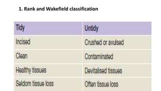
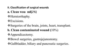
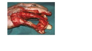
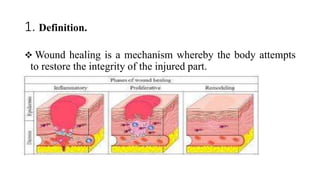
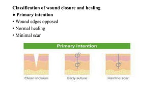
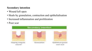
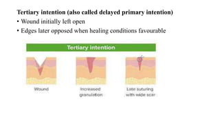
SlideShare a Scribd company logo
Surgical wound and wound
healing
Wound Definition
A wound is a break in the integrity of the skin or tissues often,
which may be associated with disruption of the structure and
function.
1. Rank and Wakefield classification
II. Classification of surgical wounds
a. Clean wou nd(2%)
Herniorrhaphy.
Excisions.
Surgeries of the brain, joints, heart, transplant.
b. Clean contaminated wound (10%)
Appendicectomy.
Bowel surgeries, gastrojejunostomy.
Gallbladder, biliary and pancreatic surgeries.
c. Contaminated wound (15–30%)
Acute abdominal conditions.
Open fresh accidental wounds.
d. Dirty infected wound (40–70%)
Abscess drainage.
Pyocele.
Empyema gallbladder.
Faecal peritonitis.
Classification based on the type of the wound
1. Clean incised wound: It is a tidy, simple, clean cut wound with
• linear cut edges; usually due to a sharp object like blade, glass
• piece or knife. It is treated by primary suturing.
Lacerated wound:
It has ragged edges with some part of the tissues getting devitalized;
viability of the tissues may be impaired;
depth of the injury and tissue damage should be carefully assessed.
Proper adequate wound excision,.
Bruise or contusion:
 It is due to blow or blunt force to the skin and tissues underneath
wherein blood vessels or capillaries are damaged underneath.
There is skin discoloration without breaking of the skin; broken
vessels cause seepage of blood underneath; minor soft tissue injury
crushes small vessels without breaking the skin accumulating trapped
blood underneath.
 It is more on the skin over the bones; lax area like face, scrotum,
eyes; vascular areas; children, old aged, fair skin people.
Haematoma:
 It is a localized collection of blood after blunt trauma or after surgery.
Collected fluid blood in few minutes to hours gets clotted; later
eventually it gets liquefied and shows discoloured fluid.
Large haematoma needs drainage under general or regional
anaesthesia.
 Small haematoma usually gets absorbed (like scalp haematoma).
Abrasion:
 It is superficial injury (scratch/graze/pressure/ contact) and is due to
shearing of the skin where the surface is rubbed off.
This tangential force causes loss of epidermis exposing dermal
vessels and nerves leading into profuse painful oozing.
Abrasion heals by epithelialization.
Traction and avulsion injuries:
 These are complex injuries with tissues getting displaced from its
normal anatomical position and alignment.
Degloving
occurs when the skin and subcutaneous fat are stripped by avulsion
from the underlying fascia, leaving neurovascular structures, tendon or
bone exposed.
1. Definition.
 Wound healing is a mechanism whereby the body attempts
to restore the integrity of the injured part.
NORMAL WOUND HEALING
This is variously described as taking place in three or four phases, the
most commonly agreed being:
1 . Hemostatic and inflammatory phase;
2 . Proliferative phase;
3 . Remodelling phase (maturing phase).
1 . Hemostasis and inflammatory phase
Begins immediately after wounding and lasts 2–3 days.
 Bleeding is followed by vasoconstriction and thrombus
formation to limit blood loss.
Platelets stick to the damaged endothelial lining of vessels,
releasing adenosine diphosphate (ADP), which causes
thrombocytic aggregates to fill the wound.
When bleeding stops, the platelets then release several cytokines from
their alpha granules.
These are platelet-derived growth factor (PDGF), platelet factor IV
and transforming growth factor beta (TGFβ).
These attract inflammatory cells such as polymorphonuclear
leukocytes (PMN) and macrophages
Platelets and the local injured tissue release vasoactive amines,
such as histamine, serotonin and prostaglandins, which increase
vascular permeability, thereby aiding infiltration of these
inflammatory cells.
Macrophages remove devitalised tissue and microorganisms
while regulating fibroblast activity in the proliferative phase of
healing.
The initial framework for structural support of cells is provided
by fibrin produced by fibrinogen.
PMNs
 are the first infiltrating cells to enter the wound site, peaking at 24
to 48 hours.
Functions:
 Primary role is phagocytosis of bacteria and tissue debris.
Major source of cytokines early during inflammation, especially
TNF-α3
TNF-α3: angiogenesis and collagen synthesis.
Release proteases (collagenases), which participate in matrix
and ground substance degradation in the early phase of wound
healing.
not appear to play a role in collagen deposition or acquisition of
mechanical wound strength
Macrophages,
participate in wound débridement via phagocytosis
The macrophage’s most pivotal function is activation and recruitment
of other cells via mediators such as cytokines and growth factors.
By releasing such mediators as TGF-β, vascular endothelial growth
factor (VEGF), insulin-like growth factor (IGF), epithelial growth
factor (EGF), and lactate, macrophages regulate cell proliferation,
matrix synthesis, and angiogenesis.
 Macrophages also play a significant role in regulating angiogenesis
and matrix deposition and remodeling
Clinicaly in inflammatory phase patient present with
1. rubor (redness),
2. Tumor (swelling),
3. calor (heat)
4. dolor (pain).
2. Proliferative phase.
Lasts from the third day to the third week,
Consisting mainly of
 fibroblast activity with the production of collagen and ground
substance(glycosaminoglycans and proteoglycans),
the growth of new blood vessels as capillary loops (angioneogenesis)
 the re-epithelialization of the wound surface.
The wound tissue formed in the early part of this phase is called
granulation tissue.
In the latter part of this phase, there is an increase in the tensile strength
of the wound due to increased collagen, which is at first deposited in a
random fashion and consists of type III collagen.
This proliferative phase with its increase of collagen deposition is
associated with wound contraction, which can considerably reduce the
surface area of a wound over the first 3 weeks of healing.
3. The remodelling phase
Characterized by maturation of collagen (type I replacing type III until a
ratio of 4:1 is achieved).
There is a realignment of collagen fibres along the lines of tension,
decreased wound vascularity, and wound contraction due to fibroblast
and myofibroblast activity.
This maturation of collagen leads to increased tensile strength in the
wound which is maximal at the 12th week post injury and represents
approximately 80% of the uninjured skin strength.
Factors influencing healing of a wound
Site of the wound
Structures involved
 Mechanism of wounding
Incision Crush
Crush avulsion
Contamination (foreign bodies/bacteria)a
Loss of tissue
Other local factors
Vascular insufficiency (arterial or venous)
Previous radiation
Pressure
● Systemic factors
Malnutrition or vitamin and mineral deficiencies
Disease (e.g. diabetes mellitus)
Medications (e.g. steroids)
Immune deficiencies (e.g. chemotherapy, AIDS)
Smoking
Bone
The phases are as above,
but periosteal and endosteal proliferation leads to the formation of callus, which is
immature bone consisting of osteoid (mineralised by hydroxyapatite and laid down
by osteoblasts).
In the remodelling phase, cortical structure and the medullary cavity are restored.
If fracture ends are accurately opposed and rigidly fixed, callus formation is minimal
and primary healing occurs.
 If a gap exists, then secondary healing may lead to delayed union, non-union or
malunion.
Nerve
Distal to the wound, Wallerian degeneration occurs.
Proximally, the nerve suffers traumatic degeneration as far as the last
node of Ranvier.
The regenerating nerve fibres are attracted to their receptors by
neurotrophism, which is mediated by growth factors, hormones and
other extracellular matrix trophins.
Nerve regeneration is characterized by profuse growth of new nerve
fibres which sprout from the cut proximal end.
Overgrowth of these, coupled with poor approximation, may lead to
neuroma formation.
Tendon
Repair follows the normal pattern of wound healing,
There are two main mechanisms whereby nutrients, cells and new
vessels reach the severed tendon.
 These are intrinsic, which consists of vincular blood flow and
synovial diffusion,
And extrinsic, which depends on the formation of fibrous
adhesions between the tendon and the tendon sheath.
The random nature of the initial collagen produced means that the
tendon lacks tensile strength for the first 3–6 weeks.
Active mobilisation prevents adhesions limiting range of motion, but
the tendon must be protected by splintage in order to avoid rupture of
the repair.
Classification of wound closure and healing
● Primary intention
• Wound edges opposed
• Normal healing
• Minimal scar
Secondary intention
• Wound left open
• Heals by granulation, contraction and epithelialisation
• Increased inflammation and proliferation
• Poor scar
Tertiary intention (also called delayed primary intention)
• Wound initially left open
• Edges later opposed when healing conditions favourable

More Related Content

Similar to wound healing [Autosaved].pptx

wound healing/cosmetic dentistry courses
wound healing/cosmetic dentistry courseswound healing/cosmetic dentistry courses
wound healing/cosmetic dentistry courses
Indian dental academy
 
Karya ilmiah ummu fix
Karya ilmiah ummu fixKarya ilmiah ummu fix
Karya ilmiah ummu fix
Dzatun Nithaqain
 
Wound healing
Wound healingWound healing
Wound healing
Harish Kumawat
 
1. WOUND & WOUND HEALING III BHMS SURGERY.ppsx
1. WOUND & WOUND HEALING III BHMS SURGERY.ppsx1. WOUND & WOUND HEALING III BHMS SURGERY.ppsx
1. WOUND & WOUND HEALING III BHMS SURGERY.ppsx
drjapu
 
4.Wound Healing.pptx
4.Wound Healing.pptx4.Wound Healing.pptx
4.Wound Healing.pptx
PradipChauhan27
 
Wound healing
Wound healingWound healing
Wound healing
zenebe teklu
 
Healing of-oral-wounds - copy
Healing of-oral-wounds - copyHealing of-oral-wounds - copy
Healing of-oral-wounds - copy
Gaurav Salunkhe
 
WOUND HEALING ZEY edited copy copy.pptx
WOUND HEALING ZEY edited copy copy.pptxWOUND HEALING ZEY edited copy copy.pptx
WOUND HEALING ZEY edited copy copy.pptx
ZeytunSomo1
 
Cuidado de heridas_en_emergencia_i
Cuidado de heridas_en_emergencia_iCuidado de heridas_en_emergencia_i
Cuidado de heridas_en_emergencia_i
Andres Felipe Jimenez Lopez
 
Wound healing
Wound healingWound healing
Wound healing
Ali Waqar Hasan
 
Wound healing
Wound healing Wound healing
Wound healing
Amphetaminesd
 
repair 2.pptx
repair 2.pptxrepair 2.pptx
repair 2.pptx
DunsonNampaso
 
Types of wounds and management.
Types of wounds and management.Types of wounds and management.
Types of wounds and management.
Abdul Wahab
 
LEC 9 Healing process.pptx
LEC 9 Healing process.pptxLEC 9 Healing process.pptx
LEC 9 Healing process.pptx
SOLOMONKIPSEREK
 
phato healing.pptx
phato healing.pptxphato healing.pptx
phato healing.pptx
Dawitashebr
 
Wound healing
Wound healingWound healing
Wound healing
Dr Alok Kumar
 
woundhealing-2.pdf
woundhealing-2.pdfwoundhealing-2.pdf
woundhealing-2.pdf
mohamedebrahim179815
 
Healing
HealingHealing
Healing
Khalid Yousuf
 
Wound repair
Wound repairWound repair
Wound repair
Avneet Soni
 
Wound healing
Wound healingWound healing
Wound healing
Asif Ansari
 

Similar to wound healing [Autosaved].pptx (20)

wound healing/cosmetic dentistry courses
wound healing/cosmetic dentistry courseswound healing/cosmetic dentistry courses
wound healing/cosmetic dentistry courses
 
Karya ilmiah ummu fix
Karya ilmiah ummu fixKarya ilmiah ummu fix
Karya ilmiah ummu fix
 
Wound healing
Wound healingWound healing
Wound healing
 
1. WOUND & WOUND HEALING III BHMS SURGERY.ppsx
1. WOUND & WOUND HEALING III BHMS SURGERY.ppsx1. WOUND & WOUND HEALING III BHMS SURGERY.ppsx
1. WOUND & WOUND HEALING III BHMS SURGERY.ppsx
 
4.Wound Healing.pptx
4.Wound Healing.pptx4.Wound Healing.pptx
4.Wound Healing.pptx
 
Wound healing
Wound healingWound healing
Wound healing
 
Healing of-oral-wounds - copy
Healing of-oral-wounds - copyHealing of-oral-wounds - copy
Healing of-oral-wounds - copy
 
WOUND HEALING ZEY edited copy copy.pptx
WOUND HEALING ZEY edited copy copy.pptxWOUND HEALING ZEY edited copy copy.pptx
WOUND HEALING ZEY edited copy copy.pptx
 
Cuidado de heridas_en_emergencia_i
Cuidado de heridas_en_emergencia_iCuidado de heridas_en_emergencia_i
Cuidado de heridas_en_emergencia_i
 
Wound healing
Wound healingWound healing
Wound healing
 
Wound healing
Wound healing Wound healing
Wound healing
 
repair 2.pptx
repair 2.pptxrepair 2.pptx
repair 2.pptx
 
Types of wounds and management.
Types of wounds and management.Types of wounds and management.
Types of wounds and management.
 
LEC 9 Healing process.pptx
LEC 9 Healing process.pptxLEC 9 Healing process.pptx
LEC 9 Healing process.pptx
 
phato healing.pptx
phato healing.pptxphato healing.pptx
phato healing.pptx
 
Wound healing
Wound healingWound healing
Wound healing
 
woundhealing-2.pdf
woundhealing-2.pdfwoundhealing-2.pdf
woundhealing-2.pdf
 
Healing
HealingHealing
Healing
 
Wound repair
Wound repairWound repair
Wound repair
 
Wound healing
Wound healingWound healing
Wound healing
 

Recently uploaded

Clinic ^%[+27633867063*Abortion Pills For Sale In Tembisa Central
Clinic ^%[+27633867063*Abortion Pills For Sale In Tembisa CentralClinic ^%[+27633867063*Abortion Pills For Sale In Tembisa Central
Clinic ^%[+27633867063*Abortion Pills For Sale In Tembisa Central
19various
 
Chapter 11 Nutrition and Chronic Diseases.pptx
Chapter 11 Nutrition and Chronic Diseases.pptxChapter 11 Nutrition and Chronic Diseases.pptx
Chapter 11 Nutrition and Chronic Diseases.pptx
Earlene McNair
 
Diabetic nephropathy diagnosis treatment
Diabetic nephropathy diagnosis treatmentDiabetic nephropathy diagnosis treatment
Diabetic nephropathy diagnosis treatment
arahmanzai5
 
Netter's Atlas of Human Anatomy 7.ed.pdf
Netter's Atlas of Human Anatomy 7.ed.pdfNetter's Atlas of Human Anatomy 7.ed.pdf
Netter's Atlas of Human Anatomy 7.ed.pdf
BrissaOrtiz3
 
Cardiac Assessment for B.sc Nursing Student.pdf
Cardiac Assessment for B.sc Nursing Student.pdfCardiac Assessment for B.sc Nursing Student.pdf
Cardiac Assessment for B.sc Nursing Student.pdf
shivalingatalekar1
 
Muscles of Mastication by Dr. Rabia Inam Gandapore.pptx
Muscles of Mastication by Dr. Rabia Inam Gandapore.pptxMuscles of Mastication by Dr. Rabia Inam Gandapore.pptx
Muscles of Mastication by Dr. Rabia Inam Gandapore.pptx
Dr. Rabia Inam Gandapore
 
Ear and its clinical correlations By Dr. Rabia Inam Gandapore.pptx
Ear and its clinical correlations By Dr. Rabia Inam Gandapore.pptxEar and its clinical correlations By Dr. Rabia Inam Gandapore.pptx
Ear and its clinical correlations By Dr. Rabia Inam Gandapore.pptx
Dr. Rabia Inam Gandapore
 
Does Over-Masturbation Contribute to Chronic Prostatitis.pptx
Does Over-Masturbation Contribute to Chronic Prostatitis.pptxDoes Over-Masturbation Contribute to Chronic Prostatitis.pptx
Does Over-Masturbation Contribute to Chronic Prostatitis.pptx
walterHu5
 
Vestibulocochlear Nerve by Dr. Rabia Inam Gandapore.pptx
Vestibulocochlear Nerve by Dr. Rabia Inam Gandapore.pptxVestibulocochlear Nerve by Dr. Rabia Inam Gandapore.pptx
Vestibulocochlear Nerve by Dr. Rabia Inam Gandapore.pptx
Dr. Rabia Inam Gandapore
 
Osteoporosis - Definition , Evaluation and Management .pdf
Osteoporosis - Definition , Evaluation and Management .pdfOsteoporosis - Definition , Evaluation and Management .pdf
Osteoporosis - Definition , Evaluation and Management .pdf
Jim Jacob Roy
 
Role of Mukta Pishti in the Management of Hyperthyroidism
Role of Mukta Pishti in the Management of HyperthyroidismRole of Mukta Pishti in the Management of Hyperthyroidism
Role of Mukta Pishti in the Management of Hyperthyroidism
Dr. Jyothirmai Paindla
 
Abortion PG Seminar Power point presentation
Abortion PG Seminar Power point presentationAbortion PG Seminar Power point presentation
Abortion PG Seminar Power point presentation
AksshayaRajanbabu
 
Aortic Association CBL Pilot April 19 – 20 Bern
Aortic Association CBL Pilot April 19 – 20 BernAortic Association CBL Pilot April 19 – 20 Bern
Aortic Association CBL Pilot April 19 – 20 Bern
suvadeepdas911
 
Ketone bodies and metabolism-biochemistry
Ketone bodies and metabolism-biochemistryKetone bodies and metabolism-biochemistry
Ketone bodies and metabolism-biochemistry
Dhayanithi C
 
Tests for analysis of different pharmaceutical.pptx
Tests for analysis of different pharmaceutical.pptxTests for analysis of different pharmaceutical.pptx
Tests for analysis of different pharmaceutical.pptx
taiba qazi
 
CHEMOTHERAPY_RDP_CHAPTER 4_ANTI VIRAL DRUGS.pdf
CHEMOTHERAPY_RDP_CHAPTER 4_ANTI VIRAL DRUGS.pdfCHEMOTHERAPY_RDP_CHAPTER 4_ANTI VIRAL DRUGS.pdf
CHEMOTHERAPY_RDP_CHAPTER 4_ANTI VIRAL DRUGS.pdf
rishi2789
 
CBL Seminar 2024_Preliminary Program.pdf
CBL Seminar 2024_Preliminary Program.pdfCBL Seminar 2024_Preliminary Program.pdf
CBL Seminar 2024_Preliminary Program.pdf
suvadeepdas911
 
REGULATION FOR COMBINATION PRODUCTS AND MEDICAL DEVICES.pptx
REGULATION FOR COMBINATION PRODUCTS AND MEDICAL DEVICES.pptxREGULATION FOR COMBINATION PRODUCTS AND MEDICAL DEVICES.pptx
REGULATION FOR COMBINATION PRODUCTS AND MEDICAL DEVICES.pptx
LaniyaNasrink
 
LOOPS in orthodontics t loop bull loop vertical loop mushroom loop stop loop
LOOPS in orthodontics t loop bull loop vertical loop mushroom loop stop loopLOOPS in orthodontics t loop bull loop vertical loop mushroom loop stop loop
LOOPS in orthodontics t loop bull loop vertical loop mushroom loop stop loop
debosmitaasanyal1
 
Histopathology of Rheumatoid Arthritis: Visual treat
Histopathology of Rheumatoid Arthritis: Visual treatHistopathology of Rheumatoid Arthritis: Visual treat
Histopathology of Rheumatoid Arthritis: Visual treat
DIVYANSHU740006
 

Recently uploaded (20)

Clinic ^%[+27633867063*Abortion Pills For Sale In Tembisa Central
Clinic ^%[+27633867063*Abortion Pills For Sale In Tembisa CentralClinic ^%[+27633867063*Abortion Pills For Sale In Tembisa Central
Clinic ^%[+27633867063*Abortion Pills For Sale In Tembisa Central
 
Chapter 11 Nutrition and Chronic Diseases.pptx
Chapter 11 Nutrition and Chronic Diseases.pptxChapter 11 Nutrition and Chronic Diseases.pptx
Chapter 11 Nutrition and Chronic Diseases.pptx
 
Diabetic nephropathy diagnosis treatment
Diabetic nephropathy diagnosis treatmentDiabetic nephropathy diagnosis treatment
Diabetic nephropathy diagnosis treatment
 
Netter's Atlas of Human Anatomy 7.ed.pdf
Netter's Atlas of Human Anatomy 7.ed.pdfNetter's Atlas of Human Anatomy 7.ed.pdf
Netter's Atlas of Human Anatomy 7.ed.pdf
 
Cardiac Assessment for B.sc Nursing Student.pdf
Cardiac Assessment for B.sc Nursing Student.pdfCardiac Assessment for B.sc Nursing Student.pdf
Cardiac Assessment for B.sc Nursing Student.pdf
 
Muscles of Mastication by Dr. Rabia Inam Gandapore.pptx
Muscles of Mastication by Dr. Rabia Inam Gandapore.pptxMuscles of Mastication by Dr. Rabia Inam Gandapore.pptx
Muscles of Mastication by Dr. Rabia Inam Gandapore.pptx
 
Ear and its clinical correlations By Dr. Rabia Inam Gandapore.pptx
Ear and its clinical correlations By Dr. Rabia Inam Gandapore.pptxEar and its clinical correlations By Dr. Rabia Inam Gandapore.pptx
Ear and its clinical correlations By Dr. Rabia Inam Gandapore.pptx
 
Does Over-Masturbation Contribute to Chronic Prostatitis.pptx
Does Over-Masturbation Contribute to Chronic Prostatitis.pptxDoes Over-Masturbation Contribute to Chronic Prostatitis.pptx
Does Over-Masturbation Contribute to Chronic Prostatitis.pptx
 
Vestibulocochlear Nerve by Dr. Rabia Inam Gandapore.pptx
Vestibulocochlear Nerve by Dr. Rabia Inam Gandapore.pptxVestibulocochlear Nerve by Dr. Rabia Inam Gandapore.pptx
Vestibulocochlear Nerve by Dr. Rabia Inam Gandapore.pptx
 
Osteoporosis - Definition , Evaluation and Management .pdf
Osteoporosis - Definition , Evaluation and Management .pdfOsteoporosis - Definition , Evaluation and Management .pdf
Osteoporosis - Definition , Evaluation and Management .pdf
 
Role of Mukta Pishti in the Management of Hyperthyroidism
Role of Mukta Pishti in the Management of HyperthyroidismRole of Mukta Pishti in the Management of Hyperthyroidism
Role of Mukta Pishti in the Management of Hyperthyroidism
 
Abortion PG Seminar Power point presentation
Abortion PG Seminar Power point presentationAbortion PG Seminar Power point presentation
Abortion PG Seminar Power point presentation
 
Aortic Association CBL Pilot April 19 – 20 Bern
Aortic Association CBL Pilot April 19 – 20 BernAortic Association CBL Pilot April 19 – 20 Bern
Aortic Association CBL Pilot April 19 – 20 Bern
 
Ketone bodies and metabolism-biochemistry
Ketone bodies and metabolism-biochemistryKetone bodies and metabolism-biochemistry
Ketone bodies and metabolism-biochemistry
 
Tests for analysis of different pharmaceutical.pptx
Tests for analysis of different pharmaceutical.pptxTests for analysis of different pharmaceutical.pptx
Tests for analysis of different pharmaceutical.pptx
 
CHEMOTHERAPY_RDP_CHAPTER 4_ANTI VIRAL DRUGS.pdf
CHEMOTHERAPY_RDP_CHAPTER 4_ANTI VIRAL DRUGS.pdfCHEMOTHERAPY_RDP_CHAPTER 4_ANTI VIRAL DRUGS.pdf
CHEMOTHERAPY_RDP_CHAPTER 4_ANTI VIRAL DRUGS.pdf
 
CBL Seminar 2024_Preliminary Program.pdf
CBL Seminar 2024_Preliminary Program.pdfCBL Seminar 2024_Preliminary Program.pdf
CBL Seminar 2024_Preliminary Program.pdf
 
REGULATION FOR COMBINATION PRODUCTS AND MEDICAL DEVICES.pptx
REGULATION FOR COMBINATION PRODUCTS AND MEDICAL DEVICES.pptxREGULATION FOR COMBINATION PRODUCTS AND MEDICAL DEVICES.pptx
REGULATION FOR COMBINATION PRODUCTS AND MEDICAL DEVICES.pptx
 
LOOPS in orthodontics t loop bull loop vertical loop mushroom loop stop loop
LOOPS in orthodontics t loop bull loop vertical loop mushroom loop stop loopLOOPS in orthodontics t loop bull loop vertical loop mushroom loop stop loop
LOOPS in orthodontics t loop bull loop vertical loop mushroom loop stop loop
 
Histopathology of Rheumatoid Arthritis: Visual treat
Histopathology of Rheumatoid Arthritis: Visual treatHistopathology of Rheumatoid Arthritis: Visual treat
Histopathology of Rheumatoid Arthritis: Visual treat
 

wound healing [Autosaved].pptx

  • 1. Surgical wound and wound healing
  • 2. Wound Definition A wound is a break in the integrity of the skin or tissues often, which may be associated with disruption of the structure and function.
  • 3. 1. Rank and Wakefield classification
  • 4. II. Classification of surgical wounds a. Clean wou nd(2%) Herniorrhaphy. Excisions. Surgeries of the brain, joints, heart, transplant. b. Clean contaminated wound (10%) Appendicectomy. Bowel surgeries, gastrojejunostomy. Gallbladder, biliary and pancreatic surgeries.
  • 5. c. Contaminated wound (15–30%) Acute abdominal conditions. Open fresh accidental wounds. d. Dirty infected wound (40–70%) Abscess drainage. Pyocele. Empyema gallbladder. Faecal peritonitis.
  • 6. Classification based on the type of the wound 1. Clean incised wound: It is a tidy, simple, clean cut wound with • linear cut edges; usually due to a sharp object like blade, glass • piece or knife. It is treated by primary suturing.
  • 7. Lacerated wound: It has ragged edges with some part of the tissues getting devitalized; viability of the tissues may be impaired; depth of the injury and tissue damage should be carefully assessed. Proper adequate wound excision,.
  • 8. Bruise or contusion:  It is due to blow or blunt force to the skin and tissues underneath wherein blood vessels or capillaries are damaged underneath. There is skin discoloration without breaking of the skin; broken vessels cause seepage of blood underneath; minor soft tissue injury crushes small vessels without breaking the skin accumulating trapped blood underneath.  It is more on the skin over the bones; lax area like face, scrotum, eyes; vascular areas; children, old aged, fair skin people.
  • 9. Haematoma:  It is a localized collection of blood after blunt trauma or after surgery. Collected fluid blood in few minutes to hours gets clotted; later eventually it gets liquefied and shows discoloured fluid. Large haematoma needs drainage under general or regional anaesthesia.  Small haematoma usually gets absorbed (like scalp haematoma).
  • 10.
  • 11. Abrasion:  It is superficial injury (scratch/graze/pressure/ contact) and is due to shearing of the skin where the surface is rubbed off. This tangential force causes loss of epidermis exposing dermal vessels and nerves leading into profuse painful oozing. Abrasion heals by epithelialization.
  • 12. Traction and avulsion injuries:  These are complex injuries with tissues getting displaced from its normal anatomical position and alignment. Degloving occurs when the skin and subcutaneous fat are stripped by avulsion from the underlying fascia, leaving neurovascular structures, tendon or bone exposed.
  • 13.
  • 14. 1. Definition.  Wound healing is a mechanism whereby the body attempts to restore the integrity of the injured part.
  • 15. NORMAL WOUND HEALING This is variously described as taking place in three or four phases, the most commonly agreed being: 1 . Hemostatic and inflammatory phase; 2 . Proliferative phase; 3 . Remodelling phase (maturing phase).
  • 16. 1 . Hemostasis and inflammatory phase Begins immediately after wounding and lasts 2–3 days.  Bleeding is followed by vasoconstriction and thrombus formation to limit blood loss. Platelets stick to the damaged endothelial lining of vessels, releasing adenosine diphosphate (ADP), which causes thrombocytic aggregates to fill the wound.
  • 17. When bleeding stops, the platelets then release several cytokines from their alpha granules. These are platelet-derived growth factor (PDGF), platelet factor IV and transforming growth factor beta (TGFβ). These attract inflammatory cells such as polymorphonuclear leukocytes (PMN) and macrophages
  • 18. Platelets and the local injured tissue release vasoactive amines, such as histamine, serotonin and prostaglandins, which increase vascular permeability, thereby aiding infiltration of these inflammatory cells. Macrophages remove devitalised tissue and microorganisms while regulating fibroblast activity in the proliferative phase of healing. The initial framework for structural support of cells is provided by fibrin produced by fibrinogen.
  • 19. PMNs  are the first infiltrating cells to enter the wound site, peaking at 24 to 48 hours. Functions:  Primary role is phagocytosis of bacteria and tissue debris. Major source of cytokines early during inflammation, especially TNF-α3 TNF-α3: angiogenesis and collagen synthesis. Release proteases (collagenases), which participate in matrix and ground substance degradation in the early phase of wound healing. not appear to play a role in collagen deposition or acquisition of mechanical wound strength
  • 20. Macrophages, participate in wound débridement via phagocytosis The macrophage’s most pivotal function is activation and recruitment of other cells via mediators such as cytokines and growth factors. By releasing such mediators as TGF-β, vascular endothelial growth factor (VEGF), insulin-like growth factor (IGF), epithelial growth factor (EGF), and lactate, macrophages regulate cell proliferation, matrix synthesis, and angiogenesis.  Macrophages also play a significant role in regulating angiogenesis and matrix deposition and remodeling
  • 21. Clinicaly in inflammatory phase patient present with 1. rubor (redness), 2. Tumor (swelling), 3. calor (heat) 4. dolor (pain).
  • 22. 2. Proliferative phase. Lasts from the third day to the third week, Consisting mainly of  fibroblast activity with the production of collagen and ground substance(glycosaminoglycans and proteoglycans), the growth of new blood vessels as capillary loops (angioneogenesis)  the re-epithelialization of the wound surface.
  • 23. The wound tissue formed in the early part of this phase is called granulation tissue. In the latter part of this phase, there is an increase in the tensile strength of the wound due to increased collagen, which is at first deposited in a random fashion and consists of type III collagen. This proliferative phase with its increase of collagen deposition is associated with wound contraction, which can considerably reduce the surface area of a wound over the first 3 weeks of healing.
  • 24. 3. The remodelling phase Characterized by maturation of collagen (type I replacing type III until a ratio of 4:1 is achieved). There is a realignment of collagen fibres along the lines of tension, decreased wound vascularity, and wound contraction due to fibroblast and myofibroblast activity. This maturation of collagen leads to increased tensile strength in the wound which is maximal at the 12th week post injury and represents approximately 80% of the uninjured skin strength.
  • 25. Factors influencing healing of a wound Site of the wound Structures involved  Mechanism of wounding Incision Crush Crush avulsion Contamination (foreign bodies/bacteria)a Loss of tissue Other local factors Vascular insufficiency (arterial or venous) Previous radiation Pressure
  • 26. ● Systemic factors Malnutrition or vitamin and mineral deficiencies Disease (e.g. diabetes mellitus) Medications (e.g. steroids) Immune deficiencies (e.g. chemotherapy, AIDS) Smoking
  • 27. Bone The phases are as above, but periosteal and endosteal proliferation leads to the formation of callus, which is immature bone consisting of osteoid (mineralised by hydroxyapatite and laid down by osteoblasts). In the remodelling phase, cortical structure and the medullary cavity are restored. If fracture ends are accurately opposed and rigidly fixed, callus formation is minimal and primary healing occurs.  If a gap exists, then secondary healing may lead to delayed union, non-union or malunion.
  • 28. Nerve Distal to the wound, Wallerian degeneration occurs. Proximally, the nerve suffers traumatic degeneration as far as the last node of Ranvier. The regenerating nerve fibres are attracted to their receptors by neurotrophism, which is mediated by growth factors, hormones and other extracellular matrix trophins. Nerve regeneration is characterized by profuse growth of new nerve fibres which sprout from the cut proximal end. Overgrowth of these, coupled with poor approximation, may lead to neuroma formation.
  • 29. Tendon Repair follows the normal pattern of wound healing, There are two main mechanisms whereby nutrients, cells and new vessels reach the severed tendon.  These are intrinsic, which consists of vincular blood flow and synovial diffusion, And extrinsic, which depends on the formation of fibrous adhesions between the tendon and the tendon sheath.
  • 30. The random nature of the initial collagen produced means that the tendon lacks tensile strength for the first 3–6 weeks. Active mobilisation prevents adhesions limiting range of motion, but the tendon must be protected by splintage in order to avoid rupture of the repair.
  • 31. Classification of wound closure and healing ● Primary intention • Wound edges opposed • Normal healing • Minimal scar
  • 32. Secondary intention • Wound left open • Heals by granulation, contraction and epithelialisation • Increased inflammation and proliferation • Poor scar
  • 33. Tertiary intention (also called delayed primary intention) • Wound initially left open • Edges later opposed when healing conditions favourable