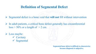
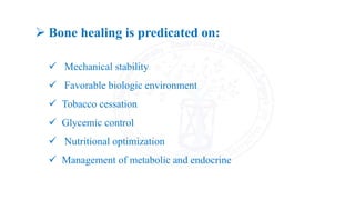
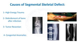
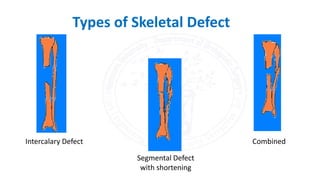
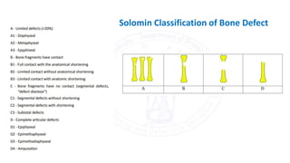
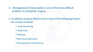
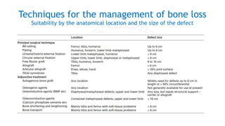
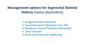
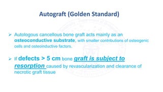
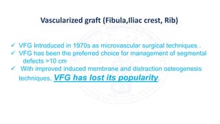
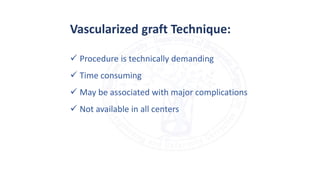
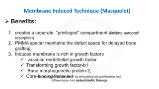
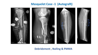
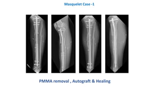
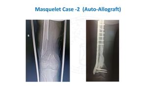
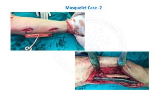
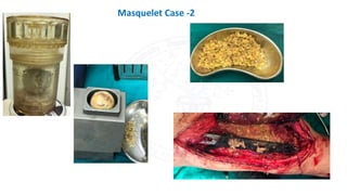
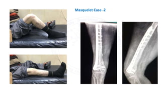
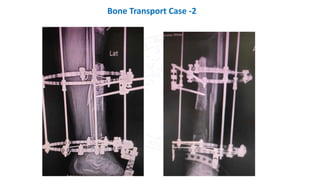
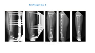
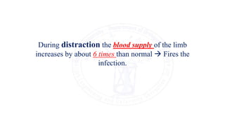
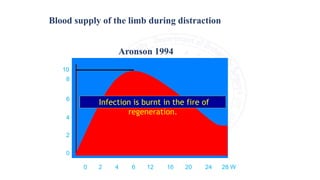
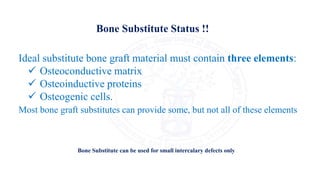
Management of Bone Defects
Abdallah El-Azanki MBBS, MSc
Trauma & Orthopedic Surgery
Clinical Fellow Limb Lengthening & Deformity Correction
Department of Orthopedic Surgery
Mansoura University
Cedar Tree of
Lebanon
 Segmental defect is a bone void that will not fill without intervention
 In adult patients, a critical bone defect generally has circumferential
loss > 50% or a length of > 2 cm.
 Loss maybe:
 Cavitary
 Segmental
Segmental bone defect is difficult to characterize
because diagnosis is subjective.
Definition of Segmental Defect
 Bone healing is predicated on:
 Mechanical stability
 Favorable biologic environment
 Tobacco cessation
 Glycemic control
 Nutritional optimization
 Management of metabolic and endocrine
4- Congenital Anomalies
Causes of Segmental Skeletal Defect:
1- High Energy Trauma
2- Debridement of bone
after infection
3- Tumor Resection
Types of Skeletal Defect
Intercalary Defect
Segmental Defect
with shortening
Combined
Solomin Classification of Bone Defect
 Management of bone defect is one of the most difficult
problem in orthopedic surgery.
 In addition to bone defect one or more of the following factors
are usually involved:
 Limb shortening
 Deformity
Infection
Soft tissue deficiency
Neurovascular insufficiency
Techniques for the management of bone loss
Suitability by the anatomical location and the size of the defect
Management options for Segmental Skeletal
Defects (status dependent)
1. Autograft (Golden Standard)
2. Vascularized graft (Fibula,Iliac crest, Rib)
3. Membrane Induced Technique (Masquelet)
4. Bone Transport
5. Acute shortening and Lengthening
 Autologous cancellous bone graft acts mainly as an
osteoconductive substrate, with smaller contributions of osteogenic
cells and osteoinductive factors.
 If defects > 5 cm bone graft is subject to
resorption caused by revascularization and clearance of
necrotic graft tissue
Autograft (Golden Standard)
Open diaphyseal fracture of the femur with comminution following
debridement and intramedullary Nailing then bone grafting and union.
Iliac Autograft
Vascularized graft (Fibula,Iliac crest, Rib)
 VFG Introduced in 1970s as microvascular surgical techniques .
 VFG has been the preferred choice for management of segmental
defects >10 cm
 With improved induced membrane and distraction osteogenesis
techniques, VFG has lost its popularity.
 Procedure is technically demanding
 Time consuming
 May be associated with major complications
 Not available in all centers
Vascularized graft Technique:
Membrane Induced Technique (Masquelet)
1. creates a separate “privileged” compartment (limiting autograft
resorption)
2. PMMA spacer maintains the defect space for delayed bone
grafting.
3. Induced membrane is rich in growth factors
 vascular endothelial growth factor
 Transforming growth factor-b1
 Bone morphogenetic protein-2,
 Core-binding factor a-1
 Benefits:
Improve graft consolidation by stimulating cell proliferation and
differentiation into osteoblastic lineage
 Bone defect is stabilized with external fixation (originally) or
internal fixation
 Second stage is typically completed 6 to 8 weeks later
 Allows for reconstruction of large segmental bone defects (5-24
cm) with a minimal number of interventions compared with other
reconstructive techniques.
Needs sufficient Autograft and in Allo-Autograft state must be in
ratio of 3:1.
The use of allograft avoids potential donor site morbidity and
overcomes the limitations in size and shape of the autograft.
 Infection
 Refracture
 Disease transmission
Debridement , Nailing & PMMA
Masquelet Case -1 (Autograft)
PMMA removal , Autograft & Healing
Masquelet Case -1
Masquelet Case -2 (Auto-Allograft)
Masquelet Case -2
Masquelet Case -2
Masquelet Case -2
Bone Transport
Ilizarov discovered this method while treating nonunions with a
fine-wire circular frame and carefully refined the technique in a
series of animal studies.
Transport can be done by many devises (Ilizarov, TSF, LRS,
Nails…)
 Transport at a rate of 1mm per day
 Once the transport segment reaches the far end of the
defect (docking site) the segment is compressed for
several weeks until fracture callus forms unless graft
needed.
 Advantages of using distraction osteogenesis:
 Reliability
 Minimal risk of further injury to soft tissues
 Ability to bear weight during the reconstruction
 No limits for the size of the defect reconstruction
 The main disadvantage is the length of time required for reconstruction (an average
of 10 to 12 months for a defect 10 cm in size) and the resultant psychological burden
on patients.
 Pintract infection in 80% of cases.
Bone Transport Case - 1
Bone Transport Case -2
Bone Transport Case -3
During distraction the blood supply of the limb
increases by about 6 times than normal  Fires the
infection.
0 2 4 6 12 16 20 24 28 W
10
8
6
4
2
0
Aronson 1994
Infection is burnt in the fire of
regeneration.
Blood supply of the limb during distraction
Ideal substitute bone graft material must contain three elements:
 Osteoconductive matrix
 Osteoinductive proteins
 Osteogenic cells.
Most bone graft substitutes can provide some, but not all of these elements
Bone Substitute Status !!
Bone Substitute can be used for small intercalary defects only
Thank You

Management of Bone Defects

  • 1.
    Management of BoneDefects Abdallah El-Azanki MBBS, MSc Trauma & Orthopedic Surgery Clinical Fellow Limb Lengthening & Deformity Correction Department of Orthopedic Surgery Mansoura University Cedar Tree of Lebanon
  • 2.
     Segmental defectis a bone void that will not fill without intervention  In adult patients, a critical bone defect generally has circumferential loss > 50% or a length of > 2 cm.  Loss maybe:  Cavitary  Segmental Segmental bone defect is difficult to characterize because diagnosis is subjective. Definition of Segmental Defect
  • 3.
     Bone healingis predicated on:  Mechanical stability  Favorable biologic environment  Tobacco cessation  Glycemic control  Nutritional optimization  Management of metabolic and endocrine
  • 4.
    4- Congenital Anomalies Causesof Segmental Skeletal Defect: 1- High Energy Trauma 2- Debridement of bone after infection 3- Tumor Resection
  • 5.
    Types of SkeletalDefect Intercalary Defect Segmental Defect with shortening Combined
  • 6.
  • 7.
     Management ofbone defect is one of the most difficult problem in orthopedic surgery.  In addition to bone defect one or more of the following factors are usually involved:  Limb shortening  Deformity Infection Soft tissue deficiency Neurovascular insufficiency
  • 8.
    Techniques for themanagement of bone loss Suitability by the anatomical location and the size of the defect
  • 9.
    Management options forSegmental Skeletal Defects (status dependent) 1. Autograft (Golden Standard) 2. Vascularized graft (Fibula,Iliac crest, Rib) 3. Membrane Induced Technique (Masquelet) 4. Bone Transport 5. Acute shortening and Lengthening
  • 10.
     Autologous cancellousbone graft acts mainly as an osteoconductive substrate, with smaller contributions of osteogenic cells and osteoinductive factors.  If defects > 5 cm bone graft is subject to resorption caused by revascularization and clearance of necrotic graft tissue Autograft (Golden Standard)
  • 11.
    Open diaphyseal fractureof the femur with comminution following debridement and intramedullary Nailing then bone grafting and union.
  • 12.
  • 13.
    Vascularized graft (Fibula,Iliaccrest, Rib)  VFG Introduced in 1970s as microvascular surgical techniques .  VFG has been the preferred choice for management of segmental defects >10 cm  With improved induced membrane and distraction osteogenesis techniques, VFG has lost its popularity.
  • 14.
     Procedure istechnically demanding  Time consuming  May be associated with major complications  Not available in all centers Vascularized graft Technique:
  • 15.
    Membrane Induced Technique(Masquelet) 1. creates a separate “privileged” compartment (limiting autograft resorption) 2. PMMA spacer maintains the defect space for delayed bone grafting. 3. Induced membrane is rich in growth factors  vascular endothelial growth factor  Transforming growth factor-b1  Bone morphogenetic protein-2,  Core-binding factor a-1  Benefits: Improve graft consolidation by stimulating cell proliferation and differentiation into osteoblastic lineage
  • 16.
     Bone defectis stabilized with external fixation (originally) or internal fixation  Second stage is typically completed 6 to 8 weeks later  Allows for reconstruction of large segmental bone defects (5-24 cm) with a minimal number of interventions compared with other reconstructive techniques. Needs sufficient Autograft and in Allo-Autograft state must be in ratio of 3:1.
  • 17.
    The use ofallograft avoids potential donor site morbidity and overcomes the limitations in size and shape of the autograft.  Infection  Refracture  Disease transmission
  • 18.
    Debridement , Nailing& PMMA Masquelet Case -1 (Autograft)
  • 19.
    PMMA removal ,Autograft & Healing Masquelet Case -1
  • 20.
    Masquelet Case -2(Auto-Allograft)
  • 21.
  • 22.
  • 23.
  • 24.
    Bone Transport Ilizarov discoveredthis method while treating nonunions with a fine-wire circular frame and carefully refined the technique in a series of animal studies. Transport can be done by many devises (Ilizarov, TSF, LRS, Nails…)  Transport at a rate of 1mm per day  Once the transport segment reaches the far end of the defect (docking site) the segment is compressed for several weeks until fracture callus forms unless graft needed.
  • 25.
     Advantages ofusing distraction osteogenesis:  Reliability  Minimal risk of further injury to soft tissues  Ability to bear weight during the reconstruction  No limits for the size of the defect reconstruction  The main disadvantage is the length of time required for reconstruction (an average of 10 to 12 months for a defect 10 cm in size) and the resultant psychological burden on patients.  Pintract infection in 80% of cases.
  • 27.
  • 28.
  • 29.
  • 30.
  • 31.
    During distraction theblood supply of the limb increases by about 6 times than normal  Fires the infection.
  • 32.
    0 2 46 12 16 20 24 28 W 10 8 6 4 2 0 Aronson 1994 Infection is burnt in the fire of regeneration. Blood supply of the limb during distraction
  • 33.
    Ideal substitute bonegraft material must contain three elements:  Osteoconductive matrix  Osteoinductive proteins  Osteogenic cells. Most bone graft substitutes can provide some, but not all of these elements Bone Substitute Status !! Bone Substitute can be used for small intercalary defects only
  • 34.