
Recommended
More Related Content
What's hot
What's hot (20)
Viewers also liked
Viewers also liked (20)
Similar to Mandible
Similar to Mandible (20)
Recently uploaded
Recently uploaded (20)
Mandible
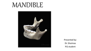
- 1. MANDIBLE Presented by: Dr. Shaimaa P.G student
- 2. CONTENTS 1. Introduction 2. Anatomy of Mandible 3. Prenatal Development of Mandible 4. Age changes in mandible 5. Postnatal growth of mandible 6. Muscle attachment 7. Nerve supply to the mandible 8. Arterial supply to the mandible 9. Developmental anomalies
- 3. Introduction • The mandible is horseshoe shaped and supports the teeth of the lower dental arch. • This bone is movable and has no bony articulation with the skull. It is the heaviest and strongest bone of the head and serves as a framework for the floor of the mouth. • It is situated immediately below the maxillary and zygomatic bone, its condyles rest in the mandibular fossa of the temporal bone. This articulation is the temporomandibular joint. • The mandible has a horizontal portion, or body, and two vertical portions, or rami. The rami join the body at an obtuse angle.
- 4. Anatomy of mandible: Mandible can be divided into 1. Body of the mandible 2. Rami 3. The alveolar process
- 5. Body consists of two lateral halves, which are joined at the median shortly after the birth, marked by a slight ridge known as symphysis. Two surfaces: - Internal - External Separated by upper and lower border. - Upper border - bears sockets for teeth - Lower border - base of mandible
- 6. Rami: Ramus ends superiorly into condyloid process, coronoid process and mandibular notch.
- 7. Internal surface of the body mandible is roughened by eminences called the superior and inferior mental spines or genial tubercles. divided into 2 portions by a well defined ridge the mylohyoid line
- 8. Alveolar process: • The border of the alveolar process outlines the alveoli of the teeth and is very thin at its anterior portion around the roots of the incisor teeth but thicker posteriorly. • The alveolar process, the superior body of the mandible, differs from the same process in the maxilla in 1 very important particular: it is not as cancellous, and instead of the facial plates being relatively thin, it is equally as heavy as the lingual plate. • Posterior to the third molar a triangular shallow fossa is outlined, it is called the retromolar triangle. The cortical plate over this fossa is not as v as the bone surrounding it, and it is more cancellous under the thin cortical plate.
- 9. Alveoli- • The central incisor alveolus is flattened on its mesial surface and is usually somewhat concave distally to accommodate the development groove on the root. • The canine alveolus is quite large and oval and, deep accommodate the root of the mandibular canine. The alveolus of the first and second premolars are similar in outline. • The socket of the first molar is divided by an inter root septum, which is strong and regular. The alveolus of the mesial root is kidney shaped
- 10. Development of the mandible: • In human beings, meckel’s cartilage has a close relationship to the developing mandible but makes no contribution to it. • At 6 weeks hyaline cartilaginous rod surrounded by a fibro cellular capsule extends from the developing ear region to the midline of the fused mandibular processes. • At 7 weeks of development, intramembraneous ossification begins in this condensation, forming the first bone of the mandible. • This spread of new bone formation occurs anteriorly along the lateral aspect of meckel’s cartilage, forming a trough that consists of lateral and medial plates that unite beneath the
- 11. • The two separate centres of ossification remains separated at the mandibular symphysis until shortly after birth. • The ramus of the mandible develops by a rapid spread of ossification posteriorly into the mesenchyme of the first arch turning away from meckel’s cartilage this point of divergence is marked by the lingual in the adult mandible, the point at which the inferior alveolar nerve enters the body of mandible. • Meckel’s cartilage has the following fate, its most posterior extremity forms the incus and malleus of the inner ear and the sphenomalleolar ligament.
- 12. The further growth of the mandible until birth is influenced strongly by the appearance of 3 secondary cartilages and the development of muscular attachments. These secondary cartilages include: • Condylar cartilage-most important • Coronoid cartilage • Symphyseal cartilage
- 13. • Develops from condylar cartilage appear as separate area of mesenchymal condensation along developing mandible around 8th week. • Cartilage fuses with mandibular ramus around 4th month. This area develop in cone- shaped cartilage around 10th week. • By the 14th week first evidence of endochondral bone formation appear in condylar region.
- 14. • Coronoid cartilage-Secondary cartilage appears in coronoid process around 10-14th week. • Cartilage grow as a response of developing temporalis muscle. • Coronoid cartilage become incorporated into expanding intramembranous bone of ramus and disappear before birth. • Symphyseal cartilage- Throughout intrauterine life left and right mandible are not fused at midline. Joined by connective tissue at midline. • On either side of symphysis, symphyseal cartilage appear between 10th & 14th week postconception. • Thus the mandible is membrane bone, developed in relation to the nerve of the first arch and almost entirely independent of meckel’s cartilage.
- 15. Age changes in the mandible At birth • The body of the bone is a mere shell, containing the sockets of the two incisor, the canine, and the two deciduous molar teeth, imperfectly partitioned off from one another. • The mandibular canal is of large size, and runs near the lower border of the bone; the mental foramen opens beneath the socket of the first deciduous molar tooth • The angle is obtuse (175°), and the condyloid portion is nearly in line with the body. • The coronoid process is of comparatively large size, and projects above the level of the condyle.
- 16. Childhood The two segments of the bone become joined at the symphysis, from below upward, in the first year; but a trace of separation may be visible in the beginning of the second year, near the alveolar margin. The body becomes elongated in its whole length, but more especially behind the mental foramen, to provide space for the three additional teeth developed in this part • thickening of the subdental portion which enables the jaw to withstand the powerful action of the masticatory muscles; • The mandibular canal, after the second dentition, is situated just above the level of the mylohyoid line; and the mental foramen occupies the position usual to it in the adult. • The angle becomes less obtuse, owing to the separation of the jaws by the teeth; about the fourth year it is 140°.
- 17. Adulthood • The alveolar and subdental portions of the body are usually of equal depth. • The mental foramen opens midway between the upper and lower borders of the bone, and the mandibular canal runs nearly parallel with the mylohyoid line. • The ramus is almost vertical in direction, the angle measuring from 110° to 120°.
- 18. Old age • The bone becomes greatly reduced in size, for with the loss of the teeth the alveolar process is absorbed, and, consequently, the chief part of the bone is below the oblique line. • The mandibular canal, with the mental foramen opening from it, is close to the alveolar border. • The ramus is oblique in direction, the angle measures about 140°, and the neck of the condyle is more or less bent backward.
- 19. Postnatal growth of mandible • Mandibular growth is combination of morphologic effect of both capsular & periosteal matrices. • Capsular matrices growth causes expansion of orofacial capsule. • Enclose macroskeletal unit (mandible) passively & secondarily translated in new position. Periosteal matrices related to mandibular microskeletal units responds to this volumetric expansion. • Such alterations in their spatial position causes them to grow. Both translation & change in form comprises totality of mandibular growth.
- 20. The main site of postnatal mandibular growth: • Condylar cartilage • Ant. & Post. Border of rami • Alveolar ridge Red arrows-bone resorption Blue arrows-bone deposition
- 21. Growth at condyle: Major site of mandibular growth. Growth of condylar cartilage increases length & height of mandible. Condylar cartilage serves as both : Articular cartilage : characterised by fibrocartilage surface. Growth cartilage : analogous to epiphyseal plate in long bone. Interstitial & appositional growth within plate produce linear movement of condyle in upward & backward direction towards temporal bone. As condylar growth cartilage moves obliquely upward & posteriorly. Entire head of condyle moves in same direction by forming new condyle behind moving cartilage. This process is continuous & condyle moves by growth. Formation of bone within condyle causes mandible rami to grow upward & backward. Displacing entire mandible in Downward &
- 22. • Condylar head is broad & neck derived from head by remodeling with marked reduction in width. • Reduction brought about by surface resorption on outer(periosteum) surface & deposition on inner(endosteum) surface • Buccal & lingual cortical plates moves inward towards each other results in reduced transverse dimension of neck.
- 23. Growth remodeling process in condylar bone follows “v” principle. Bone deposition - inner surface. Bone resorption - outer surface of V shaped neck Results in growth movement of entire V in post. & sup. direction.
- 24. Sigmoid notch: Bone deposition - post. Border of coronoid process Bone resorption - ant. Face of neck. Periosteal bone added - lingual surface of ramus just below sigmoid notch continue down from condylar head around lingual side of sigmoid notch , then extends up to apex of coronoid process.
- 25. Coronoid process: To produce backward movement of ramus : Ant. Margin of ramus & coronoid process, must undergo progressive removal. This growth change first recognized by JOHN HUNTER & later verified by HUMPHRY (1864). Forward facing ant. Border of coronoid process is resorptive around temporal crest on lingual side Coronoid process follows “v” principle. Movement of this v towards its wider ends. Bone Deposition - inner surface Bone Resorption - outer surface which bring about growth in upward & backward direction.
- 26. Growth at ramus: Bone deposition-post. border of Ramus Bone resorption-ant. border of ramus leads to AP growth of mandible • Ramus moves backward in relation to body of mandible • Buccal side-Upper part of mand. Ramus possesses a resorptive surface. Resorptive surface continuous down from neck on to upper part of ramus. Below this area deposition occur. • Lingual side- Bone deposition - part of ramus located ant. & sup. to oblique ridge extending down from neck on to ramus. Producing growth in sup. as well as in post. direction
- 27. Angle of mandible- Selective bone remodelling causes flaring of angle of mandible on age advancement. Buccal surface Bone deposition - posteroinferior surface Bone resorption - anterosuperior surface Lingual surface Bone deposition - anterosuperior surface Bone resorption - posteroinferior surface Causes flaring of angle of mandible.
- 28. Chin- • Growth of chin occurs at puberty as age advances. Chin become prominent at puberty especially in males, by selective remodelling. • Bone deposition at the mental protuberance. Cortex is thick, dense composed of slow growing type of lamellar bone.
- 29. Alveolar growth- occurs around tooth buds. As teeth develop & begin to erupt, alv. Process increases in size & height. • Continued growth of alveolar Bone increases height of mandibuar body. • Alveolar Process grows upward & outward on expanding arch. • This permits dental arch to accommodate larger permanent teeth
- 30. Muscle attachement The muscles of mastication are a group of muscles associated with movements of the jaw Embryologically, the muscles of mastication develop from the first pharyngeal arch. Consequently they are innervated by a branch of the trigeminal nerve (CN V),mandibular nerve.
- 31. Masseter ORIGIN: Ant. 2/3rd of lower border of zygomatic arch & zygomatic process of maxilla. INSERTION: Ramus & coronoid process of mandible. NERVE SUPPLY: Masseteric branch from ant. Division of mandibular nerve. ACTIONS:- Elevates mandible to close mouth. Superficial fibres Protract the mandible.
- 32. Temporalis ORIGIN: Temporal fossa INSERTION: Coronoid process & ant. Border of ramus. NERVE SUPPLY: Temporal branch from ant. Division of mandibular nerve. ACTIONS: -Elevates mandible. -Side to side grinding movement. -Post. Fibres Retract the protracted mandible.
- 33. Lateral pterygoid ORIGIN: UPPER HEAD - crest of greater wing of sphenoid LOWER HEAD - lat. surface of lat. pterygoid plate INSERTION: -Pterygoid fovea on ant. Surface of neck of mandible -Ant. Margin of articular disc & capsule of TMJ NERVE SUPPLY: - Branch of ant. division of mabdibular nerve. ACTIONS: Depresses the mandible to open mouth. Protract the mandible. Helps in grinding movement. Medial pterygoid ORIGIN: SUPERFICIAL HEAD – Tuberosity of maxilla. DEEP HEAD - Medial surface of lat. Pterygoid plate. INSERTION: postero-inferiorly to medial surface of ramus. NERVE SUPPLY- nerve to medial pterygoid. ACTIONS – Elevates mandible Protraction of the mandible. Side to side movement.
- 34. Buccinator ORIGIN: From alv. Process of maxilla & mandible, TMJ INSERTION: in the fibres of orbicularis oris NERVE SUPPLY: buccal branch of facial nerve. ACTIONS: Flattens cheek against gums &teeth. Prevents accumulatiom of food in the vestibule. aids whistling & smiling. neonates helps in suckle.
- 35. Platysma ORIGIN : subcutaneous tissue of infraclavicular & supraclavicular. INSERTION : Base of the mandible,skin of cheek & lower lip, angle of mouth. NERVE SUPPLY : Cervical branch of facial nerve. ACTIONS :- depresses mandible -Pulls angle of mouth downwards.
- 36. Mentalis ORIGIN: Incisive fossa of mandible INSERTION: skin of chin. ACTIONS: - elevates & wrinkles skin of chin. -protrude lower lip. NERVE SUPPLY: Mandibular branch of facial nerve
- 37. Mylohyoid muscle ORIGIN: Mylohyoid line of mandible. INSERTION: Post. Fibres – hyoid bone. Middle & Ant. Fibres - median raphe between mandible & hyoid bone NERVE SUPPLY : Mylohyoid nerve , from inf. Alveolar branch of mandibular nerve. . ACTIONS : Elevate floor of mouth at first stage of deglutition. depression of mandible. Elevation of hyoid bone
- 38. Nerve supply of the mandible:
- 39. Arterial supply
- 40. ANOMALIES OF DEVELOPMENT • Agnathia • Micrognathia • Macrognathia • Hemifacial macrosomia • Mandibular dyostosis
- 41. Agnathia: Agnathia (also termed hypognathous] is absence of a portion or the entirety of one or both jaws.It is a very rare condition. deficiency of neural crest tissue in lower part of face.
- 42. Micrognathia is a condition in which the jaw is undersized. It is a symptom of a variety of craniofacial conditions. Sometimes called mandibular hypoplasia, micrognathia may interfere with your child’s feeding and breathing. Micrognathia is somewhat common in infants, but often corrects itself as your child grows. In some children, micrognathia can cause abnormal tooth alignment because there is not enough room in your child’s mouth for the teeth to grow. Micrognathia can present as a birth defect in numerous syndromes, including cleft lip, cleft palate, Pierre Robin sequence or syndrome, Stickler’s syndrome, Beckwith-Wiedemann syndrome, hemifacial microsomia, Teacher Collins syndrome and others. Micrognathia can be inherited (passed on through genes)or caused by a genetic mutation.
- 43. Macrognathia Macrognathia: An abnormally large jaw. Macrognathia can be associated with pituitary gigantism, tumors, and other disorders. Macrognathia can often be corrected with surgery.
- 44. Hemifacial microsomia Hemifacial microsomia is a congenital disorder that affects the development of the lower half of the face, most commonly the ears, the mouth and the mandible. It can occur on one side of the face or both. If severe it can lead to difficulties in breathing, obstructing the trachea and requiring a tracheotomy. It is the second most common facial birth defect after clefts, with an incidence in the range of 1 in 3500 to 4500. Hemifacial microsomia shares many similarities with Treacher Collins syndrome.
- 45. Mandibular dyostosis Treacher Collins syndrome (TCS), also known as Treacher Collins–Franceschetti syndrome,[1] or mandibulofacial dysostosis,[2] is a rare autosomal dominant congenital disorder characterized by craniofacial deformities, such as absent cheekbones. Treacher Collins syndrome is found in about one in 50,000[4] births. The typical physical features include downward-slanting eyes, micrognathia (a small lower jaw), conductive hearing loss, underdeveloped zygoma, drooping part of the lateral lower eyelids, and malformed or absent ears.