The document summarizes the anatomy of the skull base. It describes the three cranial fossae - anterior, middle, and posterior. Each fossa is bounded by specific bones and contains important structures. Foramina and fissures transmitting nerves and vessels are located within the bones of each fossa. Clinical relevance is discussed for fractures of the anterior skull base, pituitary surgery involving the middle fossa, and cerebellar tonsillar herniation through the foramen magnum in the posterior fossa.
introduction to skull, parts of skull, bones involved forming skull, different views of skull, norma basalis, anterio cranial middle cranial and posterior cranial fossa, clinical aspects of cranial fossa, foramens present in the cranial fossa
4 th ventricle- Anatomical and surgical perspectivesuresh Bishokarma
4th ventricle connects the entire ventricular system of brain. Its connection with cisterns magna and cerebella pontine cistern via foramen of magenta and Luschka. CSF absorbs into the arachnoid granulation.
4 th ventricle- Anatomical and surgical perspectivesuresh Bishokarma
4th ventricle connects the entire ventricular system of brain. Its connection with cisterns magna and cerebella pontine cistern via foramen of magenta and Luschka. CSF absorbs into the arachnoid granulation.
Join live classes, download study aids, sell your documents, join or host your own classes online, get tutoring, tutor students, take practices tests and more at Examville.com
Lateral skull base anatomy and applied science by Dr, bomkar bamBomkar Bam
the lateral skull base is complex anatomy that is usually students finds difficult to understand. here concise literature is made to understand the skull base more easily.
Anti ulcer drugs and their Advance pharmacology ||
Anti-ulcer drugs are medications used to prevent and treat ulcers in the stomach and upper part of the small intestine (duodenal ulcers). These ulcers are often caused by an imbalance between stomach acid and the mucosal lining, which protects the stomach lining.
||Scope: Overview of various classes of anti-ulcer drugs, their mechanisms of action, indications, side effects, and clinical considerations.
TEST BANK for Operations Management, 14th Edition by William J. Stevenson, Ve...kevinkariuki227
TEST BANK for Operations Management, 14th Edition by William J. Stevenson, Verified Chapters 1 - 19, Complete Newest Version.pdf
Tom Selleck Health: A Comprehensive Look at the Iconic Actor’s Wellness Journeygreendigital
Tom Selleck, an enduring figure in Hollywood. has captivated audiences for decades with his rugged charm, iconic moustache. and memorable roles in television and film. From his breakout role as Thomas Magnum in Magnum P.I. to his current portrayal of Frank Reagan in Blue Bloods. Selleck's career has spanned over 50 years. But beyond his professional achievements. fans have often been curious about Tom Selleck Health. especially as he has aged in the public eye.
Follow us on: Pinterest
Introduction
Many have been interested in Tom Selleck health. not only because of his enduring presence on screen but also because of the challenges. and lifestyle choices he has faced and made over the years. This article delves into the various aspects of Tom Selleck health. exploring his fitness regimen, diet, mental health. and the challenges he has encountered as he ages. We'll look at how he maintains his well-being. the health issues he has faced, and his approach to ageing .
Early Life and Career
Childhood and Athletic Beginnings
Tom Selleck was born on January 29, 1945, in Detroit, Michigan, and grew up in Sherman Oaks, California. From an early age, he was involved in sports, particularly basketball. which played a significant role in his physical development. His athletic pursuits continued into college. where he attended the University of Southern California (USC) on a basketball scholarship. This early involvement in sports laid a strong foundation for his physical health and disciplined lifestyle.
Transition to Acting
Selleck's transition from an athlete to an actor came with its physical demands. His first significant role in "Magnum P.I." required him to perform various stunts and maintain a fit appearance. This role, which he played from 1980 to 1988. necessitated a rigorous fitness routine to meet the show's demands. setting the stage for his long-term commitment to health and wellness.
Fitness Regimen
Workout Routine
Tom Selleck health and fitness regimen has evolved. adapting to his changing roles and age. During his "Magnum, P.I." days. Selleck's workouts were intense and focused on building and maintaining muscle mass. His routine included weightlifting, cardiovascular exercises. and specific training for the stunts he performed on the show.
Selleck adjusted his fitness routine as he aged to suit his body's needs. Today, his workouts focus on maintaining flexibility, strength, and cardiovascular health. He incorporates low-impact exercises such as swimming, walking, and light weightlifting. This balanced approach helps him stay fit without putting undue strain on his joints and muscles.
Importance of Flexibility and Mobility
In recent years, Selleck has emphasized the importance of flexibility and mobility in his fitness regimen. Understanding the natural decline in muscle mass and joint flexibility with age. he includes stretching and yoga in his routine. These practices help prevent injuries, improve posture, and maintain mobilit
Ozempic: Preoperative Management of Patients on GLP-1 Receptor Agonists Saeid Safari
Preoperative Management of Patients on GLP-1 Receptor Agonists like Ozempic and Semiglutide
ASA GUIDELINE
NYSORA Guideline
2 Case Reports of Gastric Ultrasound
Pulmonary Thromboembolism - etilogy, types, medical- Surgical and nursing man...VarunMahajani
Disruption of blood supply to lung alveoli due to blockage of one or more pulmonary blood vessels is called as Pulmonary thromboembolism. In this presentation we will discuss its causes, types and its management in depth.
Lung Cancer: Artificial Intelligence, Synergetics, Complex System Analysis, S...Oleg Kshivets
RESULTS: Overall life span (LS) was 2252.1±1742.5 days and cumulative 5-year survival (5YS) reached 73.2%, 10 years – 64.8%, 20 years – 42.5%. 513 LCP lived more than 5 years (LS=3124.6±1525.6 days), 148 LCP – more than 10 years (LS=5054.4±1504.1 days).199 LCP died because of LC (LS=562.7±374.5 days). 5YS of LCP after bi/lobectomies was significantly superior in comparison with LCP after pneumonectomies (78.1% vs.63.7%, P=0.00001 by log-rank test). AT significantly improved 5YS (66.3% vs. 34.8%) (P=0.00000 by log-rank test) only for LCP with N1-2. Cox modeling displayed that 5YS of LCP significantly depended on: phase transition (PT) early-invasive LC in terms of synergetics, PT N0—N12, cell ratio factors (ratio between cancer cells- CC and blood cells subpopulations), G1-3, histology, glucose, AT, blood cell circuit, prothrombin index, heparin tolerance, recalcification time (P=0.000-0.038). Neural networks, genetic algorithm selection and bootstrap simulation revealed relationships between 5YS and PT early-invasive LC (rank=1), PT N0—N12 (rank=2), thrombocytes/CC (3), erythrocytes/CC (4), eosinophils/CC (5), healthy cells/CC (6), lymphocytes/CC (7), segmented neutrophils/CC (8), stick neutrophils/CC (9), monocytes/CC (10); leucocytes/CC (11). Correct prediction of 5YS was 100% by neural networks computing (area under ROC curve=1.0; error=0.0).
CONCLUSIONS: 5YS of LCP after radical procedures significantly depended on: 1) PT early-invasive cancer; 2) PT N0--N12; 3) cell ratio factors; 4) blood cell circuit; 5) biochemical factors; 6) hemostasis system; 7) AT; 8) LC characteristics; 9) LC cell dynamics; 10) surgery type: lobectomy/pneumonectomy; 11) anthropometric data. Optimal diagnosis and treatment strategies for LC are: 1) screening and early detection of LC; 2) availability of experienced thoracic surgeons because of complexity of radical procedures; 3) aggressive en block surgery and adequate lymph node dissection for completeness; 4) precise prediction; 5) adjuvant chemoimmunoradiotherapy for LCP with unfavorable prognosis.
ARTIFICIAL INTELLIGENCE IN HEALTHCARE.pdfAnujkumaranit
Artificial intelligence (AI) refers to the simulation of human intelligence processes by machines, especially computer systems. It encompasses tasks such as learning, reasoning, problem-solving, perception, and language understanding. AI technologies are revolutionizing various fields, from healthcare to finance, by enabling machines to perform tasks that typically require human intelligence.
These lecture slides, by Dr Sidra Arshad, offer a quick overview of physiological basis of a normal electrocardiogram.
Learning objectives:
1. Define an electrocardiogram (ECG) and electrocardiography
2. Describe how dipoles generated by the heart produce the waveforms of the ECG
3. Describe the components of a normal electrocardiogram of a typical bipolar leads (limb II)
4. Differentiate between intervals and segments
5. Enlist some common indications for obtaining an ECG
Study Resources:
1. Chapter 11, Guyton and Hall Textbook of Medical Physiology, 14th edition
2. Chapter 9, Human Physiology - From Cells to Systems, Lauralee Sherwood, 9th edition
3. Chapter 29, Ganong’s Review of Medical Physiology, 26th edition
4. Electrocardiogram, StatPearls - https://www.ncbi.nlm.nih.gov/books/NBK549803/
5. ECG in Medical Practice by ABM Abdullah, 4th edition
6. ECG Basics, http://www.nataliescasebook.com/tag/e-c-g-basics
These simplified slides by Dr. Sidra Arshad present an overview of the non-respiratory functions of the respiratory tract.
Learning objectives:
1. Enlist the non-respiratory functions of the respiratory tract
2. Briefly explain how these functions are carried out
3. Discuss the significance of dead space
4. Differentiate between minute ventilation and alveolar ventilation
5. Describe the cough and sneeze reflexes
Study Resources:
1. Chapter 39, Guyton and Hall Textbook of Medical Physiology, 14th edition
2. Chapter 34, Ganong’s Review of Medical Physiology, 26th edition
3. Chapter 17, Human Physiology by Lauralee Sherwood, 9th edition
4. Non-respiratory functions of the lungs https://academic.oup.com/bjaed/article/13/3/98/278874
- Video recording of this lecture in English language: https://youtu.be/lK81BzxMqdo
- Video recording of this lecture in Arabic language: https://youtu.be/Ve4P0COk9OI
- Link to download the book free: https://nephrotube.blogspot.com/p/nephrotube-nephrology-books.html
- Link to NephroTube website: www.NephroTube.com
- Link to NephroTube social media accounts: https://nephrotube.blogspot.com/p/join-nephrotube-on-social-media.html
Couples presenting to the infertility clinic- Do they really have infertility...Sujoy Dasgupta
Dr Sujoy Dasgupta presented the study on "Couples presenting to the infertility clinic- Do they really have infertility? – The unexplored stories of non-consummation" in the 13th Congress of the Asia Pacific Initiative on Reproduction (ASPIRE 2024) at Manila on 24 May, 2024.
Title: Sense of Taste
Presenter: Dr. Faiza, Assistant Professor of Physiology
Qualifications:
MBBS (Best Graduate, AIMC Lahore)
FCPS Physiology
ICMT, CHPE, DHPE (STMU)
MPH (GC University, Faisalabad)
MBA (Virtual University of Pakistan)
Learning Objectives:
Describe the structure and function of taste buds.
Describe the relationship between the taste threshold and taste index of common substances.
Explain the chemical basis and signal transduction of taste perception for each type of primary taste sensation.
Recognize different abnormalities of taste perception and their causes.
Key Topics:
Significance of Taste Sensation:
Differentiation between pleasant and harmful food
Influence on behavior
Selection of food based on metabolic needs
Receptors of Taste:
Taste buds on the tongue
Influence of sense of smell, texture of food, and pain stimulation (e.g., by pepper)
Primary and Secondary Taste Sensations:
Primary taste sensations: Sweet, Sour, Salty, Bitter, Umami
Chemical basis and signal transduction mechanisms for each taste
Taste Threshold and Index:
Taste threshold values for Sweet (sucrose), Salty (NaCl), Sour (HCl), and Bitter (Quinine)
Taste index relationship: Inversely proportional to taste threshold
Taste Blindness:
Inability to taste certain substances, particularly thiourea compounds
Example: Phenylthiocarbamide
Structure and Function of Taste Buds:
Composition: Epithelial cells, Sustentacular/Supporting cells, Taste cells, Basal cells
Features: Taste pores, Taste hairs/microvilli, and Taste nerve fibers
Location of Taste Buds:
Found in papillae of the tongue (Fungiform, Circumvallate, Foliate)
Also present on the palate, tonsillar pillars, epiglottis, and proximal esophagus
Mechanism of Taste Stimulation:
Interaction of taste substances with receptors on microvilli
Signal transduction pathways for Umami, Sweet, Bitter, Sour, and Salty tastes
Taste Sensitivity and Adaptation:
Decrease in sensitivity with age
Rapid adaptation of taste sensation
Role of Saliva in Taste:
Dissolution of tastants to reach receptors
Washing away the stimulus
Taste Preferences and Aversions:
Mechanisms behind taste preference and aversion
Influence of receptors and neural pathways
Impact of Sensory Nerve Damage:
Degeneration of taste buds if the sensory nerve fiber is cut
Abnormalities of Taste Detection:
Conditions: Ageusia, Hypogeusia, Dysgeusia (parageusia)
Causes: Nerve damage, neurological disorders, infections, poor oral hygiene, adverse drug effects, deficiencies, aging, tobacco use, altered neurotransmitter levels
Neurotransmitters and Taste Threshold:
Effects of serotonin (5-HT) and norepinephrine (NE) on taste sensitivity
Supertasters:
25% of the population with heightened sensitivity to taste, especially bitterness
Increased number of fungiform papillae
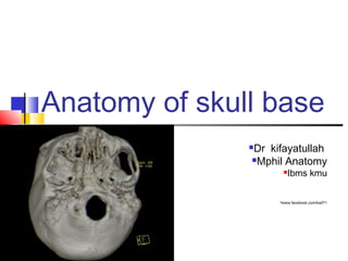
1. Anatomy of skull base
Dr kifayatullah
Mphil Anatomy
Ibms kmu
www.facebook.com/kaif71
2. Learning objectives
To identifies the bones of the skull
base as well as their boundaries.
To know the important anatomic
structures passing in and out of
base of skull
To analyze the different pathologies
which can affect this region.
3. 1) General consedration
skull with
Anterior view
Lateral view
Posterior view
Superior view
Inferior view
Cranial cavity with
Roof ( skullcap or calvaria)
Floor (base of skull)
4. 2) Structural peculiarities of the
skull base.
The skull base forms the floor of
the cranial cavity and separates
the brain from other facial
structures.
It has two views
Superior
Inferior
5. Cont……
It is formed by
2 paired bones, frontal and
temporal,
3 unpaired ethmoid,
sphenoid and occipital.
Skull base is divided into
anterior cranial fossa
Middle cranial fossa
posterior cranial fossa
6. A) ANTERIOR CRANIAL
FOSSA
a) Boundaries
It is bounded as follows:
Anteriorly and laterally anterior
limit of the anterior skull base is the
posterior wall of the frontal sinus.
Posteriorly it is bounded by the
lesser wings and body of the sphenoid
bone,
medially horizontal part (cribriform
plate), vertical part (perpendicular
plate, crista galli)
The floor consists of the frontal
bone(orbital plates), ethmoid bone and
the anterior aspects of the body and
lesser wings of the sphenoid bone
7. b) relations
Anterior cranial fossa lies above the nasal cavity seaparted from it by cribriform
plate and orbits separated from skull cavity by orbital plates of frontal bone
c) Contents
ACF lodges frontal lobes of cerebral hemisphere
It has several bony landmarks
The frontal bone has a ridge the frontal crest. It gives attachment for the
falx cerebri (a sheet of dura mater separating cerebral hemisphere )
In the midline of the ethmoid bone, the crista galli is situated.it also gives
attachment to falx cerebri.
The anterior aspect of the sphenoid bone lies within the anterior cranial
fossa. From the central body of sphenoid bone the lesser wings arise.its
rounded ends known as the anterior clinoid processes gives attachment to
tentorium cerebelli (a sheet of dura mater that divides the cerebrum from the
cerebellum).
8. d) foramen in ACF
On either side of the crista galli is thecribriform plate. It is a sheet of bone
which contains numerous small foramina – these transmit olfactory nerve
fibres (CN I) into the nasal cavity. It also contains two larger foramen:
Anterior ethmoidal foramentransmits the anterior ethmoidal artery,
nerve and vein.
Posterior ethmoidal foramentransmits the posterior ethmoidal artery,
nerve and vein.
Foramen ceacum lies between frontal and ethmoid bones transmit
emissary vein
9. d) clinical relevance
The cribriform plate of the ethmoid is
the thinnest part of the anterior cranial
fossa, and therefore most likely to
fracture. There are two major
consequences of cribriform plate
fracture:
Anosmia – the olfactory nerve fibres run
through the cribriform plate, and can be
‘sheared’, resulting in loss of sense of
smell.
CSF rhinorrhoea - the fragments of
bone can tear the meningeal coverings
of the brain, causing the leakage of
cerebrospinal fluid into the nasal cavity.
This is visible as a clear fluid.
10. A) MIDDLE CRANIAL FOSSA
It’s the central portion of cranial floor and is “butterfly
shaped”
The middle cranial fossa consists of three bones –
the sphenoid bone and the two temporal bones.
a) Boundries
Anteriorly and laterally it is bounded by the
lesser wings of the sphenoid bone.
Anteriorly and medially it is bounded by the
limbus of the sphenoid bone
Posteriorly and laterally it is bounded by the
superior border of the petrous part of the
temporal bone.
Posteriorly and medially it is bounded by the
dorsum sellae of the sphenoid bone.
The floor is formed by the body and greater wing
of the sphenoid, and the squamous and petrous
parts of the temporal bone.
11. b) Contents
The middle cranial fossa accommodates
pituitary gland and the temporal
lobes of the brain.
MCF is marked by numerous bony
landmarks
Central Part
The central part is formed by the
body of the sphenoid bone. It
contains the sella turcica, which is
a saddle-shaped area and support
the pituitary gland, it consists of
three parts:
The tuberculum sellae
The hypophysial fossa
or pituitary fossa
The dorsum sellae
12. The sella turcica is surrounded by
the anterior and posterior clinoid
processes. The anterior clinoid
processes arise from the sphenoidal
lesser wings, while the posterior
clinoid processes are the
superolateral projections of the
dorsum sellae. They serve as
attachment points for the
tentorium cerebelli.
Lateral Parts
The depressed lateral parts of
the middle cranial fossa are
formed by the greater
wings of the sphenoid bone,
and the
squamous and petrous parts
of the temporal bones
13. c) Foramen and fissures
1) Sphenoid bone
The optic canals are situated anteriorly in
the MCF.
they transmit the optic nerves (CN II) and
ophthalmic arteries into the orbital cavities.
The superior orbital fissure opens
anteriorly into the orbit. It transmits the
oculomotor nerve (CN III), trochlear nerve
(CN IV), opthalmic branch of the trigeminal
nerve (CN V1), abducens nerve (CN VI),
opthalmic veins and sympathetic fibres.
14. The foramen rotundum opens
into the pterygopalatine fossa and
transmits the maxillary branch of the
trigeminal nerve (CN V2).
The foramen ovale opens into the
infratemporal fossa, transmitting the
mandibular branch of the trigeminal
nerve (CN V3) and accessory
meningeal artery.
The foramen spinosum also
opens into the infratemporal fossa. It
transmits the middle meningeal
artery, middle meningeal vein and a
meningeal branch of CN V3.
15. 2) Temporal Bone
The temporal bone has 3 major foramina:
Hiatus of the greater petrosal
nerve – transmits the greater petrosal
nerve (facial nerve
Hiatus of the lesser petrosal nerve –
transmits the lesser petrosal nerve
(glossopharyngeal nerve).
Carotid canal – located posteriorly and
medially to the foramen ovale. This is
traversed by the internal carotid artery.
At the junction of the sphenoid, temporal
and occipital bones is the foramen
lacerum,which is pierced only by small
blood vessels.
16. C) Clinical Relevance: Pituitary Surgery
The pituitary gland lies in the sella turcica of the sphenoid bone,within the
middle cranial fossa. In cases of a pituitary tumour, it may need to removed
surgically usually endoscopic transsphenoidal surgery. An endoscope
is inserted through the nostrils, or more rarely through an incision into either
the upper lip or nasal septum. It is then advanced through the nasal cavity.
The sphenoid sinus is opened and the endoscope passes through to the
pituitary gland where it lies on the sella turcica. The tumour can then be
removed in sections.
Complications of pituitary surgery include CSF rhinorrhoea, meningitis,
diabetes insipidis, haemorrhage and visual disturbances.
17. B) POSTERIOR CRANIAL
FOSSA
The posterior cranial fossa is the most posterior
and deep of the three cranial fossae.
A) Boundaries
The posterior cranial fossa is comprised of
three bones: the occipital bone and two
temporal bones.
Anteriorly and medially it is bounded by
the dorsum sellae of the sphenoid bone.
Anteriorly and laterally it is bounded by
the superior border of the petrous part of
the temporal bone.
Posteriorly it is bounded by the internal
surface of the squamous part of the
occipital bone.
The floor consists of the mastoid part of
the temporal bone and the squamous,
condylar and basilar parts of the occipital
bone.
18. B) Contents
It accommodates the brainstem
and cerebellum..
There are several bony
landmarks and foramina
present in the posterior cranial
fossa
Temporal Bone
The internal acoustic
meatus is an oval opening in
the posterior aspect of the
petrous part of the temporal
bone. It transmits the facial
nerve (CN VII),
vestibulocochlear nerve (CN
VIII) and labrynthine artery.
19. Occipital Bone
A large opening, the foramen magnum, lies
centrally in the floor of the posterior cranial fossa
It transmits the medulla of the brain, meninges,
vertebral arteries, spinal accessory nerve
(ascending), dural veins and anterior and posterior
spinal arteries.
The jugular foramina are situated either side of
the foramen magnum.Each transmits the
glossopharyngeal nerve, vagus nerve, spinal
accessory nerve (descending), internal jugular
vein, inferior petrosal sinus, sigmoid sinus and
meningeal branches of the ascending pharyngeal
and occipital arteries.
Anterolaterally is the hypoglossal canal.
It transmits the hypoglossal nerve through the
occipital bone.
20. C) Clinical Relevance:
Cerebellar Tonsillar Herniation
Cerebellar tonsillar herniation is the
downward displacement of
the cerebellar tonsils through
the foramen magnum. It is also
known as ‘coning’.
It is produced by a raised intracranial
pressure.
Cerebellar tonsillar herniation results in
the compression of
the pons and medulla, which contain
the cardiac and respiratory centres.
Thus, a herniation of this type
ultimately results in death from
cardiorespiratory arrest.