Muscles of mastication
•Download as PPTX, PDF•
360 likes•66,819 views

The document discusses the muscles of mastication. It describes the four primary muscles - masseter, temporalis, lateral pterygoid, and medial pterygoid. It details the origin, insertion, nerve supply, blood supply, actions and functions of each muscle. The document also briefly discusses secondary muscles like the suprahyoid muscles. Clinical considerations related to the muscles of mastication like tetanus, bruxism, and myofascial pain dysfunction syndrome are mentioned at the end.

Recommended
More Related Content
What's hot
What's hot (20)
Similar to Muscles of mastication
Similar to Muscles of mastication (20)
More from DrSyed Asif
More from DrSyed Asif (6)
Recently uploaded
Recently uploaded (20)
Muscles of mastication
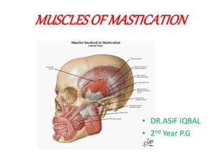
- 1. MUSCLES OF MASTICATION • DR.ASiF IQBAL • 2nd Year P.G
- 2. CONTENTS • INTRODUCTION • DEFINITIONS • MUSCLES OF MASTICATION • CLINICAL CONSIDERATIONS OF MUSCLES • REFERENCES
- 5. MASTICATION : • Rhythmic opposition and separation of jaws with the involvement of teeth ,lips ,cheeks and tongue for chewing of food in order to prepare it for swallowing and digestion. • Main purpose of mastication is to reduce the size of food particles to a size that is convenient for swallowing (bolus formation) with the help of saliva.
- 6. MUSCLE OF MASTICATION • The muscles which are required for mastication are known as the muscles of mastication, These muscles help mainly in the movement of the mandible and not the maxilla as maxilla is an integral part of the skull and the mandible being the only movable bone in the skull. • There are many muscles which help in the process of mastication but the main muscles which take part in the process are
- 7. PRIMARY MUSCLES OF MASTICATION • Masseter • Temporalis • Lateral pterygoid • Medial pterygoid
- 8. SECONDARY MUSCLES OF MASTICATION • Suprahyoid • Digastric • Stylohyoid • Mylohyoid • Geniohyoid • Infrahyoid muscles • Sternohyoid • Thyrohyoid • Omohyoid
- 9. THESE MUSCLES ATTACHED TO MANDIBLE ARE PRIMARILY RESPONSIBLE FOR : • ELEVATING • DEPRESSING • LATERAL MOVEMENT • RETRUDING
- 10. • They are funtionally classified as: Jaw elevator 1. Masseter 2. Medial pterigoid 3. Temporalis Jaw depresser 1. Lateral pterigoid 2. Digastric 3. Geniohyoid 4. Mylohyoid
- 12. LATERAL VIEW OF A FOUR WEEK EMBRYO SHOWING MUSCLES DERIVED FROM BRANCHIAL ARCHES
- 13. COMMEN CHARATERSTIC OF ALL MUSCLE OF MASTICATION All are inserted to the mandible. All are innervated by the mandibular division of the trigeminal nerve. All are concerned for biting and chewing. FUNCTIONS To move the mandible. To secure then stabilize the mandibular positions. To determine the direction of mandibular movements .
- 14. Masseter • The masseter is a thick, somewhat quadrilateral muscle, consisting of two parts, superficial and deep. The fibers of the two portions are continuous at their insertion. The masseter muscle is sometimes the target of plastic jaw reduction surgery.
- 18. • MIDDLE LAYER • Origin -anterior 2/3 of the deep surface and posterior 1/3 of the lower border of the zygomatic arch, • Insertion - middle part of ramus. • DEEP LAYER: • Origin -deep surface of the zygomatic arch, • Insertion - upper part of the ramus and into the coronoid process.
- 21. Origin : zygomatic arch and maxilla Insertion : coronoid process, ramus of mandible Artery Supply :masseteric artery Nerve supply : mandibular nerve (V3) Actions : elevation (as in closing of the mouth) and protraction of mandible
- 22. ACTIONS OF MASSETER Actions: • Elevates the mandible to close the mouth and to occlude the teeth in mastication. • Its activity in the resting position is minimal. • It has a small effect in side-to-side movement, protraction and retraction.
- 25. Palpation • The patient is asked to clench their teeth and, using both hands, the practitioner palpates the masseter muscles on both sides extraorally, making sure that the patient continues to clenchduring the procedure. • Palpate the origin of the masseter bilaterally along the zygomatic arch and continue to palpate down the body of the mandible where the masseter is attached
- 26. Palpate multiple areas of masseter muscle
- 28. Clinical Importance of Masseter Muscle of Mastication: • Masseter muscle can be palpated both intraorally and extraorally • The masseter muscle is sometimes the target of plastic jaw reduction surgery. • The muscle that commonly undergoes Hypertrophy in Bruxism is Masseter • Because of the Multipennate arrangement of fibers masseter is a very powerful muscle
- 29. TEMPORALIS
- 30. Temporalis The temporal muscle, also known as the temporalis, is one of the muscles of mastication. It covers much of the temporal bone. Structure : It arises from the temporal fossa and the deep part of temporal fascia. It passes medial to the zygomatic arch and inserts onto the coronoid process of the mandible. The temporal muscle is covered by the temporal fascia, also known as the temporal aponeurosis. The muscle is accessible on the temples, and can be seen and felt contracting while the jaw is clenching and unclenching.
- 37. Origin and Insertion: From the Parietal bone of the skull and is inserted on the coronoid process of the mandible. Arterial supply: The Deep Temporal artery supplies the large muscle. Nerve Supply: Trigeminal nerve( this nerve has been associated with being the cause of Headache and migrane. Embryology :The temporalis is derived from the first pharyngeal arch in development.
- 38. Functions: • Elevation of the mandible • Retraction of the mandible. • Crushing of food between the molars. • Posterior fibers draw the mandible backwards after it has been protruded. • It is also a contributor to side to side grinding movement.
- 39. ACTIONS OF TEMPORALIS • Elevates the mandible, this movement requires both the upward pull of anterior fibers and backward pull of the posterior fibers. • Posterior fibers draw the mandible backwards after it has been protruded. • It is also a contributory to side to side grinding movement.
- 40. SIDE TO SIDE GRINDING MOVEMENT
- 41. Palpation
- 42. Palpation • To locate the muscle ,have the patient clench. • Apply two pounds of pressure
- 44. Clinical Importance of Temporalis Muscle: • Sudden contraction of temporalis muscle will result in coronoid fracture, which is rare.
- 46. LATERAL PTERYGOID ATTACHMENTS It is a short thick muscle with two parts or head • UPPER head arise from infratemporal surface and infratemporal crest of greater wing of sphenoid bone • LOWER head arise from lateral surface of lateral pterygoid plate. • Its fibers pass backwards and laterally to be inserted into a depression (pterygoid fovea)on the front of the neck of the mandible and into the articular capsule and disc of the temporomandibular articulation.
- 50. BLOOD SUPPLY Pterygoid branch of 2nd part of maxillary artery NERVE SUPPLY Nerve to lateral pterigoid branch anterior division of trigiminal nerve
- 51. ACTIONS OF LATERAL PTERYGOID • Assists in opening the mouth with suprahyoid muscles. • Right lateral pterygoid and right medial pterygoid turns the chin to left side as a part of grinding movement. • When the medial and lateral pterygoids of two sides act together they protrude the mandible so that the lower incisors project in front of the other. • The upper (superior) head being involved in chewing
- 52. The combinded efforts of the Digastrics and Lateral Pterygoids provide for natural jaw opening.
- 53. SIDE TO SIDE GRINDING MOVEMENT
- 54. Medial and lateral pterygoid act together to protrude the mandible
- 55. Palpation of Lateral pterygoid
- 58. Medial Pterygoid muscle: • It is a thick muscle of mastication. Origin and Insertion : • It Arises lateral pterygoid plate, and from the maxillary tuberosity. • Insertion is seen on the Medial angle of the Mandible
- 60. NERVE SUPPLY • Branch of the main trunk of the mandibular nerve. BLOOD SUPPLY • Pterygoid branch of 2nd part of maxillary artery
- 61. • Functions: • Elevates the mandible. • Closes the jaw. • Helps in side to side movement.
- 64. Palpation of medial pterigoid
- 65. Palpation of medial pterygoid
- 66. • gently palpate them on the medial aspect of the jaw, • simultaneously from both inside and outside the mouth
- 67. Clinical Importance of Medial Pterygoid Muscle: • Medial Pterygoid muscle can be palpated only intraorally • Most commonly involved in MPDS • Trismus following inferior alveolar nerve block is mostly due to involvement of medial pterygoid muscle
- 68. The 4 primary muscles of mastication are in turn supported or supplemented by few secondary muscles known as SUPRAHYOID GROUP of muscles they are • DIGASTRIC • MYLOHYOID • GENIOHYOID
- 69. DIAGASTRIC MUSCLE • Two bellies united by tendon • The muscle has secondary role in mastication as a depressor muscle adding to the action of lateral pterygoid muscle when mouth is to be opened against resistance.
- 70. MYLOHYOID MUSLE • Flat triangular • The secondary role of this muscle is evident as a depressor seen in action when mouth is to be opened against resistance. • It elevates the floor of mouth to help in deglutition.
- 72. GENIOHYOID • Short and narrow musle lies above mylohyoid • When the hyoid bone is fixed, it depresses the mandible
- 74. IMPORTANT FACTS ABOUT MASTICATION • There are about 15 chews in a series from the time of food entry until swallowing • Average jaw opening during chewing is between 16-20mm • Average lateral displacement on chewing is between 3 and 5mm • Men chew faster and have a shorter occlusal phase than women, it also depends on the type of food
- 76. TETANUS(LOCK JAW) • Caused by exotoxins of gram positive bacillus Clostridium tetani. • Disease of the nervous system characterized by intense activity of motor neuron and resulting in severe muscle spasm CLINICAL FEATURES • Pain and stiffness in the jaws and neck muscles ,with muscle rigidity producing trismus and dysphagia
- 77. TREATMENT • All patients should receive antimicrobial drugs • Active and passive immunization. • Surgical wound care • Anticonvulsant if indicated
- 78. BRUXISM Bruxism : Jaw clenching, with or without forcible excursive movements, where the intensity of the clenching dictates the severity (or lack of) grinding . Clenching- It can occur as a brief rhythmic strong contractions of the jaw muscles during eccentric lateral jaw movements, or in maximum intercuspation, Causes 1) Associated with stressful events 2)Non stress related or hereditary
- 79. • Bruxism may lead to -tooth wear -fracture of the teeth or restoratrion -uncosmetic muscle hypertrophy • Treatment -coronoplasty -maxillary stabalization appliance
- 81. MYOFACIAL PAIN DYSFUNCTION SYNDROME • Pain • Muscle tenderness • Clicking in the joint • Limitation in the mouth opening TREATMENT • Physiotherapy and Myotherapeutic exercises • Transcutaneous Electronic Nerve Stimulation • Muscle relaxants • surgery
- 84. REFERENCES • B.D.Chaurasias, Human anatomy • Shafer,Hine,Textbook of oral pathology • Human anatomy A K Dutta • Grays Anatomy • Journal Refernces
- 85. THANK YOU