The document summarizes the growth and development of the mandible from prenatal to postnatal stages. During prenatal development, the mandibular arch forms and fuses in the midline to form the mandible. Ossification begins from centers on each side and spreads. The condyle and coronoid process show endochondral bone formation. Postnatally, remodeling occurs throughout the mandible through bone deposition and resorption to accommodate tooth eruption, muscle growth, and maintain articulation with the cranial base as the face grows. Growth centers like the condyle, ramus, and coronoid process contribute to mandibular lengthening and shaping through adolescence.
MANDIBULAR ANATOMICAL LANDMARKS
PRESENTED BY
ROSHALMARIA THOMAS
IV/II
THE ANATOMY OF EDENTULOUS RIDGES IN THE MAXILLA AND MANDIBLE IS VERY IMPORTANT FOR THE DESIGN OF THE COMPLETE DENTURE
THE TOTAL AREA OF SUPPORT FROM THE MANDIBLE IS SIGNIFICANTLY LESS THAN FROM THE MAXILLA.
THE AVERAGE AVAILABLE DENTURE BEARING AREA FOR AN EDENTULOUS MANDIBLE IS 14cm2 , WHEREAS FOR EDENTULOS MAXILLA IT IS 24cm2. THEREFORE THE MANDIBLE IS LESS CAPABLE OF RESISTING OCCLUSAL FORCES THAN THE MAXILLA.
Labial frenum
Fibrous band
Muscles incisivus and orbicularis oris
Active
Labial vestibule
Space between residual alveolar ridge and lips
Length and thickness of labial flange-influences lip support and retention
Buccal frenum
Overlies depressor anguli oris
Fibers of buccinators attached
Buccal vestibule
Extends- posteriorly from buccal frenum to retromolar pad region
Residual alveolar ridge on one side and buccinators on other
Influenced by action of masseter
Lingual frenum
Should be relieved
High lingual frenum is called tongue tie –affects stability
Alveololingual sulcus
Extends from lingual frenum to retromylohyoid curtain
Divided into 3 parts- anterior, middle and posterior
Anterior region- from lingual frenum to premylohyoid fossa
Flange is shorter anteriorly and should touch the floorof the mouth whentip of tongue touches upper incisors
Middle- extends from premylohyoid fossa to distal end of mylohyoid ridge
Shallower due to prominence of mylohyoid ridge and action of mylohyoid muscle
Posterior- retromylohyoid fossa
Typical S form of lingual sulcus
Retromolar pad
Posterior seal of mandibular denture
Pear shaped
Triangular keratinized soft pad of tissue at distal end of ridge
Bounded posteriorly by tendons of temporalis, laterally by buccinators and medially by pterygomandibular raphe and superior constrictor
Denture should extend one half to two thirds of retromolar pad
Buccal shelf area
Area between buccal frenum and anterior border of masseter
Width increases as resorption continues
Lies at right angles to occlusal forces- primary stress bearing area
Residual alveolar ridge
Edentulous mandible may become flat with concave denture bearing surface
In such cases, structures attaching on lingual side of ridge attach over the ridge
Due to resorption mandible inclines outwards and becomes progressively wider
Mylohyoid ridge
Runs along lingual surface of mandible
Anteriorly lies close to inferior border of mandible, posteriorly lies flush along the ridge
Thin mucosa- easily traumatized- hence should be relieved
Undercut present under the ridge
Mental foramen
Between first and second premolar region
Relieved- as pressure may cause paresthesia
Genial tubercles
Pair of bony tubercles
Present anteriorly on lingual side of body of mandible
Due to resorption may become increasingly prominent- denture usage difficult
Indian Dental Academy: will be one of the most relevant and exciting training center with best faculty and flexible training programs for dental professionals who wish to advance in their dental practice,Offers certified courses in Dental implants,Orthodontics,Endodontics,Cosmetic Dentistry, Prosthetic Dentistry, Periodontics and General Dentistry.
Neural crest cells / dental implant courses by Indian dental academy Indian dental academy
The Indian Dental Academy is the Leader in continuing dental education , training dentists in all aspects of dentistry and
offering a wide range of dental certified courses in different formats.for more details please visit
www.indiandentalacademy.com
It is a presentation in detail about the strongest structure of the oral cavity "ENAMEL". It is a simple topic but people find it difficult to learn about it. I hope my presentation is a simple method to learn about it. I would like to thank my professors for assign me this project and i learn't a lot from it and still learning my basics daily.
PHYSICAL PROPERTIES
CHEMICAL PROPERTIES
STRUCTURE OF ENAMEL
DEVELOPMENT OF ENAMEL
EPITHELIAL ENAMEL ORGAN
AMELOGENESIS
LIFE CYCLE OF AMELOBLASTS
AGE CHANGES IN ENAMEL
DEFECTS OF AMELOGENESIS
CLINICAL IMPLICATIONS
The Indian Dental Academy is the Leader in continuing dental education , training dentists in all aspects of dentistry and
offering a wide range of dental certified courses in different formats.for more details please visit
www.indiandentalacademy.com
detailed ppt on mandible, covering aspects such as anatomy, development, age changes, growth, muscle attachment, nerve and arterial supply and anomalies.
The Indian Dental Academy is the Leader in continuing dental education , training dentists in all aspects of dentistry and
offering a wide range of dental certified courses in different formats.for more details please visit
www.indiandentalacademy.com
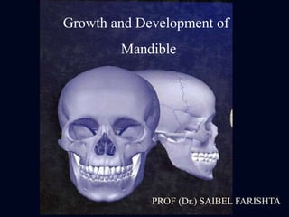
The mandible or lower jaw, is the largest & strongest bone of the face. The word “Mandible” is derived from Greek word
“mandere” – to masticate or chew. The Latin word “ mandibula” – lower jaw. It is horse-shoe shaped & the only movable bone of skull. Growth and development of an individual is divided into two periods Prenatal period and Post natal period. The first structure to develop in the primodium of the lower jaw is the mandibular division of trigeminal nerve that precedes the mesenchymal condensation forming the first [mandibular] arch. Endrocondral bone formation is seen in The condylar process, The coronoid process and The mental process. OUTER SURFACE OF MANDIBLE
1. External oblique line - origin to buccinator, depressor inferioris, depressor anguli oris.
2. Incisive fossa - origin of mentalis, mental slips of orbicularis oris.
3. Lateral surface of ramus - insertion for masseter.
4. Lower border - deep cervical fascia and platysma.
5. Postero-superior lateral surface of ramus - parotid gland.
6. Lateral surface of neck - attachment to lateral ligament of temperomandibular joint , parotid gland.
INNER SURFACE OF MANDIBLE
1. Mylohyoid line - origin to mylohyoid muscle , attachment to superior constrictor of pharynx, pterygomandibular raphae.
2. Medial surface of ramus - medial pterygoid muscle attachment.
Superior genial tubercles – genioglossus.
3. Inferior genial tubercles – origin to geniohyoid.
4. Lingula - sphenomandibular ligament.
5. Apex of coronoid process - temporalis attachment.
6. Pterygoid fovea - lateral pterygoid muscle.
7. Diagastric fossa - anterior belly of diagastric.
ARTERIAL SUPPLY OF MANDIBLE:
It is mainly divided into 2 categories :
1. Endosteal/ Central blood supply
2. Periosteal/ Peripheral blood supply
Central blood supply is via Inferior Alveolar Artery except the coronoid process which is supplied by Temporalis muscle vessels.
Inferior alveolar artery arises from maxillary artery which in turn is a branch of External carotid artery.
Inferior alveolar artery branches :
Lingual branch
Mylohyoid branch
Incisive branch
Mental branch
Peripheral blood supply is mainly via Periosteum via the nutrient vessels those penetrate the cortical bone and anastamose with the branches of Inferior alveolar artery.
VENOUS SUPPLY OF MANDIBLE
Drains into Internal Jugular vein and External Jugular vein through Maxillary vein, Facial vein and pterygoid plexus.
Growth and development of mandible in childrenDr. Harsh Shah
a brief idea about the development of mandible for indian students looking for a quick review from dentistry department
all the best to students
Presented by : Harsh SHah
Dept. of Orthodontics
SDDCH PBN
The Indian Dental Academy is the Leader in continuing dental education , training dentists in all aspects of dentistry and
offering a wide range of dental certified courses in different formats.for more details please visit
www.indiandentalacademy.com
Growth and development of mandible / dental crown & bridge coursesIndian dental academy
Description :
The Indian Dental Academy is the Leader in continuing dental education , training dentists in all aspects of dentistry and
offering a wide range of dental certified courses in different formats.for more details please visit
www.indiandentalacademy.com
PRENATAL GROWTH OF MANDIBLE
Occurs between the 4th and 7th week of intrauterine life.
4th week of intrauterine life
Formation of the head fold
Following which the developing brain and the pericardium form 2 prominent bulges on the ventral aspect of the embryo.
The 2 bulges are separated from each other by a shallow depression called stomatoedum (corresponding to the primitive mouth).
Floor of the stomatodeum is formed by the Buccopharyngeal membrane, which separates the stomatodeum from the foregut.Soon, mesoderm covering the developing forebrain proliferates, and forms a downward projection that overlaps the upper part of the stomatodeum – this downward projection is called frontonasal process.
This is a presentation by Dada Robert in a Your Skill Boost masterclass organised by the Excellence Foundation for South Sudan (EFSS) on Saturday, the 25th and Sunday, the 26th of May 2024.
He discussed the concept of quality improvement, emphasizing its applicability to various aspects of life, including personal, project, and program improvements. He defined quality as doing the right thing at the right time in the right way to achieve the best possible results and discussed the concept of the "gap" between what we know and what we do, and how this gap represents the areas we need to improve. He explained the scientific approach to quality improvement, which involves systematic performance analysis, testing and learning, and implementing change ideas. He also highlighted the importance of client focus and a team approach to quality improvement.
Ethnobotany and Ethnopharmacology:
Ethnobotany in herbal drug evaluation,
Impact of Ethnobotany in traditional medicine,
New development in herbals,
Bio-prospecting tools for drug discovery,
Role of Ethnopharmacology in drug evaluation,
Reverse Pharmacology.
Unit 8 - Information and Communication Technology (Paper I).pdfThiyagu K
This slides describes the basic concepts of ICT, basics of Email, Emerging Technology and Digital Initiatives in Education. This presentations aligns with the UGC Paper I syllabus.
Students, digital devices and success - Andreas Schleicher - 27 May 2024..pptxEduSkills OECD
Andreas Schleicher presents at the OECD webinar ‘Digital devices in schools: detrimental distraction or secret to success?’ on 27 May 2024. The presentation was based on findings from PISA 2022 results and the webinar helped launch the PISA in Focus ‘Managing screen time: How to protect and equip students against distraction’ https://www.oecd-ilibrary.org/education/managing-screen-time_7c225af4-en and the OECD Education Policy Perspective ‘Students, digital devices and success’ can be found here - https://oe.cd/il/5yV
Read| The latest issue of The Challenger is here! We are thrilled to announce that our school paper has qualified for the NATIONAL SCHOOLS PRESS CONFERENCE (NSPC) 2024. Thank you for your unwavering support and trust. Dive into the stories that made us stand out!
Synthetic Fiber Construction in lab .pptxPavel ( NSTU)
Synthetic fiber production is a fascinating and complex field that blends chemistry, engineering, and environmental science. By understanding these aspects, students can gain a comprehensive view of synthetic fiber production, its impact on society and the environment, and the potential for future innovations. Synthetic fibers play a crucial role in modern society, impacting various aspects of daily life, industry, and the environment. ynthetic fibers are integral to modern life, offering a range of benefits from cost-effectiveness and versatility to innovative applications and performance characteristics. While they pose environmental challenges, ongoing research and development aim to create more sustainable and eco-friendly alternatives. Understanding the importance of synthetic fibers helps in appreciating their role in the economy, industry, and daily life, while also emphasizing the need for sustainable practices and innovation.
2. Growth and development of
mandible
• Pre-natal embryology of mandible
• Post-natal growth of mandible
3. Pre – natal embryology of mandible
• Around the fourth
week of intra uterine
life, a prominent bilge
appears on the ventral
aspect of embryo
corresponding to the
developing brain.
4. • Below the bulge a
shallow depression
which corresponds to
the primitive mouth
appears called
Stomodeum.
5. • The floor of the
stomodeum is formed
by the
Buccopharyngeal
membrane which
seperates the
stomodeum from the
foregut.
6. • By around the fourth
week of intra-uterine
life, Five branchial
arches form in the
region of the future
head and neck.
7. • The first pharyngeal
arch is called the
Mandibular arch and
plays an important
role in the
development of naso-
maxillary region.
8. • The mesoderm
covering the
developing forebrain
proliferates and forms
a downward projection
called Fronto-nasal
Process.
9. The mandibular arches of both the sides form the
lateral walls of the stomodeum.
• The mandibular arch
gives of a bud from its
dorsal end called the
Maxillary process.
10. • The maxillary process
grows ventro-medio-
cranial to the main
part of the mandibular
arch which is now
called the
Mandibular process.
11. • Thus at this stage the
primitive mouth is
overlapped from
above by the fronto-
nasal process, below
by the mandibular
process and on either
side by the maxillry
process.
12. • The two mandibular processes grow medially and
fuse to form the lower lip and the lower jaw i,e the
Mandible.
13. Meckel’s Cartilage
• The Meckel’s cartilage is derived from the first
branchial arch around the 41st – 45th day of intra-
uterine life. It extends from the cartilaginous otic
capsule to the midline or symphysis and provides
template for guiding the growth of the mandible.
14. • A major portion of the Meckel’s Cartilage
disappears during growth and the remaining part
develops into the following structures:
1) The mental ossicles
2) Incus and malleus
3) Spine of sphenoid bone
4) Anterior ligament of malleus
5) Spheno-mandibular ligament
15. • The first structure to develop in the primordium of
the lower jaw is the mandibular division of the
trigeminal nerve. This is followed by the
mesenchymal condenstion forming the first
branchial arch.
• Neurotrophic factors produced by the nerve induce
osteogenesis in the ossification centers.
16. • A single ossification center for each half of the
mandible arises in the 6th week of intra-uterine life
in the region of the bifurcation of the inferior
alveolar nerve into mental and incisive branches.
17. • The ossifying membrane is located lateral to the
Meckel’s Cartilage and its accompanying neuro-
vascular bundle.
• From this primary center, ossification spreads
below and around the inferior alveolar nerve and
its incisive branch and upwards to form trough for
accommodating the developing tooth buds.
• Spread of the intramembranous ossification
dorsally and ventrally forms the body and ramus
of the mandible.
18. • As ossification continues, the meckel’s cartilage
becomes surrounded and invaded by the bone.
• Ossification stops at the site that will later become
the mandibular lingula from where the Meckel’s
cartilage continues into the middle ear and
develops into the auditory ossicles i,e Malleus and
Incus.
• The spheno-mandibular ligament which extends
from the lingula of the mandible to the sphenoid
bone also forms a remnant of the Meckel’s
cartilage.
19. Endochondral bone formation
• Endochondral bone formation is seen only in 3
areas of the mandible.
1) The Condylar Process.
2) The Coronoid Process.
3) The Mental Region.
20. Condylar Process
• At about the 5th week of intra-uterine life, an area
of mesenchymal condensation can be seen above
the ventral part of the developing mandible.
• This develops into a cone – shaped cartilage by
about 10th week and starts ossification by 14th
week.
21. • It then migrates inferiorly and fuses with the
mandibular ramus by about 4 months.
• Much of the cone-shaped cartilage is replaced by
bone by the middle of fetal life but its upper end
persists into adulthood acting as a growth cartilage
and an articular cartilage.
22. Coronoid Process
• Secondary accessory cartilages appear in the
region of the coronoid process by about the 10th-
14th week of intra-uterine life. This secondary
cartilage of coronoid process is believed to grow
as a response to the developing temporalis muscle.
• The coronoid accessory cartilage becomes
incorporated into the expanding intramembranous
bone of the ramus and disappears before birth.
23. The Mental Region
• In the mental region, on either side of the
symphysis, one or two small cartilages appear and
ossify in the 7th month of intra-uterine life to form
the variable numbers of mental ossicles in the
fibrous tissues of the symphysis.
• These ossicles become incorporated into the
intramembranous bone when the sympysis ossifies
completely during the first year of post-natal life.
24. Post - natal growth of mandible
• Of the facial bones, the mandible undergoes
largest amount of growth post-natally and also
exhibits the largest variability in morphology.
• While the mandible appears in the adult as a single
bone, it is developmentally and functionally
divisible into several skeletal sub-units.
• Thus the study of post-natal growth of the
mandible is made easier and more meaningful
when each of the developmental and functional
parts are considered separate.
25. The Ramus of Mandible
• The Ramus moves progressively posterior by a
combination of deposition and resorption.
• Resorption occurs on the anterior Part of the
ramus while bone deposition occurs on the
posterior region.
• This results in a DRIFT of the ramus in a posterior
direction.
26. • The functions of remodeling of the ramus are :
1) To accommodate the increasing mass of
masticatory muscles inserted into it.
2) To accommodate the enlarged breadth of the
the pharyngeal space.
3) To facilitate the lengthening of the mandibular
body, which in turn accommodates the erupting
molars.
27. Corpus or Body of Mandible
• Displacement of the ramus in a posterior direction
by remodeling process results in lengthening of
the body of the mandible.
• Thus the additional space made available by
means of resorption of the anterior border of the
ramus is made use of to accommodate the erupting
permanent molars.
28. Angle of the Mandible
• On the lingual side of
the angle of the
mandible, resorption
takes place on the
posterio-inferior
aspect while
deposition occurs on
the antero-superior
aspect.
29. • On the buccal side, resorption occurs on the
antero-superior part while deposition takes place
on the postero-inferior part
30. The Lingual Tuberosity
• The Lingual tuberosity is a direct equivalent of the
maxillary tuberosity, which forms a major site of
growth for the lower bony arch. It forms the
boundary between the ramus and the body.
• The lingual tuberosity moves posteriorly by
deposition on its posteriorly facing surface.
31. • It can be noticed that the lingual tuberosity
protrudes noticeably in a lingual direction and that
it lies well towards the midline of the ramus.
• The prominence of the tuberosity is increased by
the presence of a large resorption field just below
it. This resorption field produces a sizable
depression, the lingual fossa.
32. The Alveolar Process
• This develops in response to the presence of the
tooth buds. As the tooth erupt the alveolar process
develops and increases in height by bone
deposition at the margins.
• The alveolar bone adds to the height and the
thickness of the body of the mandible and is
particularly manifested as a ledge extending
lingual to the ramus to accommodate the third
molars.
33. The Chin
• The chin is a specific human characteristic and is
found in its fully developed form in recent man
only.
• In infancy, the chin is usually underdeveloped. As
age advances the growth of the chin becomes
significant.
• It is influenced by sexual and specific genetic
factors. Usually males are seen to have prominent
chins as compared to females.
34. • The mental protuberance forms by bone
deposition during childhood. Its prominence is
accentuated by bone resorption that occurs in the
alveolar region above it, creating a concavity.
• The deepest point in this concavity is known as
“Point B” in cephalometric terminology.
35. The Condyle
• The mandibular condyle has been recognised as an
important growth site. The head of the condyle is
covered by a thin layer of cartilage called the
Condylar cartilage.
• The presence of this cartilage is an adaptation to
withstand the compression that occurs at the joint.
36. • The role of the condyle in the growth of mandible
has remained a controversy. There are two schools
of thought regarding the role of the condyle.
1) It was earlier believed that the growth occurs at
the surface of the condylar cartilage by means
of bone deposition. Thus the Condyle grows
towards the cranial base, the entire mandible
gets displaced forwards and downwards.
37. 2) It is now believed that the growth of soft tissues
including the muscles and connective tissue
carries the mandible forwards away from the
cranial base. Bone growth follows secondarily at
the condyle to maintain constant contact with the
cranial base.
38. • The condylar growth rate increases at puberty
reaching a peak between 12 1/2 – 14 years. The
growth ceases around 20 years of age.
39. The Coronoid process
• The growth of the coronoid process follows the
enlarging “V” principle.
• Viewing, the longitudinal section of the coronoid
process from the posterior aspect, it can be seen
that deposition occurs on the lingual (medial)
surfaces of the left and right coronoid process.
40. • Viewing it from the occlusal aspect, the deposition
on the lingual of the coronoid process brings about
a posterior growth movement in the “V” pattern.
• Briefly the coronoid process has a propeller like
twist, so that its lingual side faces three general
directions all at once i,e posteriorly,superiorly and
medially.