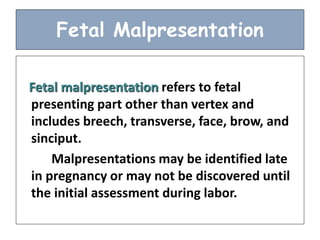
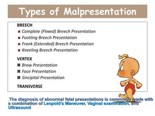
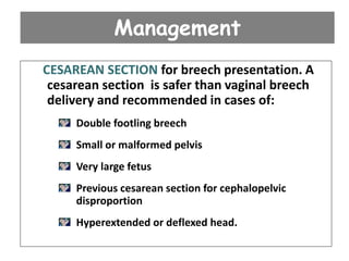
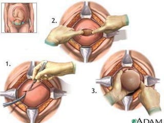
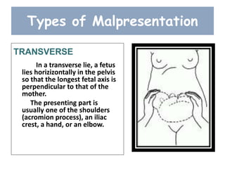
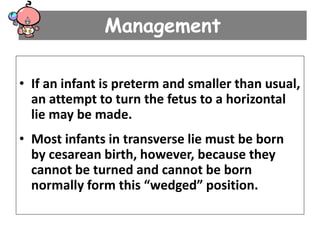
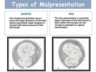
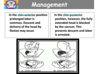
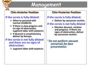
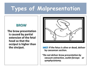
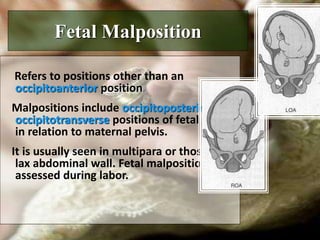
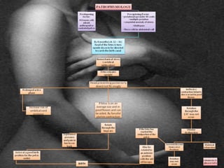
FETAL MALPRESENTATION
and
MALPOSITION
Fetal Malpresentation
Fetal malpresentation refers to fetal
presenting part other than vertex and
includes breech, transverse, face, brow, and
sinciput.
Malpresentations may be identified late
in pregnancy or may not be discovered until
the initial assessment during labor.
Related Factors
• The woman has had more than one
pregnancy
• There is more than one fetus in the
uterus
• The uterus has too much or too little
amniotic fluid
• The uterus is not normal in shape or
has abnormal growths, such as
fibroids
• placenta previa
• The baby is preterm
Types of Malpresentation
BREECH
Complete (Flexed) Breech Presentation
Footling Breech Presentation
Frank (Extended) Breech Presentation
Kneeling Breech Presentation
VERTEX
Brow Presentation
Face Presentation
Sincipital Presentation
TRANSVERSE
The diagnosis of abnormal fetal presentations is commonly made with
a combination of Leopold’s Maneuver, Vaginal examination, and
Ultrasound
Types of Malpresentation
BREECH
Breech presentation means that either the buttocks
or the feet are the first body parts that will contact the
cervix.
Breech presentations occurs in approximately 3% of
the births and are affected by fetal attitude.
Breech presentations can be difficult births, with the
presenting point influencing the degree of difficulty.
Types of Breech Presentation
Frank breech
The baby's bottom comes
first, and the legs are flexed at
the hip and extended at the
knees (with feet near the ears).
65-70% of breech babies are
in the frank breech position.
Complete Breech
The baby's hips and knees are
flexed so that the baby is sitting
crosslegged, with feet beside the
bottom.
Types of Breech
Presentation
Footling Breech
One or both feet come first,
with the bottom at a higher
position. This is rare at term
but relatively common with
premature fetuses.
Kneeling Breech
The baby is in a kneeling
position, with one or both
legs extended at the hips
and flexed at the knees.
This is extremely rare.
Maternal Risks
Prolonged labor r/t decreased pressure exerted
by the breech on the cervix.
PROM may expose client to infection.
Cesarean or forceps delivery.
Trauma to birth canal during delivery from
manipulation and forceps to free the fetal
head.
Intrapartum or postpartum hemorrhage.
Fetal Risks:
Compression or prolapse of umbilical cord.
Entrapment of fetal head in incompletely
dilated cervix.
Aspiration and asphyxia at birth.
Birth trauma from manipulation and forceps
to free the fetal head.
Management
If the woman is in early labor and the
membranes are intact, attempt External
Cephalic Version.
Tocolytics, such as Terbutaline 0.25 mg IM,
can be used before ECV to help relax the
uterus.
If ECV is successful, proceed with normal
childbirth. If EVC fails or is not advisable,
deliver by caesarean section.
Management
Attempt external version if:
Breech presentation is present at or after 37 weeks
(before 37 weeks, a successful version is more likely
spontaneously revert back to breech presentation)
Vaginal delivery is possible
Membranes are intact and amniotic fluid is adequate;
There are no complications (e.g. fetal growth restriction,
uterine bleeding, previous caesarean delivery, fetal
abnormalities, twin pregnancy, HPN, fetal death).
Management
VAGINAL BREECH DELIVERY. A vaginal breech
delivery by a skilled health care provider is safe
and feasible under the following conditions:
- complete or frank breech
- adequate clinical pelvimetry
- fetus is not too large
- no previous caesarean section for
cephalopelvic disproportion
- flexed head.
Management
CESAREAN SECTION for breech presentation. A
cesarean section is safer than vaginal breech
delivery and recommended in cases of:
Double footling breech
Small or malformed pelvis
Very large fetus
Previous cesarean section for cephalopelvic
disproportion
Hyperextended or deflexed head.
Types of Malpresentation
TRANSVERSE
In a transverse lie, a fetus
lies horizizontally in the pelvis
so that the longest fetal axis is
perpendicular to that of the
mother.
The presenting part is
usually one of the shoulders
(acromion process), an iliac
crest, a hand, or an elbow.
Management
• If an infant is preterm and smaller than usual,
an attempt to turn the fetus to a horizontal
lie may be made.
• Most infants in transverse lie must be born
by cesarean birth, however, because they
cannot be turned and cannot be born
normally form this “wedged” position.
Types of Malpresentation
SINCIPUT
The sinciput presentation occurs
when the larger diameter of the fetal
head is presented. Labor progress is
slowed with slower descent of the
fetal head.
FACE
The face presentation is caused by
hyper-extension of the fetal head so
that neither the occiput nor the
sinciput is palpable on vaginal
examination.
Management
In the chin-anterior position
prolonged labor is
common. Descent and
delivery of the head by
flexion may occur.
In the chin-posterior
position, however, the fully
extended head is blocked
by the sacrum. This
prevents descent and labor
is arrested.
Management
Chin-Anterior Position
If the cervix is fully dilated:
 Allow to proceed with
normal childbirth;
 If there is slow progress and
no sign of obstruction,
augment labor with oxytocin;
 If descent is unsatisfactory,
deliver by forceps.
If the cervix is not fully dilated
and there are no signs of
obstruction:
 augment labor with oxytocin.
Chin-Posterior Position
If the cervix is fully dilated:
 Deliver by caesarean section.
If the cervix is not fully dilated
 Monitor descent, rotation
and progress. If there are
signs of obstruction, deliver
by caesarean section.
*Do not perform vacuum
extraction for face
presentation.
Types of Malpresentation
BROW
The brow presentation
is caused by partial
extension of the fetal
head so that the
occiput is higher than
the sinciput. MGT: If the fetus is alive or dead, deliver
by caesarean section.
*Do not deliver brow presentation by
vacuum extraction, outlet forceps or
symphysiotomy.
Nursing Care of Clients with
Malpresentations
• Observe closely for abnormal labor patterns.
• Monitor fetal heart beat and contractions continuously.
• Anticipate forceps-assisted birth.
• Anticipate cesarean birth for incomplete breech or shoulder
presentation.
• Be prepared for childbirth emergencies such as cesarean
section, forceps-assisted delivery, and neonatal-
resuscitation.
• Position pt. in Trendelenburg or knee-chest position.
• Manually raise the presenting part aseptically
Fetal Malpresentation
Pathophysiology
Anxiety
Provide client and family teaching,
Be available to client for listening and talking
Provide client support and encouragement.
Encourage client to acknowledge and express feelings.
Encourage breathing exercises to relieve anxiety.
Fear
Provide client and family teaching,
Note for degree of incapacitation.
Stay with the client or make arrangements to have someone
else be there.
Provide opportunity for questions and answer honestly.
Explain procedures within level of client’s ability to
understand and handle.
Risk for Injury
Observe closely for abnormal labor patterns.
Monitor fetal heart beat and contractions continuously
Be prepared for childbirth emergencies such as cesarean section,
forceps-assisted delivery, and neonatal-resuscitation.
Maintain sterility of equipments
Anticipate forceps-assisted birth.
Anticipate cesarean birth for incomplete breech or shoulder
presentation.
Risk for infection
Stress proper hand washing techniques of all caregivers.
Maintain sterile technique.
Cleanse incision site daily and prn.
Change dressings as needed.
Encourage early ambulation, deep breathing, coughing, and
position change.
Fetal Malposition
Refers to positions other than an
occipitoanterior position.
Malpositions include occipitoposterior and
occipitotransverse positions of fetal head
in relation to maternal pelvis.
It is usually seen in multipara or those with
lax abdominal wall. Fetal malpositions are
assessed during labor.
Left Occipitoanterior Rotation
• (A) A fetus in cephalic presentation, LOA position. View is from
outlet. The fetus rotates 90 degrees from this position. (B) Descent
and flexion (C) Internal rotation complete. (D) Extension; the face
and chin are born
Types of Fetal Malposition
Occipitoposterior Position
Arrested labor may occur when
the head does not rotate and/or
descend. Delivery may be
complicated by perineal tears or
extension of an episiotomy.
Occipitotransverse Position
It is the incomplete rotation
of OP to OA results in the fetal
head being in a horizontal or
transverse position (OT).
Left Occipitoposterior Rotation
• (A) Fetus in cephalic
presentation LOP
position. View is from
outlet. The fetus rotates
135 degrees from this
position. (B) Descent and
flewion. (C) In ternal
rotation beginning.
Because of the posterior
position, the head will
rotate in a longer arc
than if it were in an
anterior position. (D)
Internal rotation
complete. (E) Extension;
the face and the chin are
born. (F) External
rotation; the fetus
rotates to place the
shoulder in an
anteroposterior position
Diagnosis:
Abdominal examination – the lower part of the abdomen is flattened,
fetal limbs are palpable anteriorly and the fetal flank.
Vaginal examination – the posterior fontanelle is toward the sacrum and
the anterior fontanelle may be easily felt if the head is deflexed
Ultrasound
Maternal risks:
• prolonged labor
• potential for operative
delivery
• extension of episiotomy,
• 3rd or 4th degree
laceration of the
perineum.
Maternal symptoms:
• Intense back pain in labor
• Dysfunctional labor
pattern
• prolonged active phase
• secondary arrest of
dilatation
• arrest of descent
Nursing MGT
Encourage the mother to lie on her side from the fetal back, which may
help with rotation.
Pelvic – rocking may Knee – chest position
help with rotation. may facilitate rotation.
Apply sacral counter – pressure with heel of hand to relieve back pain.
Continue support and encouragement:
Keep client and family informed progress.
Praise client’s efforts to maintain control.
Management
• If there are signs of obstruction or the fetal heart
rate is abnormal at any stage, deliver by caesarean
section.
• If the membranes are intact, rupture the membranes
with an amniotic hook or a Kocher clamp.
• If the cervix is not fully dilated and there are no signs
of obstruction, augment labor with oxytocin.
• If the cervix is fully dilated but there is no descent in
the expulsive phase, assess for signs of obstruction.
Management
If the cervix is fully dilated
and if:
• the leading bony edge of the
head is above -2 station,
perform caesarean section;
• the leading bony edge of the
head is between 0 station and
-2 station, Delivery by Vacuum
Extraction and Symphysiotomy
• If the operator is not proficient in
symphysiotomy, perform
caesarean section;
• If the bony edge of the fetal head
is at 0 station, deliver by vacuum
extraction or forceps.
Management
SYMPHYSIOTOMY
A surgical procedure in
which the cartilage of the
symphysis pubis is divided to
widen the pelvis allowing
childbirth when there is a
mechanical problem.
Currently the procedure is
rarely performed in developed
countries, but is still routine in
developing countries where
cesarean section is not always
an option.
Management
Forceps - provides traction or a
means of rotating the fetal
head.
Risks: fetal ecchymosis or edema
of the face, transient facial
paralysis, maternal lacerations,
or episiotomy extensions.
Vacuum extraction - Provides
traction to shorten the second
stage of labor.
Risks: newborn cephalhematoma,
retinal hemorrhage and
intracranial hemorrhage.
Nursing Diagnoses:
Impaired gas exchange
Encourage the mother to lie on her side from the fetal back, which may help
with rotation.
Knee – chest position may facilitate rotation.
Pelvic – rocking may help with rotation.
Monitor FHB appropriately
Be prepared for childbirth emergencies such as cesarean section, forceps-
assisted delivery, and neonatal-resuscitation.
Pain
Encourage relaxation with contractions.
Apply sacral counter – pressure with heel of hand to relieve back pain.
Provide comfortable environment.
Teach breathing exercises for use during early labor until client receives
pharmacologic relief.
Monitor physical response for example, palpitations/rapid pulse
Nursing Diagnoses:
Fatigue
Assess psychological and physical factors that may affect reports of fatigue level
Monitor physical response for example, palpitations/rapid pulse
Monitor fetal heart beat and contractions continuously.
Refraining from intervening with client during contraction.
Anxiety
Keep client and family informed progress.
Provide support during labor through personal touch and contact. These
methods convey concern.
Continue support and encouragement.
Make the client feel she is somewhat in control of her situation.
Provide client and family teaching.
Identify client’s perception of the threat presented by the situation.
Fetal Malposition
Pathophysiology
QUIZ TIME!!!!!!!!!!!!

Malpresentation

  • 1.
  • 2.
    Fetal Malpresentation Fetal malpresentationrefers to fetal presenting part other than vertex and includes breech, transverse, face, brow, and sinciput. Malpresentations may be identified late in pregnancy or may not be discovered until the initial assessment during labor.
  • 3.
    Related Factors • Thewoman has had more than one pregnancy • There is more than one fetus in the uterus • The uterus has too much or too little amniotic fluid • The uterus is not normal in shape or has abnormal growths, such as fibroids • placenta previa • The baby is preterm
  • 4.
    Types of Malpresentation BREECH Complete(Flexed) Breech Presentation Footling Breech Presentation Frank (Extended) Breech Presentation Kneeling Breech Presentation VERTEX Brow Presentation Face Presentation Sincipital Presentation TRANSVERSE The diagnosis of abnormal fetal presentations is commonly made with a combination of Leopold’s Maneuver, Vaginal examination, and Ultrasound
  • 5.
    Types of Malpresentation BREECH Breechpresentation means that either the buttocks or the feet are the first body parts that will contact the cervix. Breech presentations occurs in approximately 3% of the births and are affected by fetal attitude. Breech presentations can be difficult births, with the presenting point influencing the degree of difficulty.
  • 6.
    Types of BreechPresentation Frank breech The baby's bottom comes first, and the legs are flexed at the hip and extended at the knees (with feet near the ears). 65-70% of breech babies are in the frank breech position. Complete Breech The baby's hips and knees are flexed so that the baby is sitting crosslegged, with feet beside the bottom.
  • 7.
    Types of Breech Presentation FootlingBreech One or both feet come first, with the bottom at a higher position. This is rare at term but relatively common with premature fetuses. Kneeling Breech The baby is in a kneeling position, with one or both legs extended at the hips and flexed at the knees. This is extremely rare.
  • 8.
    Maternal Risks Prolonged laborr/t decreased pressure exerted by the breech on the cervix. PROM may expose client to infection. Cesarean or forceps delivery. Trauma to birth canal during delivery from manipulation and forceps to free the fetal head. Intrapartum or postpartum hemorrhage.
  • 9.
    Fetal Risks: Compression orprolapse of umbilical cord. Entrapment of fetal head in incompletely dilated cervix. Aspiration and asphyxia at birth. Birth trauma from manipulation and forceps to free the fetal head.
  • 10.
    Management If the womanis in early labor and the membranes are intact, attempt External Cephalic Version. Tocolytics, such as Terbutaline 0.25 mg IM, can be used before ECV to help relax the uterus. If ECV is successful, proceed with normal childbirth. If EVC fails or is not advisable, deliver by caesarean section.
  • 12.
    Management Attempt external versionif: Breech presentation is present at or after 37 weeks (before 37 weeks, a successful version is more likely spontaneously revert back to breech presentation) Vaginal delivery is possible Membranes are intact and amniotic fluid is adequate; There are no complications (e.g. fetal growth restriction, uterine bleeding, previous caesarean delivery, fetal abnormalities, twin pregnancy, HPN, fetal death).
  • 13.
    Management VAGINAL BREECH DELIVERY.A vaginal breech delivery by a skilled health care provider is safe and feasible under the following conditions: - complete or frank breech - adequate clinical pelvimetry - fetus is not too large - no previous caesarean section for cephalopelvic disproportion - flexed head.
  • 14.
    Management CESAREAN SECTION forbreech presentation. A cesarean section is safer than vaginal breech delivery and recommended in cases of: Double footling breech Small or malformed pelvis Very large fetus Previous cesarean section for cephalopelvic disproportion Hyperextended or deflexed head.
  • 16.
    Types of Malpresentation TRANSVERSE Ina transverse lie, a fetus lies horizizontally in the pelvis so that the longest fetal axis is perpendicular to that of the mother. The presenting part is usually one of the shoulders (acromion process), an iliac crest, a hand, or an elbow.
  • 17.
    Management • If aninfant is preterm and smaller than usual, an attempt to turn the fetus to a horizontal lie may be made. • Most infants in transverse lie must be born by cesarean birth, however, because they cannot be turned and cannot be born normally form this “wedged” position.
  • 18.
    Types of Malpresentation SINCIPUT Thesinciput presentation occurs when the larger diameter of the fetal head is presented. Labor progress is slowed with slower descent of the fetal head. FACE The face presentation is caused by hyper-extension of the fetal head so that neither the occiput nor the sinciput is palpable on vaginal examination.
  • 19.
    Management In the chin-anteriorposition prolonged labor is common. Descent and delivery of the head by flexion may occur. In the chin-posterior position, however, the fully extended head is blocked by the sacrum. This prevents descent and labor is arrested.
  • 20.
    Management Chin-Anterior Position If thecervix is fully dilated:  Allow to proceed with normal childbirth;  If there is slow progress and no sign of obstruction, augment labor with oxytocin;  If descent is unsatisfactory, deliver by forceps. If the cervix is not fully dilated and there are no signs of obstruction:  augment labor with oxytocin. Chin-Posterior Position If the cervix is fully dilated:  Deliver by caesarean section. If the cervix is not fully dilated  Monitor descent, rotation and progress. If there are signs of obstruction, deliver by caesarean section. *Do not perform vacuum extraction for face presentation.
  • 21.
    Types of Malpresentation BROW Thebrow presentation is caused by partial extension of the fetal head so that the occiput is higher than the sinciput. MGT: If the fetus is alive or dead, deliver by caesarean section. *Do not deliver brow presentation by vacuum extraction, outlet forceps or symphysiotomy.
  • 22.
    Nursing Care ofClients with Malpresentations • Observe closely for abnormal labor patterns. • Monitor fetal heart beat and contractions continuously. • Anticipate forceps-assisted birth. • Anticipate cesarean birth for incomplete breech or shoulder presentation. • Be prepared for childbirth emergencies such as cesarean section, forceps-assisted delivery, and neonatal- resuscitation. • Position pt. in Trendelenburg or knee-chest position. • Manually raise the presenting part aseptically
  • 23.
  • 25.
    Anxiety Provide client andfamily teaching, Be available to client for listening and talking Provide client support and encouragement. Encourage client to acknowledge and express feelings. Encourage breathing exercises to relieve anxiety. Fear Provide client and family teaching, Note for degree of incapacitation. Stay with the client or make arrangements to have someone else be there. Provide opportunity for questions and answer honestly. Explain procedures within level of client’s ability to understand and handle.
  • 26.
    Risk for Injury Observeclosely for abnormal labor patterns. Monitor fetal heart beat and contractions continuously Be prepared for childbirth emergencies such as cesarean section, forceps-assisted delivery, and neonatal-resuscitation. Maintain sterility of equipments Anticipate forceps-assisted birth. Anticipate cesarean birth for incomplete breech or shoulder presentation. Risk for infection Stress proper hand washing techniques of all caregivers. Maintain sterile technique. Cleanse incision site daily and prn. Change dressings as needed. Encourage early ambulation, deep breathing, coughing, and position change.
  • 27.
    Fetal Malposition Refers topositions other than an occipitoanterior position. Malpositions include occipitoposterior and occipitotransverse positions of fetal head in relation to maternal pelvis. It is usually seen in multipara or those with lax abdominal wall. Fetal malpositions are assessed during labor.
  • 28.
    Left Occipitoanterior Rotation •(A) A fetus in cephalic presentation, LOA position. View is from outlet. The fetus rotates 90 degrees from this position. (B) Descent and flexion (C) Internal rotation complete. (D) Extension; the face and chin are born
  • 29.
    Types of FetalMalposition Occipitoposterior Position Arrested labor may occur when the head does not rotate and/or descend. Delivery may be complicated by perineal tears or extension of an episiotomy. Occipitotransverse Position It is the incomplete rotation of OP to OA results in the fetal head being in a horizontal or transverse position (OT).
  • 30.
    Left Occipitoposterior Rotation •(A) Fetus in cephalic presentation LOP position. View is from outlet. The fetus rotates 135 degrees from this position. (B) Descent and flewion. (C) In ternal rotation beginning. Because of the posterior position, the head will rotate in a longer arc than if it were in an anterior position. (D) Internal rotation complete. (E) Extension; the face and the chin are born. (F) External rotation; the fetus rotates to place the shoulder in an anteroposterior position
  • 31.
    Diagnosis: Abdominal examination –the lower part of the abdomen is flattened, fetal limbs are palpable anteriorly and the fetal flank. Vaginal examination – the posterior fontanelle is toward the sacrum and the anterior fontanelle may be easily felt if the head is deflexed Ultrasound Maternal risks: • prolonged labor • potential for operative delivery • extension of episiotomy, • 3rd or 4th degree laceration of the perineum. Maternal symptoms: • Intense back pain in labor • Dysfunctional labor pattern • prolonged active phase • secondary arrest of dilatation • arrest of descent
  • 32.
    Nursing MGT Encourage themother to lie on her side from the fetal back, which may help with rotation. Pelvic – rocking may Knee – chest position help with rotation. may facilitate rotation. Apply sacral counter – pressure with heel of hand to relieve back pain. Continue support and encouragement: Keep client and family informed progress. Praise client’s efforts to maintain control.
  • 33.
    Management • If thereare signs of obstruction or the fetal heart rate is abnormal at any stage, deliver by caesarean section. • If the membranes are intact, rupture the membranes with an amniotic hook or a Kocher clamp. • If the cervix is not fully dilated and there are no signs of obstruction, augment labor with oxytocin. • If the cervix is fully dilated but there is no descent in the expulsive phase, assess for signs of obstruction.
  • 34.
    Management If the cervixis fully dilated and if: • the leading bony edge of the head is above -2 station, perform caesarean section; • the leading bony edge of the head is between 0 station and -2 station, Delivery by Vacuum Extraction and Symphysiotomy • If the operator is not proficient in symphysiotomy, perform caesarean section; • If the bony edge of the fetal head is at 0 station, deliver by vacuum extraction or forceps.
  • 35.
    Management SYMPHYSIOTOMY A surgical procedurein which the cartilage of the symphysis pubis is divided to widen the pelvis allowing childbirth when there is a mechanical problem. Currently the procedure is rarely performed in developed countries, but is still routine in developing countries where cesarean section is not always an option.
  • 36.
    Management Forceps - providestraction or a means of rotating the fetal head. Risks: fetal ecchymosis or edema of the face, transient facial paralysis, maternal lacerations, or episiotomy extensions. Vacuum extraction - Provides traction to shorten the second stage of labor. Risks: newborn cephalhematoma, retinal hemorrhage and intracranial hemorrhage.
  • 37.
    Nursing Diagnoses: Impaired gasexchange Encourage the mother to lie on her side from the fetal back, which may help with rotation. Knee – chest position may facilitate rotation. Pelvic – rocking may help with rotation. Monitor FHB appropriately Be prepared for childbirth emergencies such as cesarean section, forceps- assisted delivery, and neonatal-resuscitation. Pain Encourage relaxation with contractions. Apply sacral counter – pressure with heel of hand to relieve back pain. Provide comfortable environment. Teach breathing exercises for use during early labor until client receives pharmacologic relief. Monitor physical response for example, palpitations/rapid pulse
  • 38.
    Nursing Diagnoses: Fatigue Assess psychologicaland physical factors that may affect reports of fatigue level Monitor physical response for example, palpitations/rapid pulse Monitor fetal heart beat and contractions continuously. Refraining from intervening with client during contraction. Anxiety Keep client and family informed progress. Provide support during labor through personal touch and contact. These methods convey concern. Continue support and encouragement. Make the client feel she is somewhat in control of her situation. Provide client and family teaching. Identify client’s perception of the threat presented by the situation.
  • 39.
  • 41.