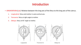
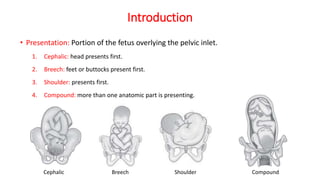
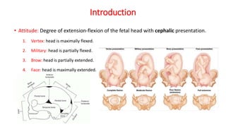
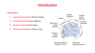
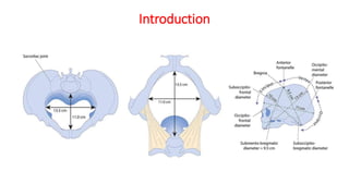
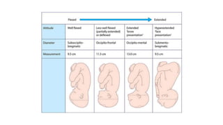
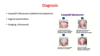
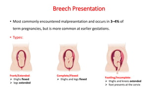
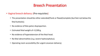
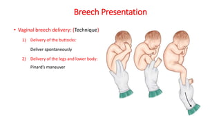
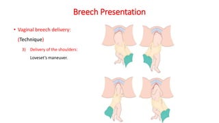
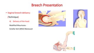
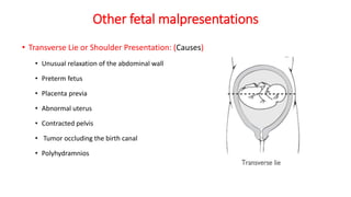
Malpresentations & Malpositions
By: Abdulrahman Hussein Karaki
Supervisors: Dr. Osama Alhasany
Dr. Thana’ Abu Fara
Referances:
•Kaplan 2021
•2017 Obstetrics by ten teachers 20th ed
•ALSO 2017
Outline:
1. Introduction.
2. Diagnosis.
3. Normal position and presentation.
4. OP Position.
5. Face Presentation.
6. Brow Presentation.
7. Breech Presentation.
8. Other fetal malpresentations.
Introduction
• ORIENTATION (Lie): Relation between the long axis of the fetus to the long axis of the uterus.
1. Longitudinal: fetus and mother in same vertical axis.
2. Transverse: fetus at right angle to mother.
3. Oblique: fetus at 45° angle to mother.
Introduction
• Presentation: Portion of the fetus overlying the pelvic inlet.
1. Cephalic: head presents first.
2. Breech: feet or buttocks present first.
3. Shoulder: presents first.
4. Compound: more than one anatomic part is presenting.
Cephalic Breech Shoulder Compound
Introduction
Introduction
Introduction
• Position: Relationship of a definite presenting fetal part to the maternal bony pelvis.
1. Occiput: with a flexed head (cephalic presentation).
2. Sacrum: with a breech presentation.
3. Mentum (chin): with an extended head (face presentation).
Introduction
• Station: Degree of descent of
the presenting part through
the birth canal; expressed in
centimeters above or below
the maternal ischial spine.
Introduction
• Attitude: Degree of extension-flexion of the fetal head with cephalic presentation.
1. Vertex: head is maximally flexed.
2. Military: head is partially flexed.
3. Brow: head is partially extended.
4. Face: head is maximally extended.
Introduction
• Diameters:
1. Suboccipito-bregmatic (9.5cm): Vertex.
2. Occipito-frontal (11.5cm): Military.
3. Occipito-mental (13cm): Brow.
4. Subhmento-bregmatic (9.5cm): Face.
Introduction
Diagnosis
• Leopold’s Maneuvers (abdominal palpation).
• Vaginal examination.
• Imaging: ultrasound.
Normal
• ORIENTATION (Lie): Longitudinal.
• Presentation: Cephalic.
• Position: Occiput anterior.
• Attitude: Vertex.
• Diameter: Suboccipito-bregmatic.
OP Position
• The fetus lies with its occiput toward the woman’s
spine and its face toward the woman’s symphysis
and abdomen.
• The exact cause of persistent OP positioning is
unknown, but transverse narrowing of the pelvis
plays a role.
• All OP fetuses are somewhat deflexed because the
vertex drops back to fill the hollow of the sacrum.
OP Position
• Diagnosis: (difficult)
Back pain, or back labor (hallmark).
Examination.
 Palpation of the anterior fontanel.
Ultrasound.
OP Position
• Labor is often prolonged and there is an increased incidence of operative vaginal
delivery, cesarean delivery, and anal sphincter lacerations.
• Mode of Delivery:
1) Spontaneous Delivery (45%).
2) Manual Rotation:
a. Flex the fetal head placing a hand in the posterior pelvis behind the occiput.
b. Apply rotatory force, the rotation should be attempted at the same time as a contraction.
3) Operative vaginal delivery:
 OP is not itself an indication for operative vaginal delivery.
Face Presentation
• The head is hyperextended so the occiput is in contact with the fetal back.
• 1 in 500 labours.
• Presenting part: Face.
• Diameter: Subhmento-bregmatic (9.5cm).
• Risk factors:
 Fetus is large or the pelvis is contracted.
 Enlargement of the neck.
 coils of cord around the neck
 Anencephalic
Face Presentation
• Diagnosis: vaginal examination.
• The mouth, nose, and the malar prominences may be palpated.
• Mode of Delivery:
The fetus must rotate to a mentum anterior position → Spontaneous vaginal delivery / Forceps
Persistent mentum posterior → cesarean delivery.
Vacuum extractor is absolutely contraindicated.
Brow Presentation
• The portion of the fetal head between the orbital ridge and the anterior
fontanel presents at the pelvic inlet.
• occurring in 1 in 2,000 labours
• Diameter: Occipito-mental (13cm).
• Diagnosis: vaginal examination.
• The frontal sutures, anterior fontanel, orbital ridges, eyes, and root of the nose may
be felt.
• Mode of Delivery:
 If it converts to vertex or face → according to their respective mechanisms.
 Persistent brow → cesarean delivery.
Breech Presentation
• Most commonly encountered malpresentation and occurs in 3–4% of
term pregnancies, but is more common at earlier gestations.
• Types:
Frank/Extended:
 thighs flexed
 legs extended
Complete/Flexed:
 thighs and legs flexed
Footling/Incomplete:
 thighs and knees extended
 foot presents at the cervix
Breech Presentation
• Predisposing factors:
Fetal/placental
 Multiple gestation.
 Prematurity.
 Placenta previa.
 Abnormality (e.g. anencephaly or hydrocephalus).
 Fetal neuromuscular condition.
 Oligohydramnios.
 Polyhydramnios.:
Maternal:
Fibroids.
Congenital uterine abnormalities
(e.g. bicornuate uterus).
Uterine surgery.
Breech Presentation
• Prenatal Management:
• If a breech presentation is clinically suspected at or after 36 weeks:
 Fetal biometry
 Amniotic fluid volume
 Placental site
 Position of the fetal legs
 Any anomalies previously undetected
• Mode of Delivery:
1) Elective caesarean section (best method)
2) External cephalic version (ECV)
3) Vaginal breech delivery
Breech Presentation
• External cephalic version:
• procedure is performed at or after 37 completed weeks’ gestation.
• Performed with a tocolytic (e.g. nifedipine).
• Success rates 50% (experience, multiparous women).
• Mildly uncomfortable for the mother.
• Should last no more than 10 mints.
• A fetal heart rate trace must be performed before and after the procedure.
• Rh- → anti-D.
Breech Presentation
• External cephalic version: (Contraindications)
• Fetal abnormality (e.g. hydrocephalus).
• Placenta previa.
• Oligohydramnios or polyhydramnios.
• History of antepartum hemorrhage.
• Previous caesarean or myomectomy scar on the uterus.
• Multiple gestation.
• Pre-eclampsia or hypertension.
• Plan to deliver by caesarean section anyway.
Breech Presentation
• External cephalic version: (Complications)
• Placental abruption.
• Premature rupture of the membranes.
• Cord accident.
• Transplacental hemorrhage.
• Fetal bradycardia or decelerations, usually transient and self‐limited
Breech Presentation
• External cephalic version: (Technique)
Elevate breech with
suprapubic hand
Push breech into iliac fossa
while flexes fetal head
Breech Presentation
• External cephalic version: (Technique)
fetus becomes
transverse lie
The fetal heart rate should be
checked after the external
version has been completed
Breech Presentation
• Vaginal breech delivery: (Pre-requisites)
• The presentation should be either extended/frank or flexed/complete (but feet not below the
fetal buttocks).
• No evidence of feto-pelvic disproportion.
• Estimated fetal weight of <3,500 g.
• No evidence of hyperextension of the fetal head.
• No fetal abnormalities (e.g. severe hydrocephalus).
• Operating room accessibility (for urgent cesarean delivery).
Breech Presentation
• Vaginal breech delivery: (Technique)
1) Delivery of the buttocks:
Deliver spontaneously
2) Delivery of the legs and lower body:
Pinard’s maneuver
Breech Presentation
• Vaginal breech delivery:
(Technique)
3) Delivery of the shoulders:
Loveset’s maneuver.
Breech Presentation
• Vaginal breech delivery:
(Technique)
4) Delivery of the head:
Modified Mauriceau‐
Smellie‐Veit (MSV) Maneuver
Breech Presentation
• Vaginal breech delivery: (Complications)
• Nuchal arm(s)
• Head entrapment
• Umbilical cord prolapse
Other fetal malpresentations
• Transverse Lie or Shoulder Presentation: (Causes)
• Unusual relaxation of the abdominal wall
• Preterm fetus
• Placenta previa
• Abnormal uterus
• Contracted pelvis
• Tumor occluding the birth canal
• Polyhydramnios
Other fetal malpresentations
• Transverse Lie or Shoulder Presentation:
• Incidence: occurring in 1 in 300 pregnancies.
• Diagnosis: Vaginal examination.
• Mode of Delivery:
• Spontaneous birth in transverse lie is impossible.
• Cesarean delivery is mandatory in most cases.
• Before the onset of labor or in early labor with intact membranes → attempt ECV.
Other fetal malpresentations
• Compound Presentation:
• Often no cause is found.
• More common with premature infants.
• Diagnosis: Vaginal examination.
• Mode of Delivery:
• Most commonly the prolapsed limb will deliver spontaneously along with the head, or sometimes the
fetus will retract its limb spontaneously.
• If the prolapsed arm appears to be impeding descent, it should be gently elevated upward.
• Occasionally cesarean delivery will be necessary.

Malpresentations & Malpositions copy.pptx

  • 1.
    Malpresentations & Malpositions By:Abdulrahman Hussein Karaki Supervisors: Dr. Osama Alhasany Dr. Thana’ Abu Fara
  • 2.
    Referances: •Kaplan 2021 •2017 Obstetricsby ten teachers 20th ed •ALSO 2017
  • 3.
    Outline: 1. Introduction. 2. Diagnosis. 3.Normal position and presentation. 4. OP Position. 5. Face Presentation. 6. Brow Presentation. 7. Breech Presentation. 8. Other fetal malpresentations.
  • 4.
    Introduction • ORIENTATION (Lie):Relation between the long axis of the fetus to the long axis of the uterus. 1. Longitudinal: fetus and mother in same vertical axis. 2. Transverse: fetus at right angle to mother. 3. Oblique: fetus at 45° angle to mother.
  • 5.
    Introduction • Presentation: Portionof the fetus overlying the pelvic inlet. 1. Cephalic: head presents first. 2. Breech: feet or buttocks present first. 3. Shoulder: presents first. 4. Compound: more than one anatomic part is presenting. Cephalic Breech Shoulder Compound
  • 6.
  • 7.
  • 8.
    Introduction • Position: Relationshipof a definite presenting fetal part to the maternal bony pelvis. 1. Occiput: with a flexed head (cephalic presentation). 2. Sacrum: with a breech presentation. 3. Mentum (chin): with an extended head (face presentation).
  • 9.
    Introduction • Station: Degreeof descent of the presenting part through the birth canal; expressed in centimeters above or below the maternal ischial spine.
  • 10.
    Introduction • Attitude: Degreeof extension-flexion of the fetal head with cephalic presentation. 1. Vertex: head is maximally flexed. 2. Military: head is partially flexed. 3. Brow: head is partially extended. 4. Face: head is maximally extended.
  • 11.
    Introduction • Diameters: 1. Suboccipito-bregmatic(9.5cm): Vertex. 2. Occipito-frontal (11.5cm): Military. 3. Occipito-mental (13cm): Brow. 4. Subhmento-bregmatic (9.5cm): Face.
  • 12.
  • 14.
    Diagnosis • Leopold’s Maneuvers(abdominal palpation). • Vaginal examination. • Imaging: ultrasound.
  • 15.
    Normal • ORIENTATION (Lie):Longitudinal. • Presentation: Cephalic. • Position: Occiput anterior. • Attitude: Vertex. • Diameter: Suboccipito-bregmatic.
  • 16.
    OP Position • Thefetus lies with its occiput toward the woman’s spine and its face toward the woman’s symphysis and abdomen. • The exact cause of persistent OP positioning is unknown, but transverse narrowing of the pelvis plays a role. • All OP fetuses are somewhat deflexed because the vertex drops back to fill the hollow of the sacrum.
  • 17.
    OP Position • Diagnosis:(difficult) Back pain, or back labor (hallmark). Examination.  Palpation of the anterior fontanel. Ultrasound.
  • 18.
    OP Position • Laboris often prolonged and there is an increased incidence of operative vaginal delivery, cesarean delivery, and anal sphincter lacerations. • Mode of Delivery: 1) Spontaneous Delivery (45%). 2) Manual Rotation: a. Flex the fetal head placing a hand in the posterior pelvis behind the occiput. b. Apply rotatory force, the rotation should be attempted at the same time as a contraction. 3) Operative vaginal delivery:  OP is not itself an indication for operative vaginal delivery.
  • 19.
    Face Presentation • Thehead is hyperextended so the occiput is in contact with the fetal back. • 1 in 500 labours. • Presenting part: Face. • Diameter: Subhmento-bregmatic (9.5cm). • Risk factors:  Fetus is large or the pelvis is contracted.  Enlargement of the neck.  coils of cord around the neck  Anencephalic
  • 20.
    Face Presentation • Diagnosis:vaginal examination. • The mouth, nose, and the malar prominences may be palpated. • Mode of Delivery: The fetus must rotate to a mentum anterior position → Spontaneous vaginal delivery / Forceps Persistent mentum posterior → cesarean delivery. Vacuum extractor is absolutely contraindicated.
  • 21.
    Brow Presentation • Theportion of the fetal head between the orbital ridge and the anterior fontanel presents at the pelvic inlet. • occurring in 1 in 2,000 labours • Diameter: Occipito-mental (13cm). • Diagnosis: vaginal examination. • The frontal sutures, anterior fontanel, orbital ridges, eyes, and root of the nose may be felt. • Mode of Delivery:  If it converts to vertex or face → according to their respective mechanisms.  Persistent brow → cesarean delivery.
  • 22.
    Breech Presentation • Mostcommonly encountered malpresentation and occurs in 3–4% of term pregnancies, but is more common at earlier gestations. • Types: Frank/Extended:  thighs flexed  legs extended Complete/Flexed:  thighs and legs flexed Footling/Incomplete:  thighs and knees extended  foot presents at the cervix
  • 23.
    Breech Presentation • Predisposingfactors: Fetal/placental  Multiple gestation.  Prematurity.  Placenta previa.  Abnormality (e.g. anencephaly or hydrocephalus).  Fetal neuromuscular condition.  Oligohydramnios.  Polyhydramnios.: Maternal: Fibroids. Congenital uterine abnormalities (e.g. bicornuate uterus). Uterine surgery.
  • 24.
    Breech Presentation • PrenatalManagement: • If a breech presentation is clinically suspected at or after 36 weeks:  Fetal biometry  Amniotic fluid volume  Placental site  Position of the fetal legs  Any anomalies previously undetected • Mode of Delivery: 1) Elective caesarean section (best method) 2) External cephalic version (ECV) 3) Vaginal breech delivery
  • 25.
    Breech Presentation • Externalcephalic version: • procedure is performed at or after 37 completed weeks’ gestation. • Performed with a tocolytic (e.g. nifedipine). • Success rates 50% (experience, multiparous women). • Mildly uncomfortable for the mother. • Should last no more than 10 mints. • A fetal heart rate trace must be performed before and after the procedure. • Rh- → anti-D.
  • 26.
    Breech Presentation • Externalcephalic version: (Contraindications) • Fetal abnormality (e.g. hydrocephalus). • Placenta previa. • Oligohydramnios or polyhydramnios. • History of antepartum hemorrhage. • Previous caesarean or myomectomy scar on the uterus. • Multiple gestation. • Pre-eclampsia or hypertension. • Plan to deliver by caesarean section anyway.
  • 27.
    Breech Presentation • Externalcephalic version: (Complications) • Placental abruption. • Premature rupture of the membranes. • Cord accident. • Transplacental hemorrhage. • Fetal bradycardia or decelerations, usually transient and self‐limited
  • 28.
    Breech Presentation • Externalcephalic version: (Technique) Elevate breech with suprapubic hand Push breech into iliac fossa while flexes fetal head
  • 29.
    Breech Presentation • Externalcephalic version: (Technique) fetus becomes transverse lie The fetal heart rate should be checked after the external version has been completed
  • 30.
    Breech Presentation • Vaginalbreech delivery: (Pre-requisites) • The presentation should be either extended/frank or flexed/complete (but feet not below the fetal buttocks). • No evidence of feto-pelvic disproportion. • Estimated fetal weight of <3,500 g. • No evidence of hyperextension of the fetal head. • No fetal abnormalities (e.g. severe hydrocephalus). • Operating room accessibility (for urgent cesarean delivery).
  • 31.
    Breech Presentation • Vaginalbreech delivery: (Technique) 1) Delivery of the buttocks: Deliver spontaneously 2) Delivery of the legs and lower body: Pinard’s maneuver
  • 32.
    Breech Presentation • Vaginalbreech delivery: (Technique) 3) Delivery of the shoulders: Loveset’s maneuver.
  • 33.
    Breech Presentation • Vaginalbreech delivery: (Technique) 4) Delivery of the head: Modified Mauriceau‐ Smellie‐Veit (MSV) Maneuver
  • 34.
    Breech Presentation • Vaginalbreech delivery: (Complications) • Nuchal arm(s) • Head entrapment • Umbilical cord prolapse
  • 35.
    Other fetal malpresentations •Transverse Lie or Shoulder Presentation: (Causes) • Unusual relaxation of the abdominal wall • Preterm fetus • Placenta previa • Abnormal uterus • Contracted pelvis • Tumor occluding the birth canal • Polyhydramnios
  • 36.
    Other fetal malpresentations •Transverse Lie or Shoulder Presentation: • Incidence: occurring in 1 in 300 pregnancies. • Diagnosis: Vaginal examination. • Mode of Delivery: • Spontaneous birth in transverse lie is impossible. • Cesarean delivery is mandatory in most cases. • Before the onset of labor or in early labor with intact membranes → attempt ECV.
  • 37.
    Other fetal malpresentations •Compound Presentation: • Often no cause is found. • More common with premature infants. • Diagnosis: Vaginal examination. • Mode of Delivery: • Most commonly the prolapsed limb will deliver spontaneously along with the head, or sometimes the fetus will retract its limb spontaneously. • If the prolapsed arm appears to be impeding descent, it should be gently elevated upward. • Occasionally cesarean delivery will be necessary.