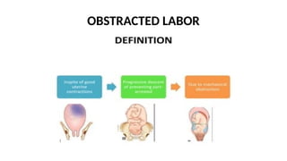
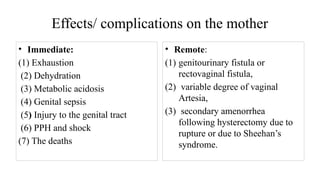
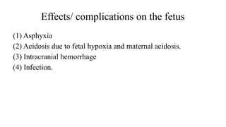
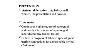
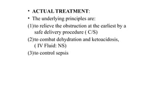
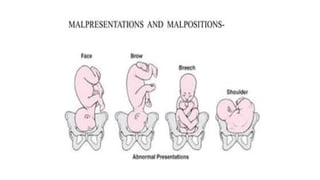
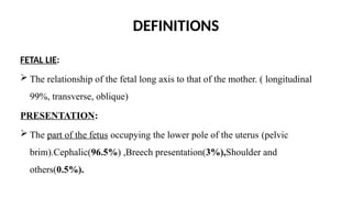
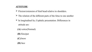
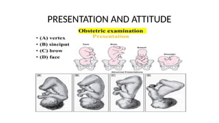
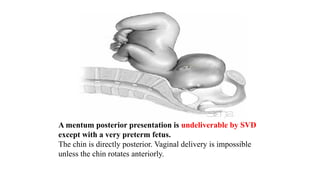
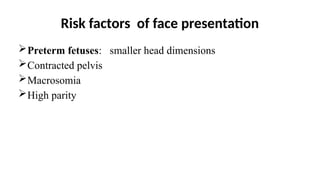
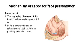
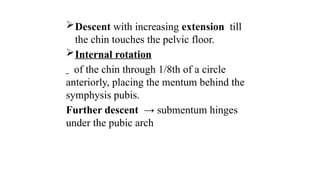
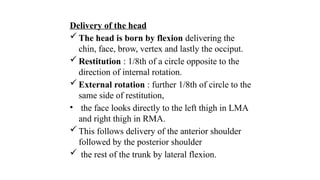
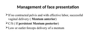
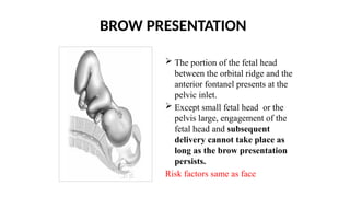
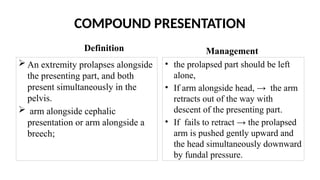
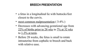
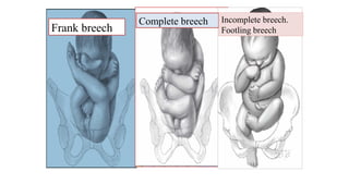
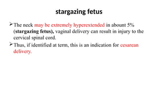
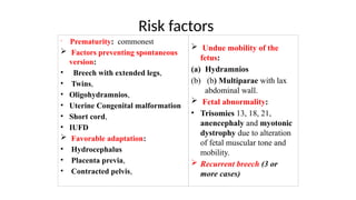
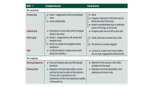
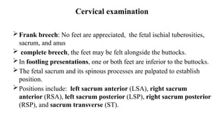
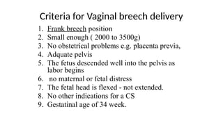
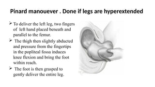
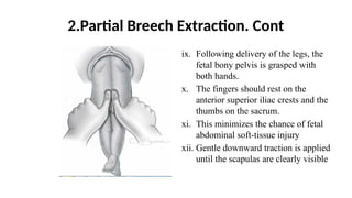
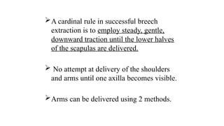
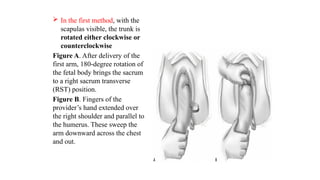
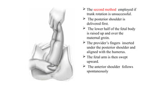
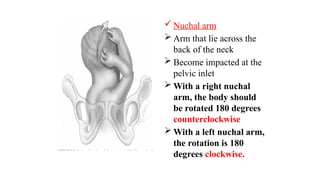
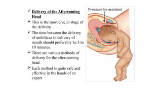
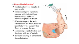
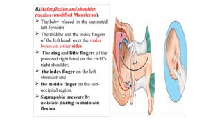
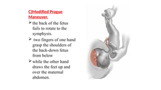
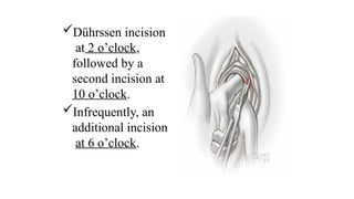
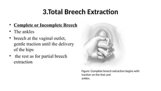
OBSTRUCTED LABOR, MAL POSITION AND
MAL PRESENTATION
MENTOR: Dr. Paul
Presented by: DR Love Lasi
OBSTRACTED LABOR
Causes
Fault in the passage:
(1) Bony:
• Cephalopelvic disproportion ,contracted pelvis,
• Secondary contracted pelvis in multiparous women.
(2) Soft tissue obstructions:
• cervical dystocia
• cervical or broad ligament fibroid,
• impacted ovarian tumor
• the nongravid horn of a bicornuate uterus
• Fault in the passenger:
(1)Transverse lie
(2) Brow presentation
(3) Congenital malformations of the
fetus:hydrocephalus (commonest),fetal
ascites, double monsters,
(4)Big baby, occipitoposterior position,
(5) Compound presentation
(6)Locked twins
Morbid anatomical changes
• Uterus:
• pathological retraction ring or Bandl’s ring
• The bladder becomes an abdominal organ and due to
compression of urethra between the presenting part and
symphysis pubis, the patient fails to empty the bladder.
• the Bandl’s ring.
The transverse depression at the junction of the superior border of the bladder and the distended lower segment is often confused with the Bandl’s ring.
• The bladder walls get traumatized:
blood stained urine,
• The base of the bladder and urethra,
nipped in between the presenting part
and symphysis pubis: pressure
necrosis.
• The devitalized tissue becomes
infected and slough off:
genitourinary fistula.
Clinical features
• Prolonged labor with severe & continuous pain.
• Abdominal examination:
• uterus smaller in size, tense and tender.
• Fetal parts neither well defined, nor fetal heart sound audible.
• Vaginal examination:
• jammed head with big caput;
• dry and edematous vagina.
Effects/ complications on the mother
• Immediate:
(1) Exhaustion
(2) Dehydration
(3) Metabolic acidosis
(4) Genital sepsis
(5) Injury to the genital tract
(6) PPH and shock
(7) The deaths
• Remote:
(1) genitourinary fistula or
rectovaginal fistula,
(2) variable degree of vaginal
Artesia,
(3) secondary amenorrhea
following hysterectomy due to
rupture or due to Sheehan’s
syndrome.
Effects/ complications on the fetus
(1) Asphyxia
(2) Acidosis due to fetal hypoxia and maternal acidosis.
(3) Intracranial hemorrhage
(4) Infection.
PREVENTION
 Antenatal detection : big baby, small
women, malpresentation and position).
Intranatal:
Continuous vigilance, use of partograph
and timely intervention of a prolonged
labor due to mechanical factors
Failure in progress of labor in spite of good
uterine contractions for a reasonable period
(2–4 hours)
• ACTUAL TREATMENT:
• The underlying principles are:
(1)to relieve the obstruction at the earliest by a
safe delivery procedure ( C/S)
(2)to combat dehydration and ketoacidosis,
( IV Fluid: NS)
(3)to control sepsis
DEFINITIONS
FETAL LIE:
 The relationship of the fetal long axis to that of the mother. ( longitudinal
99%, transverse, oblique)
PRESENTATION:
 The part of the fetus occupying the lower pole of the uterus (pelvic
brim).Cephalic(96.5%) ,Breech presentation(3%),Shoulder and
others(0.5%).
ATTITUDE:
 Flexion/extension of fetal head relative to shoulders.
 The relation of the different parts of the fetus to one another
 In longitudinal lie, Cephalic presentation. Differences in
attitude are:
(A) vertex(Normal)
(B) Sinciput
(C) brow
(D) face
PRESENTATION AND ATTITUDE
FETAL POSITION
 The relationship of an arbitrarily chosen portion
of the fetal presenting part (Denominator) to the
right or left side of the birth canal.
The pelvis is divided into equal segments of 45°
to place the denominator in each segment.
There are 8 positions with each presenting part
Figure: The position and relative
frequency of the vertex at the onset of
labor.
 2/3 of all vertex presentations : the left
occiput position, and 1/3 right occiput.
 Fetal head enters maternal pelvis and
engages in OT position.
subsequently rotates to OA position(most
cases) or OP ( small percentage of cases).
 Cephalic presentation , the denominator :
Vertex.
 In vertex it is the occiput
 Face presentation, the denominator :Mentum.
 Breech presentation, the denominator : Sacrum.
 Shoulder(Transverse lie) presentation, the
denominator :Acromion or Scapula.
FACE PRESENTATION
• the neck is hyperextended (sacral curve)
• the chin (mentum) is presenting anteriorly or posteriorly,
relative to the maternal symphysis pubis.
mentum posterior presentations persists or converts to anterior
Persistent MP causes the fetal brow (bregma) to press against
the symphysis pubis.
 This position prevents flexion of the fetal head necessary to
negotiate the birth canal.
A mentum posterior presentation is undeliverable by SVD
except with a very preterm fetus.
The chin is directly posterior. Vaginal delivery is impossible
unless the chin rotates anteriorly.
Diagnosis of face presentation
Vaginal examination
Palpation of facial features.
A breech may be mistaken for a face presentation (The anus may be
mistaken for the mouth, and the ischial tuberosities for the malar
prominences.)
Radiographically, demonstration of the hyperextended head with the
facial bones at or below the pelvic inlet is characteristic.
Risk factors of face presentation
Preterm fetuses: smaller head dimensions
Contracted pelvis
Macrosomia
High parity
Mechanism of Labor for face presentation
Engagement
 The engaging diameter of the
head is submento-bregmatic 9.5
cm
 in fully extended head or
submento-vertical 11.5 cm in
partially extended head.
Descent with increasing extension till
the chin touches the pelvic floor.
Internal rotation
of the chin through 1/8th of a circle
anteriorly, placing the mentum behind the
symphysis pubis.
Further descent → submentum hinges
under the pubic arch
Delivery of the head
 The head is born by flexion delivering the
chin, face, brow, vertex and lastly the occiput.
 Restitution : 1/8th of a circle opposite to the
direction of internal rotation.
 External rotation : further 1/8th of circle to the
same side of restitution,
• the face looks directly to the left thigh in LMA
and right thigh in RMA.
 This follows delivery of the anterior shoulder
followed by the posterior shoulder
 the rest of the trunk by lateral flexion.
Management of face presentation
If no contracted pelvis and with effective labor, successful
vaginal delivery ( Mentum anterior)
C/S ( If persistent Mentum posterior)
Low or outlet forceps delivery of a mentum
BROW PRESENTATION
 The portion of the fetal head
between the orbital ridge and the
anterior fontanel presents at the
pelvic inlet.
 Except small fetal head or the
pelvis large, engagement of the
fetal head and subsequent
delivery cannot take place as
long as the brow presentation
persists.
Risk factors same as face
Diagnosis & Management
Diagnosis
Abdominal palpation: the occiput is palpated easily
Vaginal examination : The frontal sutures, large anterior
fontanel, orbital ridges, eyes, and root of the nose are felt but
neither the mouth nor the chin is palpable.
Management
Cases with persistent brow presentation are delivered by
cesarean section.
TRANSVERSE LIE
Risk factors
(1)abdominal wall relaxation from high parity
(2) preterm fetus
(3) placenta previa
(4) abnormal uterine anatomy
(5) hydramnios
(6) contracted pelvis.
Mechanism of Labor for transverse lie
Spontaneos delivery of a fully developed newborn is
impossible
After rupture of the membranes, the fetal shoulder is forced
into the pelvis, and the corresponding arm frequently prolapses.
 the shoulder arrested by the margins of the pelvic inlet.
 then impacted firmly in the upper part of the pelvis.
Small fetus (usually <800 g) and large
pelvis, spontaneous delivery is
possible
• The head and thorax then pass through
the pelvic cavity at the same time.
• The fetus, which is doubled upon
itself, conduplicato corpore, is
expelled.
Management
Active labor in a woman with a transverse lie is an indication
for cesarean delivery.
 With the membranes intact, external version is attempted. (by
skilled person)
COMPOUND PRESENTATION
Definition
 An extremity prolapses alongside
the presenting part, and both
present simultaneously in the
pelvis.
 arm alongside cephalic
presentation or arm alongside a
breech;
Management
• the prolapsed part should be left
alone,
• If arm alongside head, → the arm
retracts out of the way with
descent of the presenting part.
• If fails to retract → the prolapsed
arm is pushed gently upward and
the head simultaneously downward
by fundal pressure.
BREECH PRESENTATION
• a fetus in a longitudinal lie with buttocks/feet
closest to the cervix.
• most common malpresentation ( 3-4% )
• Decreases with advancing gestational age from
22% of births prior to 28 wks to 7% at 32 wks
to 1-3% at term.
• Before 28 weeks, the fetus is small to rotate
intrauterine from cephalic to breech and back
with relative ease.
CLASSIFICATION OF BREECH PRESENTATIONS
 Frank:
• lower extremities flexed at the hips and extended at the knees,
• the feet lie close to the head.
 Complete breech:
• Both hips flexed,
• one or both knees also flexed.
 Incomplete breech:
• One or both hips extended.
• one or both feet or knees lie below the breech,
• A footling breech is an incomplete breech with one or both feet below the breech.
Frank breech
Complete breech Incomplete breech.
Footling breech
stargazing fetus
The neck may be extremely hyperextended in abount 5%
(stargazing fetus), vaginal delivery can result in injury to the
cervical spinal cord.
Thus, if identified at term, this is an indication for cesarean
delivery.
Risk factors

Prematurity: commonest
 Factors preventing spontaneous
version:
• Breech with extended legs,
• Twins,
• Oligohydramnios,
• Uterine Congenital malformation
• Short cord,
• IUFD
 Favorable adaptation:
• Hydrocephalus
• Placenta previa,
• Contracted pelvis,
 Undue mobility of the
fetus:
(a) Hydramnios
(b) (b) Multiparae with lax
abdominal wall.
 Fetal abnormality:
• Trisomies 13, 18, 21,
anencephaly and myotonic
dystrophy due to alteration
of fetal muscular tone and
mobility.
 Recurrent breech (3 or
more cases)
Cervical examination
Frank breech: No feet are appreciated, the fetal ischial tuberosities,
sacrum, and anus
 complete breech, the feet may be felt alongside the buttocks.
In footling presentations, one or both feet are inferior to the buttocks.
The fetal sacrum and its spinous processes are palpated to establish
position.
Positions include: left sacrum anterior (LSA), right sacrum
anterior (RSA), left sacrum posterior (LSP), right sacrum posterior
(RSP), and sacrum transverse (ST).
Criteria for Vaginal breech delivery
1. Frank breech position
2. Small enough ( 2000 to 3500g)
3. No obstetrical problems e.g. placenta previa,
4. Adquate pelvis
5. The fetus descended well into the pelvis as
labor begins
6. no maternal or fetal distress
7. The fetal head is flexed - not extended.
8. No other indications for a CS
9. Gestatinal age of 34 week.
Indications for C/s in breech
 Lack of operator experience
 Patient request for cesarean delivery
 Large fetus: >3800 to 4000 g
 Apparently healthy and viable preterm fetus (<2500 g)
 Severe fetal-growth restriction
 Fetal anomaly incompatible with vaginal delivery
 Prior perinatal death or neonatal birth trauma
 Incomplete or footling breech presentation
 Hyperextended head
 Pelvic contraction
 Prior cesarean delivery
Delivery Complications
 Maternal:
 Trauma to the genital tract,
 operative vaginal delivery (episiotomy, forceps),
 cesarean section,
 sepsis and anesthetic complications.
 Fetal:
 Perinatal death,
 Intrapartum fetal death,
 Intracranial hemorrhage,
 Birth asphyxia,
 Birth Injuries( Fractures, …).
Mechanisms of labour in breech
SACRO-ANTERIOR POSITION:
In the mechanism of breech delivery, the principal movements
occur at three places: buttocks, shoulders and the head.
Each of the three components undergo cardinal movements as those
of normal mechanism.
The first two successive parts to be born are bigger but more
compressible.
While the head because of non-moulding due to rapid descent,
presents difficulties.
1.buttocks
 engagement of the buttock is one of the oblique diameters of
the inlet.
 The engaging diameter is bi-trochanteric (10 cm or 4"), the
sacrum directed towards the ilio-pubic eminence.
 the diameter passes through the pelvic brim, the breech is
engaged.
 Descent of the buttocks until the anterior buttock touches the
pelvic floor.
Internal rotation of the anterior buttock
through 1/8th of a circle placing it behind
the symphysis pubis.
Further descent with lateral flexion of the
trunk until the anterior hip hinges under the
symphysis pubis followed by the posterior
hip.
 Delivery of the trunk and the lower limbs
follow.
 Restitution: the buttocks occupy the
original position in oblique diameter.
2. Shoulders.
Bisacromial diameter (12 cm or 4 3/4") engages oblique
diameter at brim after the delivery of the breech.
 Descent with internal rotation : the shoulders to lie in the
anteroposterior diameter of the pelvic outlet.
The trunk rotates externally : 1/8th
of a circle.
Delivery of the posterior shoulder , then anterior
one completed by anterior flexion of the
delivered trunk.
 Restitution and external rotation:
• Untwisting of the trunk : the anterior shoulder
towards the right thigh in LSA and left thigh in
RSA.
• External rotation of the shoulders to the same
direction because of internal rotation of the
occiput through 1/8th of a circle anteriorly.
• The fetal trunk positioned as dorso-anterior.
Delivery of the head
 Engagement either through the opposite oblique diameter
to buttocks or through the transverse diameter
 The engaging diameter: suboccipitofrontal (10 cm).
 Descent with increasing flexion occurs.
 Internal rotation of the occiput : anteriorly, 1/8th or
2/8th of a circle the occiput behind the symphysis pubis.
Further descent : occurs until the
subocciput hinges under the symphysis pubis
 The head is born by flexion—the chin,
mouth, nose, forehead, vertex and occiput
appearing successively.
The expulsion : the bearing down efforts
and uterine contractions.
Breech Delivery Types
1. Spontaneous Breech Delivery
2. Partial Breech Extraction
3. Total Breech Extraction
1.Spontaneous Breech Delivery
• Described in mecanisme of breech delivery
2.Partial Breech Extraction/ Assisted BD
i. Mediolateral episiotomy (only after the fetal anus is visible at the
vulva): Perfomed if necessary
ii. The breech is allowed to deliver spontaneously to the umbilicus.
iii. The posterior hip will deliver, usually from the 6 o’clock position, and
often with sufficient pressure to evoke passage of thick meconium.
iv. The anterior hip then delivers, followed by external rotation to a
sacrum anterior position.
v. The mother is encouraged to continue to push as the fetus descends
until the legs are accessible.
vi. If the lower limbs are extended when the trunk
has delivered to the level of the umbilicus, the
operator may use his/her fingers to exert
pressure on the back of the knee(Popliteal fossa)
(Pinard maneuver) and guide the thigh away
from the trunk as the trunk is rotated in the
opposite direction.
vii.This causes the knee to flex and allows
extraction of the foot and the leg.
viii.
After the legs have delivered, the cord is
checked for pulsation, and a small loop is pulled
down to prevent traction on the cord.
Pinard manouever . Done if legs are hyperextended
 To deliver the left leg, two fingers
of left hand placed beneath and
parallel to the femur.
 The thigh then slightly abducted
and pressure from the fingertips
in the popliteal fossa induces
knee flexion and bring the foot
within reach.
 The foot is then grasped to
gently deliver the entire leg.
2.Partial Breech Extraction. Cont
ix. Following delivery of the legs, the
fetal bony pelvis is grasped with
both hands.
x. The fingers should rest on the
anterior superior iliac crests and the
thumbs on the sacrum.
xi. This minimizes the chance of fetal
abdominal soft-tissue injury
xii. Gentle downward traction is applied
until the scapulas are clearly visible
A cardinal rule in successful breech
extraction is to employ steady, gentle,
downward traction until the lower halves
of the scapulas are delivered.
 No attempt at delivery of the shoulders
and arms until one axilla becomes visible.
Arms can be delivered using 2 methods.
 In the first method, with the
scapulas visible, the trunk is
rotated either clockwise or
counterclockwise
Figure A. After delivery of the
first arm, 180-degree rotation of
the fetal body brings the sacrum
to a right sacrum transverse
(RST) position.
Figure B. Fingers of the
provider’s hand extended over
the right shoulder and parallel to
the humerus. These sweep the
arm downward across the chest
and out.
 The second method employed if
trunk rotation is unsuccessful.
 The posterior shoulder is
delivered first.
 The lower half of the fetal body
is raised up and over the
maternal groin.
 The provider’s fingers inserted
under the posterior shoulder and
aligned with the humerus.
 The fetal arm is then swept
upward.
 The anterior shoulder follows
spontaneously
 Nuchal arm
 Arm that lie across the
back of the neck
 Become impacted at the
pelvic inlet
 With a right nuchal
arm, the body should
be rotated 180 degrees
counterclockwise
 With a left nuchal arm,
the rotation is 180
degrees clockwise.
 Delivery of the Aftercoming
Head
 This is the most crucial stage of
the delivery.
 The time between the delivery
of umbilicus to delivery of
mouth should preferably be 5 to
10 minutes.
 There are various methods of
delivery for the after-coming
head.
 Each method is quite safe and
effective in the hands of an
expert
a)Burns-Marshall method
 The baby allowed to hang by its
own weight.
 The assistant gives suprapubic
pressure with the flat of hand
downward and backward
direction to promote flexion.
 When the nape of the neck
visible under the pubic arch, the
grasp baby by the ankles with a
finger in between the two.
 Maintaining a steady traction and
forming a wide arc of a circle,
 the trunk is swung in upward and
forward direction
B)Malar flexion and shoulder
traction (modified Mauriceau).
 The baby placed on the supinated
left forearm
 The middle and the index fingers
of the left hand over the malar
bones on either sides
 The ring and little fingers of the
pronated right hand on the child’s
right shoulder,
 the index finger on the left
shoulder and
 the middle finger on the sub-
occipital region.
 Suprapubic pressure by
assistant during to maintain
flexion.
C)Modified Prague
Maneuver.
the back of the fetus
fails to rotate to the
symphysis.
 two fingers of one hand
grasp the shoulders of
the back-down fetus
from below
while the other hand
draws the feet up and
over the maternal
abdomen.
Dührssen incision
at 2 o’clock,
followed by a
second incision at
10 o’clock.
Infrequently, an
additional incision
at 6 o’clock.
3.Total Breech Extraction
• Complete or Incomplete Breech
• The ankles
• breech at the vaginal outlet,
gentle traction until the delivery
of the hips
• the rest as for partial breech
extraction
Figure: Complete breech extraction begins with
traction on the feet and
ankles.
Thank you

OBSTRUCTED LABOUR, MAL POSITION AND MAL PRESENTATION.pptx

  • 1.
    OBSTRUCTED LABOR, MALPOSITION AND MAL PRESENTATION MENTOR: Dr. Paul Presented by: DR Love Lasi
  • 2.
  • 3.
    Causes Fault in thepassage: (1) Bony: • Cephalopelvic disproportion ,contracted pelvis, • Secondary contracted pelvis in multiparous women. (2) Soft tissue obstructions: • cervical dystocia • cervical or broad ligament fibroid, • impacted ovarian tumor • the nongravid horn of a bicornuate uterus
  • 4.
    • Fault inthe passenger: (1)Transverse lie (2) Brow presentation (3) Congenital malformations of the fetus:hydrocephalus (commonest),fetal ascites, double monsters, (4)Big baby, occipitoposterior position, (5) Compound presentation (6)Locked twins
  • 5.
    Morbid anatomical changes •Uterus: • pathological retraction ring or Bandl’s ring • The bladder becomes an abdominal organ and due to compression of urethra between the presenting part and symphysis pubis, the patient fails to empty the bladder. • the Bandl’s ring.
  • 6.
    The transverse depressionat the junction of the superior border of the bladder and the distended lower segment is often confused with the Bandl’s ring.
  • 7.
    • The bladderwalls get traumatized: blood stained urine, • The base of the bladder and urethra, nipped in between the presenting part and symphysis pubis: pressure necrosis. • The devitalized tissue becomes infected and slough off: genitourinary fistula.
  • 8.
    Clinical features • Prolongedlabor with severe & continuous pain. • Abdominal examination: • uterus smaller in size, tense and tender. • Fetal parts neither well defined, nor fetal heart sound audible. • Vaginal examination: • jammed head with big caput; • dry and edematous vagina.
  • 9.
    Effects/ complications onthe mother • Immediate: (1) Exhaustion (2) Dehydration (3) Metabolic acidosis (4) Genital sepsis (5) Injury to the genital tract (6) PPH and shock (7) The deaths • Remote: (1) genitourinary fistula or rectovaginal fistula, (2) variable degree of vaginal Artesia, (3) secondary amenorrhea following hysterectomy due to rupture or due to Sheehan’s syndrome.
  • 10.
    Effects/ complications onthe fetus (1) Asphyxia (2) Acidosis due to fetal hypoxia and maternal acidosis. (3) Intracranial hemorrhage (4) Infection.
  • 11.
    PREVENTION  Antenatal detection: big baby, small women, malpresentation and position). Intranatal: Continuous vigilance, use of partograph and timely intervention of a prolonged labor due to mechanical factors Failure in progress of labor in spite of good uterine contractions for a reasonable period (2–4 hours)
  • 12.
    • ACTUAL TREATMENT: •The underlying principles are: (1)to relieve the obstruction at the earliest by a safe delivery procedure ( C/S) (2)to combat dehydration and ketoacidosis, ( IV Fluid: NS) (3)to control sepsis
  • 14.
    DEFINITIONS FETAL LIE:  Therelationship of the fetal long axis to that of the mother. ( longitudinal 99%, transverse, oblique) PRESENTATION:  The part of the fetus occupying the lower pole of the uterus (pelvic brim).Cephalic(96.5%) ,Breech presentation(3%),Shoulder and others(0.5%).
  • 15.
    ATTITUDE:  Flexion/extension offetal head relative to shoulders.  The relation of the different parts of the fetus to one another  In longitudinal lie, Cephalic presentation. Differences in attitude are: (A) vertex(Normal) (B) Sinciput (C) brow (D) face
  • 16.
  • 17.
    FETAL POSITION  Therelationship of an arbitrarily chosen portion of the fetal presenting part (Denominator) to the right or left side of the birth canal. The pelvis is divided into equal segments of 45° to place the denominator in each segment. There are 8 positions with each presenting part
  • 18.
    Figure: The positionand relative frequency of the vertex at the onset of labor.
  • 19.
     2/3 ofall vertex presentations : the left occiput position, and 1/3 right occiput.  Fetal head enters maternal pelvis and engages in OT position. subsequently rotates to OA position(most cases) or OP ( small percentage of cases).
  • 20.
     Cephalic presentation, the denominator : Vertex.  In vertex it is the occiput  Face presentation, the denominator :Mentum.  Breech presentation, the denominator : Sacrum.  Shoulder(Transverse lie) presentation, the denominator :Acromion or Scapula.
  • 21.
    FACE PRESENTATION • theneck is hyperextended (sacral curve) • the chin (mentum) is presenting anteriorly or posteriorly, relative to the maternal symphysis pubis. mentum posterior presentations persists or converts to anterior Persistent MP causes the fetal brow (bregma) to press against the symphysis pubis.  This position prevents flexion of the fetal head necessary to negotiate the birth canal.
  • 22.
    A mentum posteriorpresentation is undeliverable by SVD except with a very preterm fetus. The chin is directly posterior. Vaginal delivery is impossible unless the chin rotates anteriorly.
  • 23.
    Diagnosis of facepresentation Vaginal examination Palpation of facial features. A breech may be mistaken for a face presentation (The anus may be mistaken for the mouth, and the ischial tuberosities for the malar prominences.) Radiographically, demonstration of the hyperextended head with the facial bones at or below the pelvic inlet is characteristic.
  • 24.
    Risk factors offace presentation Preterm fetuses: smaller head dimensions Contracted pelvis Macrosomia High parity
  • 25.
    Mechanism of Laborfor face presentation Engagement  The engaging diameter of the head is submento-bregmatic 9.5 cm  in fully extended head or submento-vertical 11.5 cm in partially extended head.
  • 26.
    Descent with increasingextension till the chin touches the pelvic floor. Internal rotation of the chin through 1/8th of a circle anteriorly, placing the mentum behind the symphysis pubis. Further descent → submentum hinges under the pubic arch
  • 27.
    Delivery of thehead  The head is born by flexion delivering the chin, face, brow, vertex and lastly the occiput.  Restitution : 1/8th of a circle opposite to the direction of internal rotation.  External rotation : further 1/8th of circle to the same side of restitution, • the face looks directly to the left thigh in LMA and right thigh in RMA.  This follows delivery of the anterior shoulder followed by the posterior shoulder  the rest of the trunk by lateral flexion.
  • 28.
    Management of facepresentation If no contracted pelvis and with effective labor, successful vaginal delivery ( Mentum anterior) C/S ( If persistent Mentum posterior) Low or outlet forceps delivery of a mentum
  • 29.
    BROW PRESENTATION  Theportion of the fetal head between the orbital ridge and the anterior fontanel presents at the pelvic inlet.  Except small fetal head or the pelvis large, engagement of the fetal head and subsequent delivery cannot take place as long as the brow presentation persists. Risk factors same as face
  • 30.
    Diagnosis & Management Diagnosis Abdominalpalpation: the occiput is palpated easily Vaginal examination : The frontal sutures, large anterior fontanel, orbital ridges, eyes, and root of the nose are felt but neither the mouth nor the chin is palpable. Management Cases with persistent brow presentation are delivered by cesarean section.
  • 31.
    TRANSVERSE LIE Risk factors (1)abdominalwall relaxation from high parity (2) preterm fetus (3) placenta previa (4) abnormal uterine anatomy (5) hydramnios (6) contracted pelvis.
  • 32.
    Mechanism of Laborfor transverse lie Spontaneos delivery of a fully developed newborn is impossible After rupture of the membranes, the fetal shoulder is forced into the pelvis, and the corresponding arm frequently prolapses.  the shoulder arrested by the margins of the pelvic inlet.  then impacted firmly in the upper part of the pelvis.
  • 33.
    Small fetus (usually<800 g) and large pelvis, spontaneous delivery is possible • The head and thorax then pass through the pelvic cavity at the same time. • The fetus, which is doubled upon itself, conduplicato corpore, is expelled.
  • 34.
    Management Active labor ina woman with a transverse lie is an indication for cesarean delivery.  With the membranes intact, external version is attempted. (by skilled person)
  • 35.
    COMPOUND PRESENTATION Definition  Anextremity prolapses alongside the presenting part, and both present simultaneously in the pelvis.  arm alongside cephalic presentation or arm alongside a breech; Management • the prolapsed part should be left alone, • If arm alongside head, → the arm retracts out of the way with descent of the presenting part. • If fails to retract → the prolapsed arm is pushed gently upward and the head simultaneously downward by fundal pressure.
  • 36.
    BREECH PRESENTATION • afetus in a longitudinal lie with buttocks/feet closest to the cervix. • most common malpresentation ( 3-4% ) • Decreases with advancing gestational age from 22% of births prior to 28 wks to 7% at 32 wks to 1-3% at term. • Before 28 weeks, the fetus is small to rotate intrauterine from cephalic to breech and back with relative ease.
  • 37.
    CLASSIFICATION OF BREECHPRESENTATIONS  Frank: • lower extremities flexed at the hips and extended at the knees, • the feet lie close to the head.  Complete breech: • Both hips flexed, • one or both knees also flexed.  Incomplete breech: • One or both hips extended. • one or both feet or knees lie below the breech, • A footling breech is an incomplete breech with one or both feet below the breech.
  • 38.
    Frank breech Complete breechIncomplete breech. Footling breech
  • 39.
    stargazing fetus The neckmay be extremely hyperextended in abount 5% (stargazing fetus), vaginal delivery can result in injury to the cervical spinal cord. Thus, if identified at term, this is an indication for cesarean delivery.
  • 40.
    Risk factors  Prematurity: commonest Factors preventing spontaneous version: • Breech with extended legs, • Twins, • Oligohydramnios, • Uterine Congenital malformation • Short cord, • IUFD  Favorable adaptation: • Hydrocephalus • Placenta previa, • Contracted pelvis,  Undue mobility of the fetus: (a) Hydramnios (b) (b) Multiparae with lax abdominal wall.  Fetal abnormality: • Trisomies 13, 18, 21, anencephaly and myotonic dystrophy due to alteration of fetal muscular tone and mobility.  Recurrent breech (3 or more cases)
  • 42.
    Cervical examination Frank breech:No feet are appreciated, the fetal ischial tuberosities, sacrum, and anus  complete breech, the feet may be felt alongside the buttocks. In footling presentations, one or both feet are inferior to the buttocks. The fetal sacrum and its spinous processes are palpated to establish position. Positions include: left sacrum anterior (LSA), right sacrum anterior (RSA), left sacrum posterior (LSP), right sacrum posterior (RSP), and sacrum transverse (ST).
  • 43.
    Criteria for Vaginalbreech delivery 1. Frank breech position 2. Small enough ( 2000 to 3500g) 3. No obstetrical problems e.g. placenta previa, 4. Adquate pelvis 5. The fetus descended well into the pelvis as labor begins 6. no maternal or fetal distress 7. The fetal head is flexed - not extended. 8. No other indications for a CS 9. Gestatinal age of 34 week.
  • 44.
    Indications for C/sin breech  Lack of operator experience  Patient request for cesarean delivery  Large fetus: >3800 to 4000 g  Apparently healthy and viable preterm fetus (<2500 g)  Severe fetal-growth restriction  Fetal anomaly incompatible with vaginal delivery  Prior perinatal death or neonatal birth trauma  Incomplete or footling breech presentation  Hyperextended head  Pelvic contraction  Prior cesarean delivery
  • 45.
    Delivery Complications  Maternal: Trauma to the genital tract,  operative vaginal delivery (episiotomy, forceps),  cesarean section,  sepsis and anesthetic complications.  Fetal:  Perinatal death,  Intrapartum fetal death,  Intracranial hemorrhage,  Birth asphyxia,  Birth Injuries( Fractures, …).
  • 46.
    Mechanisms of labourin breech SACRO-ANTERIOR POSITION: In the mechanism of breech delivery, the principal movements occur at three places: buttocks, shoulders and the head. Each of the three components undergo cardinal movements as those of normal mechanism. The first two successive parts to be born are bigger but more compressible. While the head because of non-moulding due to rapid descent, presents difficulties.
  • 47.
    1.buttocks  engagement ofthe buttock is one of the oblique diameters of the inlet.  The engaging diameter is bi-trochanteric (10 cm or 4"), the sacrum directed towards the ilio-pubic eminence.  the diameter passes through the pelvic brim, the breech is engaged.  Descent of the buttocks until the anterior buttock touches the pelvic floor.
  • 48.
    Internal rotation ofthe anterior buttock through 1/8th of a circle placing it behind the symphysis pubis. Further descent with lateral flexion of the trunk until the anterior hip hinges under the symphysis pubis followed by the posterior hip.  Delivery of the trunk and the lower limbs follow.  Restitution: the buttocks occupy the original position in oblique diameter.
  • 49.
    2. Shoulders. Bisacromial diameter(12 cm or 4 3/4") engages oblique diameter at brim after the delivery of the breech.  Descent with internal rotation : the shoulders to lie in the anteroposterior diameter of the pelvic outlet. The trunk rotates externally : 1/8th of a circle.
  • 50.
    Delivery of theposterior shoulder , then anterior one completed by anterior flexion of the delivered trunk.  Restitution and external rotation: • Untwisting of the trunk : the anterior shoulder towards the right thigh in LSA and left thigh in RSA. • External rotation of the shoulders to the same direction because of internal rotation of the occiput through 1/8th of a circle anteriorly. • The fetal trunk positioned as dorso-anterior.
  • 51.
    Delivery of thehead  Engagement either through the opposite oblique diameter to buttocks or through the transverse diameter  The engaging diameter: suboccipitofrontal (10 cm).  Descent with increasing flexion occurs.  Internal rotation of the occiput : anteriorly, 1/8th or 2/8th of a circle the occiput behind the symphysis pubis.
  • 52.
    Further descent :occurs until the subocciput hinges under the symphysis pubis  The head is born by flexion—the chin, mouth, nose, forehead, vertex and occiput appearing successively. The expulsion : the bearing down efforts and uterine contractions.
  • 53.
    Breech Delivery Types 1.Spontaneous Breech Delivery 2. Partial Breech Extraction 3. Total Breech Extraction
  • 54.
    1.Spontaneous Breech Delivery •Described in mecanisme of breech delivery
  • 55.
    2.Partial Breech Extraction/Assisted BD i. Mediolateral episiotomy (only after the fetal anus is visible at the vulva): Perfomed if necessary ii. The breech is allowed to deliver spontaneously to the umbilicus. iii. The posterior hip will deliver, usually from the 6 o’clock position, and often with sufficient pressure to evoke passage of thick meconium. iv. The anterior hip then delivers, followed by external rotation to a sacrum anterior position. v. The mother is encouraged to continue to push as the fetus descends until the legs are accessible.
  • 56.
    vi. If thelower limbs are extended when the trunk has delivered to the level of the umbilicus, the operator may use his/her fingers to exert pressure on the back of the knee(Popliteal fossa) (Pinard maneuver) and guide the thigh away from the trunk as the trunk is rotated in the opposite direction. vii.This causes the knee to flex and allows extraction of the foot and the leg. viii. After the legs have delivered, the cord is checked for pulsation, and a small loop is pulled down to prevent traction on the cord.
  • 57.
    Pinard manouever .Done if legs are hyperextended  To deliver the left leg, two fingers of left hand placed beneath and parallel to the femur.  The thigh then slightly abducted and pressure from the fingertips in the popliteal fossa induces knee flexion and bring the foot within reach.  The foot is then grasped to gently deliver the entire leg.
  • 58.
    2.Partial Breech Extraction.Cont ix. Following delivery of the legs, the fetal bony pelvis is grasped with both hands. x. The fingers should rest on the anterior superior iliac crests and the thumbs on the sacrum. xi. This minimizes the chance of fetal abdominal soft-tissue injury xii. Gentle downward traction is applied until the scapulas are clearly visible
  • 59.
    A cardinal rulein successful breech extraction is to employ steady, gentle, downward traction until the lower halves of the scapulas are delivered.  No attempt at delivery of the shoulders and arms until one axilla becomes visible. Arms can be delivered using 2 methods.
  • 60.
     In thefirst method, with the scapulas visible, the trunk is rotated either clockwise or counterclockwise Figure A. After delivery of the first arm, 180-degree rotation of the fetal body brings the sacrum to a right sacrum transverse (RST) position. Figure B. Fingers of the provider’s hand extended over the right shoulder and parallel to the humerus. These sweep the arm downward across the chest and out.
  • 61.
     The secondmethod employed if trunk rotation is unsuccessful.  The posterior shoulder is delivered first.  The lower half of the fetal body is raised up and over the maternal groin.  The provider’s fingers inserted under the posterior shoulder and aligned with the humerus.  The fetal arm is then swept upward.  The anterior shoulder follows spontaneously
  • 62.
     Nuchal arm Arm that lie across the back of the neck  Become impacted at the pelvic inlet  With a right nuchal arm, the body should be rotated 180 degrees counterclockwise  With a left nuchal arm, the rotation is 180 degrees clockwise.
  • 63.
     Delivery ofthe Aftercoming Head  This is the most crucial stage of the delivery.  The time between the delivery of umbilicus to delivery of mouth should preferably be 5 to 10 minutes.  There are various methods of delivery for the after-coming head.  Each method is quite safe and effective in the hands of an expert
  • 64.
    a)Burns-Marshall method  Thebaby allowed to hang by its own weight.  The assistant gives suprapubic pressure with the flat of hand downward and backward direction to promote flexion.  When the nape of the neck visible under the pubic arch, the grasp baby by the ankles with a finger in between the two.  Maintaining a steady traction and forming a wide arc of a circle,  the trunk is swung in upward and forward direction
  • 65.
    B)Malar flexion andshoulder traction (modified Mauriceau).  The baby placed on the supinated left forearm  The middle and the index fingers of the left hand over the malar bones on either sides  The ring and little fingers of the pronated right hand on the child’s right shoulder,  the index finger on the left shoulder and  the middle finger on the sub- occipital region.  Suprapubic pressure by assistant during to maintain flexion.
  • 66.
    C)Modified Prague Maneuver. the backof the fetus fails to rotate to the symphysis.  two fingers of one hand grasp the shoulders of the back-down fetus from below while the other hand draws the feet up and over the maternal abdomen.
  • 67.
    Dührssen incision at 2o’clock, followed by a second incision at 10 o’clock. Infrequently, an additional incision at 6 o’clock.
  • 68.
    3.Total Breech Extraction •Complete or Incomplete Breech • The ankles • breech at the vaginal outlet, gentle traction until the delivery of the hips • the rest as for partial breech extraction Figure: Complete breech extraction begins with traction on the feet and ankles.
  • 69.