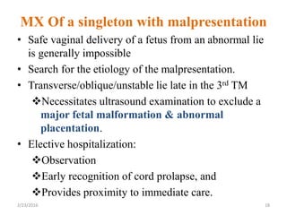
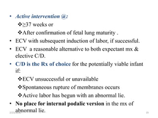
Malpresentation and Malposition
2/23/2016 1
Learning objectives
• Define malpresentation
• Mention pathogenesis for malpresentation
• List different types of malpresentation
• Understand the mode of delivery for each
malpresentation types
• Define malposition
• List types of malposition
2/23/2016 2
• Presentation is defined as:
Part of the fetus that directly overlies the pelvic
inlet, or foremost within the birth canal or in
closest proximity to it.
• Vertex presentation is normal presentation which
accounts 95-96%.
• Any presentation other than vertex are called
Malpresentation.
• Malpresentations include: Face, brow, breech,
shoulder, or compound.
• Malpresentation is often associated with increased
risk to both the mother and the fetus
2/23/2016 3
2/23/2016 4
• Factors associated with malpresentation
include:
1. Diminished vertical polarity of the
uterine cavity,
2. Increased or decreased fetal mobility,
3. Obstructed pelvic inlet, and
4. Fetal malformation
2/23/2016 5
• Diminished vertical polarity of the uterine cavity:
Parity
Myoma
Placentation
Müllerian duct fusion abnormalities such as septate
uterus or uterus didelphys
• Increased fetal mobility
Prematurity
Polyhydramnios
Multiple pregnancy
2/23/2016 6
• CPD
Severe fetal hydrocephalus
Contracted maternal pelvis
• Decreased fetal mobility
Aneuploidies,
Myotonic dystrophy,
Joint contractures from various etiologies,
Arthrogryposis,
Oligohydramnios, and
Fetal neurologic dysfunction that result in
decreased fetal muscle tone, strength, or activity
2/23/2016 7
`
• Variety of maneuvers intended to facilitate vaginal
delivery for malpresentations:
Destructive delivery
Manual or instrumental attempts
Internal podalic version(IPV)
• IPV followed by a complete breech extraction was once
advocated as a solution.
• However, it was associated with:
High fetal or maternal morbidity or mortality rate
& have been largely abandoned.
• C/D has become the recommended alternative to
manipulative vaginal techniques when normal progress
toward vaginal delivery is not observed.
2/23/2016 8
Abnormal Axial Lie
• Fetal “lie” indicates the orientation of the fetal spine
relative to the spine of the mother.
• Unstable lie if the
Fetal membranes are intact & there is great fetal
mobility resulting in frequent changes of lie or
presentation.
• Abnormal fetal lie is diagnosed:
1 in 300 cases, or 0.33% of pregnancies at term.
2% of pregnancies at 32 weeks, or six times the
rate found at term.
2/23/2016 9
• Persistence of abnormal lie >37 weeks requires a systematic clinical assessment &
a plan for mx, b/c rupture of membranes imposes high risk of:
Cord prolapse,
Fetal compromise, and
Maternal morbidity if neglected.
• Any condition that alters the normal vertical polarity of the intrauterine cavity will
predispose to abnormal lie.
2/23/2016 10
• Diagnosis of the abnormal lie: made by
Palpation or
Vaginal examination and
Verified by ultrasound.
• Sensitivity of Leopold’s maneuvers for the detection of
malpresentation to be only 28- 41% .
• Fetal loss rate of:
9.2% with an early diagnosis
27.5% with a delayed diagnosis indicates that early
diagnosis improves fetal outcome.
• Perinatal mortality rate for unstable or transverse lie
varies from 3.9% to 24%, with maternal mortality as
high as 10%.
2/23/2016 11
• Maternal deaths are usually related to:
Infection after PROM,
Hemorrhage secondary to abnormal placentation,
Complications of operative intervention for CPD , or
traumatic delivery
Uterine rupture
• Fetal loss of phenotypically & chromosomally normal is
primarily associated with:
Neglect
Prolapsed cord or
Traumatic delivery.
• Cord prolapse occurs 20 times as often with abnormal lie as
it does with a cephalic presentation
2/23/2016 12
Transverse lie
• Long axis of the fetus is perpendicular to
that of the mother
• Incidence 0.3%
• Shoulder is over the pelvic inlet
(shoulder presentation)
• Side of the mother on which the
acromion rests determines the
designation of the lie as Rt or Lt acromial
2/23/2016 13
Diagnosis
Inspection:
• Wide abdomen, the fundus extends to
only slightly above the umblicus
Palpation
• No fetal pole in the fundus
• Ballotable head found in one iliac
fossa and breech in the other.
2/23/2016 14
Vaginal Exam
 Early stage of labor: gridiron feel of
the ribs of the thorax
 Further dilatation: scapula and
clavicle
 Late labor: shoulder tightly wedged in
the pelvic canal
 Arm prolapse in to the vagina
U/S and X-ray are confirmatory
2/23/2016 15
Etiology
• Abdominal wall relaxation from high
parity
• Preterm fetus
• Placenta previa
• Abnormal Ux anatomy
• Excessive amniotic fluid
• Contracted pelvis
2/23/2016 16
Management
• Spontaneous delivery of fully developed
fetus is impossible
• C/delivery – in established labor
• ECV – before labor or early labor with
intact membrane
2/23/2016 17
MX Of a singleton with malpresentation
• Safe vaginal delivery of a fetus from an abnormal lie
is generally impossible
• Search for the etiology of the malpresentation.
• Transverse/oblique/unstable lie late in the 3rd TM
Necessitates ultrasound examination to exclude a
major fetal malformation & abnormal
placentation.
• Elective hospitalization:
Observation
Early recognition of cord prolapse, and
Provides proximity to immediate care.
2/23/2016 18
• Active intervention @:
≥37 weeks or
After confirmation of fetal lung maturity .
• ECV with subsequent induction of labor, if successful.
• ECV a reasonable alternative to both expectant mx &
elective C/D.
• C/D is the Rx of choice for the potentially viable infant
if:
ECV unsuccessful or unavailable
Spontaneous rupture of membranes occurs
Active labor has begun with an abnormal lie.
• No place for internal podalic version in the mx of
abnormal lie.
2/23/2016 19
 Attitude: position of the fetal head in relation to the neck.
 Normal attitude of the fetal vertex during labor is full flexion on the neck.
 Deflexed attitudes include various degrees of deflection
 Although safe vaginal delivery is possible in many cases, C/D is the only
appropriate alternative when arrest of progress is observed.
2/23/2016 20
INCIDENCE OF
MALPRESENTATION
• 96% of fetuses at term will present with vertex.
• 91.4% of fetuses present in vertex , occiput anterior (OA).
• Fetuses that are not in vertex presentation are considered to
have a malpresentation.
• At term, the types & estimated incidences of malpresentations
are:
Breech (1/33 deliveries)
Cephalic malpresentations (1/18 deliveries)
Face (1/600 to 1/800 deliveries)
Brow (1/500 to 1/4000 deliveries)
Compound (1/1500 deliveries)
Transverse lie (1/833 deliveries)
2/23/2016 21
FACE PRESENTATION
• Fetal neck is sharply deflexed, allowing the occiput
to touch the back and the face (from forehead to
chin) to present in the birth canal
2/23/2016 22
2/23/2016 23
Etiology and risk factors
• It is presumed to occur because of factors that:
 Favor extension or
 Prevent flexion of the fetal neck.
• A common risk factor is:
 An anomalous fetus (anencephaly, massive hydrocephalus, or an anterior
neck mass).
 Multiple nuchal cord loops
 Other factors includes:
 CPD
 Prematurity/low birth weight
 Macrosomia
 Contracted maternal pelvis & platypelloid pelvis
 polyhydramnios
 Black race and Multiparity
 Extreme laxity of the anterior abdominal wall
2/23/2016 24
Diagnosis
• Usually made late in the 1st or 2nd stage of
labor.
• On digital examination, landmarks are
palpating:
Orbital ridge and orbits,
Saddle of the nose
Mouth, and chin.
• Sonography = will show a hyper extended fetal
neck.
• Although imaging studies can be performed it is
not mandatory.
2/23/2016 25
2/23/2016 26
Course and management of labor
• At the time of diagnosis:
60 % will be in the MA position
26 %will be MP and
15 % will be MT.
• 30-50% of MP & MT positions will spontaneously
convert to the MA position during the course of labor.
• Mx requires close observation of the progress of labor
b/c CPD is more likely than with vertex presentation.
• The widest diameter of the fetal head negotiating the
pelvis in face presentation is the trachelo-bregmatic or
trachelo-parietal diameter average length 12.6 cm.
2/23/2016 27
• Despite the increased diameter, >75% of
MA are delivered vaginally,
• Whereas persistent MP & MT fetuses
require cesarean birth
• Abnormalities of the fetal heart rate
occur more frequently with face
presentations
2/23/2016 28
Mentum anterior
• Once engagement has occurred in MA, fetal
neck extends even further backward such that
the occiput touches the back.
• Internal rotation occurs b/n the level of
ischial spines & ischial tuberosities , making
the chin the actual presenting part of the face.
• As the face descends onto the perineum, the
fetal chin passes under the maternal symphysis
pubis, slight flexion of the neck occurs.
• Parturient may begin pushing at full dilatation.
2/23/2016 29
• Oxytocin augmentation:
If indicated and
Fetal heart rate pattern is reassuring.
• Outlet forceps should only be used by experienced
practitioners.
• Since engagement does not occur until the face is at +2
station, forceps should only be applied to the face that is
bulging the perineum.
• Attempts at:
Version,
Extraction, or
Midforceps delivery should be avoided, as they are
associated with unnecessary maternal trauma and
neonatal injury
2/23/2016 30
Mentum posterior
• In the MP position: the neck, head, & shoulders
must enter the pelvis simultaneously;
• However, the pelvis is usually not large enough to
accommodate.
• Also the fetal neck must extend the length of
the maternal sacrum (average 12 cm) in order to
reach the perineum.
• Lastly, an open fetal mouth may act as a fulcrum
against the sacrum preventing further descent.
• Therefore, MP will not deliver vaginally unless:
Spontaneous rotation occurs or
Fetus is very small (eg, very preterm)
2/23/2016 31
2/23/2016 32
Brow presentation
• Portion of fetal head b/n orbital ridge & the anterior
fontanel presents at the pelvic inlet
• Rarest (o.o6%)
• Presenting diameter is mento-vertical (mento-
parietal (or mento-bregmatic)) (13.5cm)
• Unstable commonly often converts to face or
vertex presentation.
• Fetal head occupies a position midway b/n flexion
(occiput) & extension (face)
• Engagement can’t take place ;unless head is small or
pelvis is very large
2/23/2016 33
Diagnosis
Abdominal palpation: occiput & chin
palpated easily
PV: frontal suture, large anterior fontanel
Orbital ridge & Eyes
Root of nose felt
Etiology
Same as face
2/23/2016 34
Mechanism of labor
• Very small fetus & large pelvis labor is
possible.
• Large fetus: engagement is impossible
until marked moulding shortens
occipitomental ø (13.5cm).
• Caput succedaneum: is over the forehead.
• Managed by C/S unless small fetus with
roomy pelvis.
2/23/2016 35
2/23/2016 36
2/23/2016 37
Compound presentation
• An extremity prolapse along side the presenting
part with both presenting in the pelvis
simultaneously
Cause:
Condition that prevents complete occlusion
of the pelvic inlet by the fetal head.
Management
The prolapsed part should be left as it is.
It will not interfere with labor
Retracts out of the way with descent of the
head
2/23/2016 38
Prognosis
Increased perinatal loss
Preterm delivery
Cord prolapse
Traumatic obstetrical procedures
2/23/2016 39
BREECH PRESENTAION
• The term derives from the same word as
britches, which described a cloth covering the
loins and thighs.
• There are three major types of breech
presentation:
Frank(50-70%)
Incomplete(10 to 40%)
Complete(5-10%)
kneeling
2/23/2016 40
Frank breech
The lower extremities are flexed at the hips and
extended at the knees.
Incomplete breech
One or both feet felt below the breech
A foot or knee is lower most in the birth
canal
Complete breech
One or both knees are flexed
The feet may be felt along side the
buttocks
2/23/2016 41
2/23/2016 43
PREVALENCE
20 to 25% of fetuses under 28 weeks are
breech,
only 7 to 16% are breech at 32 weeks, and
only 3 to 4% are breech at term
Spontaneous version may occur at any time
before delivery, even after 40 weeks of
gestation.
Likelihood of spontaneous version to
cephalic presentation after 36 weeks is
25%.
2/23/2016 44
PATHOGENESIS AND RISK
FACTORS
• Altered intrauterine contour or volume:
Uterine anomalies (eg, bicornuate or septate uterus)
Space occupying lesions (eg, uterine leiomyomata)
Placental abnormalities (eg, PP, cornual placenta)
Multiparity
Extremes of AFV (polyhydramnios, oligohydramnios)
Contracted maternal pelvis
• Altered fetal shape:
Fetal anomaly (eg, anencephaly, hydrocephaly,
sacrococcygeal teratoma, neck mass)
Extended fetal legs
2/23/2016 45
• Impaired fetal mobility:
Crowding from multiple gestation
Neurologic impairment
Short umbilical cord
Fetal asphyxia
• Other purported risk factors include:
Primiparity
Female gender
Maternal anticonvulsant therapy
Older maternal age
Fetal growth restriction, and
Previous breech presentation
2/23/2016 46
• Risk of breech presentation in a 2nd
pregnancy:
9% if the first infant was breech and
2% if the first infant was non breech .
After two consecutive breech deliveries, 21
to 28%
After 3 consecutive breech deliveries the
risk is 38%.
• Men or women who were delivered at term
from breech presentation were twice as likely
to have firstborn offspring in breech
2/23/2016 47
Diagnosis
Abdominal palpatión
• Hard , round, ballotable, fetal head occupy the
fundus
• The softer, breech in the LUs above the pelvic inlet
• FHB will be heard more easily at or above the
umbilicus
Vaginal exam
Frank breech
• Ischial tuberosity , sacrum , anus felt
• External genitalia , genital groove after further
descent
• Footling or complete breech __ foot may be felt
2/23/2016 48
Face presentation esp. in prolonged labor
• Breech:
Finger encounter muscular resistance by the
anus
stained with meconium on removal
Ischial tuberosity and anus are in straight
line
While mouth and malar eminence form
triangular shape in face.
2/23/2016 49
Imaging studies
U/S:
Confirmatory
Fetal anomaly
Type of breech, status of head
Estimation of fetal Wt (size)
X-ray:
Confirm the diagnosis (if u/s not available)
Determine the attitude
For pelvimetry
Other: CT, MRI
2/23/2016 50
Management
• Ante partum
 Follow closely for spontaneous version
 If persists ECV
• Labor and delivery
Absolute indications for C/delivery
large fetus ( EFW >3500gm)
Any degree of pelvic contracture
Hyper extended head
Footling breech
Ux dysfunction
2/23/2016 51
• Other obstetric indications for C/S
Sever IUGR
Breech with poor obstetric performance
(previous perinatal death ,Hx of infertility )
Elderly primigravida
Others:
Request for sterilization
Lack of an experienced operator
Zatuchini – Andros score <4
2/23/2016 52
If the score is 0-4, cesarean delivery is
recommended
decision regarding mode of delivery should
depend on the experience of the health care
provider
Zatuchni-Andros Breech Scoring
Add 0
Points
Add 1
Point
Add 2
Points
Parity 0 1 2
Gestationa
l age (wk)
39+ 38 <37
EFW (lb) 8lb (3.6kg)
7-8 lb(3.2-
3.6kg)
<7
lb(<3.2kg)
Previous
breech
0 1 2
Dilatation 2 3 4
Station -3 -2 -1
2/23/2016 53
Vaginal delivery
Indications
• No maternal or fetal indication for c/s
• Wt < 3500gm
• Franck breech
• Adequate pelvis
• Zatuchini – Andros score > 4
• Documented lethal fetal congenital anomalies
• Presentation of mother in advanced labor with no
maternal or fetal distress
2/23/2016 54
Mechanism of labor
Engagement and descent:
Takes place with the Bitrochanteric Ø in one of the
oblique Ø of the Pelvis
Internal rotation
Ant. hip toward the pubic arch
Bitrochanteric Ø in Antero-posterior
Lateral flexion
External rotation :
After birth of the breech
Back turning anteriorly
Shoulder brought into one of the oblique Ø of the
pelvis
2/23/2016 55
Methods of vaginal delivery
Spontaneous breech delivery
• Delivery occurs without assistance except
support of the infant
• No obstetric maneuvers are applied to the baby
Assisted vaginal breech delivery ( partial
breech extraction)
• Allow spontaneous delivery up to umbilicus
then the delivery of shoulders , arms , and
head assisted
2/23/2016 56
Total breech extraction
• Entire body is manually delivered
• Replaced by c/s except in desperate conditions
Indication:
• Fetal distress in 2nd stage of labor
• Cord prolapse or entanglement around the leg
• Need for expeditious delivery 2nd twin
2/23/2016 57
Total Breech Extraction….con
2/23/2016 58
Assisted vaginal breech delivery
• Instruct the mother to bear down with every
contraction (2nd stage).
• Episiotomy – when fetal anus is visible and
perineum distended unless perineum is well
relaxed.
• Allow the breech to be delivered and wait
without intervention till body born up to the
level of umbilicus.
2/23/2016 59
Pinnard maneuver
• If no spontaneous delivery of the legs
• Splinting the medial thigh of the fetus with the Position parallel to
the femur and exerting pressure laterally to sweep the legs away
from the mid line.
2/23/2016 60
2/23/2016 61
Delivery of the arms and shoulders
Loϋset maneuver:
 Rotate the fetus by half a circle(180) then reverse the
rotation half a circle
Delivery of the posterior arm followed by anterior
2/23/2016 62
2/23/2016 63
Delivery of the head
A. Mauriceau – smellie – veit maneuver (MSV)
• Index and middle finger of one hand over the maxillae to
flex the head
• Two fingers of the other hand hooked over the fetal neck
• Gentle suprapubic pressure→ by assistant
• Body of fetus elevated toward maternal abdomen
2/23/2016 64
B. Wigand maneuver
like MSV but differs
Assistant not needed to apply suprapubic
pressure
One hand put on the suprapubic area to provide
suprapubic pressure
C. Modified Prague maneuver
• Extraction of the head in a persistent OP
• Flex the head with in the birth canal and results in
delivery of the occiput over the perineum
2/23/2016 65
two fingers of one hand grasping the shoulders of the back-
down fetus from below while the other hand draws the
feet up over the maternal abdomen
2/23/2016 66
Head…
D. Burn’s marshall maneuver
• Allow the wt of the child’s trunk to pull the head
into the pelvis
• Trunk allowed to hang down unsupported ( one
or two min)
• The suboccipital area → if seen delivery could be
by lifting the child up towards the mothers
abdomen
E. Forceps:
 Pipers forceps
2/23/2016 67
2/23/2016 68
MALPOSITIONS
2/23/2016 69
• Position:
–Relation ship of a definite fetal presenting
presenting part o the maternal bony
pelvis.
–The most common position at delivery is
occiput anterior.
• Occiput: with a flexed head (cephalic
presentation)
• Sacrum: with breech presentation
• Mentum (chin): with an extended head (face
(face presentation)
2/23/2016 70
2/23/2016 71
Persistent occiput posterior position
• Most OP rotate to OA
• May be normal in early labor
Cause:
• Precise reason not known
Transverse narrowing of the mid pelvis
When it persists it may cause dystocia
2/3 of OP deliveries occurs with
who were OA at the beginning of
labor
2/23/2016 72
Possibilities for vaginal delivery of OP
 Spontaneous delivery__ if pelvis is roomy
 Forceps delivery as an OP
 Manual rotation to OA followed by spontaneous or
forceps delivery
 The requirements for forceps rotation must be met
before manual rotation
 Forceps rotation to OA and delivery
 Vacuum extraction for rotation, extraction or both
2/23/2016 73
Persistent occiput transverse position
• In absence of pelvic abnormality, it is frequently a
transient position
• Tends to rotate to OA
Cause
– Pelvic dystocia
– Ux dystocia
– Platypelloid or android pelvis
2/23/2016 74
Delivery
• In absence of CPD options of vaginal delivery
• Manual rotation to the occiput anterior or
posterior
• Forceps (Kiellands) in OT position
• Augmentation (if hypotonic ux dysfunction )
2/23/2016 75
2/23/2016 76

Malpresentation and Malposition.pptx

  • 1.
  • 2.
    Learning objectives • Definemalpresentation • Mention pathogenesis for malpresentation • List different types of malpresentation • Understand the mode of delivery for each malpresentation types • Define malposition • List types of malposition 2/23/2016 2
  • 3.
    • Presentation isdefined as: Part of the fetus that directly overlies the pelvic inlet, or foremost within the birth canal or in closest proximity to it. • Vertex presentation is normal presentation which accounts 95-96%. • Any presentation other than vertex are called Malpresentation. • Malpresentations include: Face, brow, breech, shoulder, or compound. • Malpresentation is often associated with increased risk to both the mother and the fetus 2/23/2016 3
  • 4.
  • 5.
    • Factors associatedwith malpresentation include: 1. Diminished vertical polarity of the uterine cavity, 2. Increased or decreased fetal mobility, 3. Obstructed pelvic inlet, and 4. Fetal malformation 2/23/2016 5
  • 6.
    • Diminished verticalpolarity of the uterine cavity: Parity Myoma Placentation Müllerian duct fusion abnormalities such as septate uterus or uterus didelphys • Increased fetal mobility Prematurity Polyhydramnios Multiple pregnancy 2/23/2016 6
  • 7.
    • CPD Severe fetalhydrocephalus Contracted maternal pelvis • Decreased fetal mobility Aneuploidies, Myotonic dystrophy, Joint contractures from various etiologies, Arthrogryposis, Oligohydramnios, and Fetal neurologic dysfunction that result in decreased fetal muscle tone, strength, or activity 2/23/2016 7
  • 8.
    ` • Variety ofmaneuvers intended to facilitate vaginal delivery for malpresentations: Destructive delivery Manual or instrumental attempts Internal podalic version(IPV) • IPV followed by a complete breech extraction was once advocated as a solution. • However, it was associated with: High fetal or maternal morbidity or mortality rate & have been largely abandoned. • C/D has become the recommended alternative to manipulative vaginal techniques when normal progress toward vaginal delivery is not observed. 2/23/2016 8
  • 9.
    Abnormal Axial Lie •Fetal “lie” indicates the orientation of the fetal spine relative to the spine of the mother. • Unstable lie if the Fetal membranes are intact & there is great fetal mobility resulting in frequent changes of lie or presentation. • Abnormal fetal lie is diagnosed: 1 in 300 cases, or 0.33% of pregnancies at term. 2% of pregnancies at 32 weeks, or six times the rate found at term. 2/23/2016 9
  • 10.
    • Persistence ofabnormal lie >37 weeks requires a systematic clinical assessment & a plan for mx, b/c rupture of membranes imposes high risk of: Cord prolapse, Fetal compromise, and Maternal morbidity if neglected. • Any condition that alters the normal vertical polarity of the intrauterine cavity will predispose to abnormal lie. 2/23/2016 10
  • 11.
    • Diagnosis ofthe abnormal lie: made by Palpation or Vaginal examination and Verified by ultrasound. • Sensitivity of Leopold’s maneuvers for the detection of malpresentation to be only 28- 41% . • Fetal loss rate of: 9.2% with an early diagnosis 27.5% with a delayed diagnosis indicates that early diagnosis improves fetal outcome. • Perinatal mortality rate for unstable or transverse lie varies from 3.9% to 24%, with maternal mortality as high as 10%. 2/23/2016 11
  • 12.
    • Maternal deathsare usually related to: Infection after PROM, Hemorrhage secondary to abnormal placentation, Complications of operative intervention for CPD , or traumatic delivery Uterine rupture • Fetal loss of phenotypically & chromosomally normal is primarily associated with: Neglect Prolapsed cord or Traumatic delivery. • Cord prolapse occurs 20 times as often with abnormal lie as it does with a cephalic presentation 2/23/2016 12
  • 13.
    Transverse lie • Longaxis of the fetus is perpendicular to that of the mother • Incidence 0.3% • Shoulder is over the pelvic inlet (shoulder presentation) • Side of the mother on which the acromion rests determines the designation of the lie as Rt or Lt acromial 2/23/2016 13
  • 14.
    Diagnosis Inspection: • Wide abdomen,the fundus extends to only slightly above the umblicus Palpation • No fetal pole in the fundus • Ballotable head found in one iliac fossa and breech in the other. 2/23/2016 14
  • 15.
    Vaginal Exam  Earlystage of labor: gridiron feel of the ribs of the thorax  Further dilatation: scapula and clavicle  Late labor: shoulder tightly wedged in the pelvic canal  Arm prolapse in to the vagina U/S and X-ray are confirmatory 2/23/2016 15
  • 16.
    Etiology • Abdominal wallrelaxation from high parity • Preterm fetus • Placenta previa • Abnormal Ux anatomy • Excessive amniotic fluid • Contracted pelvis 2/23/2016 16
  • 17.
    Management • Spontaneous deliveryof fully developed fetus is impossible • C/delivery – in established labor • ECV – before labor or early labor with intact membrane 2/23/2016 17
  • 18.
    MX Of asingleton with malpresentation • Safe vaginal delivery of a fetus from an abnormal lie is generally impossible • Search for the etiology of the malpresentation. • Transverse/oblique/unstable lie late in the 3rd TM Necessitates ultrasound examination to exclude a major fetal malformation & abnormal placentation. • Elective hospitalization: Observation Early recognition of cord prolapse, and Provides proximity to immediate care. 2/23/2016 18
  • 19.
    • Active intervention@: ≥37 weeks or After confirmation of fetal lung maturity . • ECV with subsequent induction of labor, if successful. • ECV a reasonable alternative to both expectant mx & elective C/D. • C/D is the Rx of choice for the potentially viable infant if: ECV unsuccessful or unavailable Spontaneous rupture of membranes occurs Active labor has begun with an abnormal lie. • No place for internal podalic version in the mx of abnormal lie. 2/23/2016 19
  • 20.
     Attitude: positionof the fetal head in relation to the neck.  Normal attitude of the fetal vertex during labor is full flexion on the neck.  Deflexed attitudes include various degrees of deflection  Although safe vaginal delivery is possible in many cases, C/D is the only appropriate alternative when arrest of progress is observed. 2/23/2016 20
  • 21.
    INCIDENCE OF MALPRESENTATION • 96%of fetuses at term will present with vertex. • 91.4% of fetuses present in vertex , occiput anterior (OA). • Fetuses that are not in vertex presentation are considered to have a malpresentation. • At term, the types & estimated incidences of malpresentations are: Breech (1/33 deliveries) Cephalic malpresentations (1/18 deliveries) Face (1/600 to 1/800 deliveries) Brow (1/500 to 1/4000 deliveries) Compound (1/1500 deliveries) Transverse lie (1/833 deliveries) 2/23/2016 21
  • 22.
    FACE PRESENTATION • Fetalneck is sharply deflexed, allowing the occiput to touch the back and the face (from forehead to chin) to present in the birth canal 2/23/2016 22
  • 23.
  • 24.
    Etiology and riskfactors • It is presumed to occur because of factors that:  Favor extension or  Prevent flexion of the fetal neck. • A common risk factor is:  An anomalous fetus (anencephaly, massive hydrocephalus, or an anterior neck mass).  Multiple nuchal cord loops  Other factors includes:  CPD  Prematurity/low birth weight  Macrosomia  Contracted maternal pelvis & platypelloid pelvis  polyhydramnios  Black race and Multiparity  Extreme laxity of the anterior abdominal wall 2/23/2016 24
  • 25.
    Diagnosis • Usually madelate in the 1st or 2nd stage of labor. • On digital examination, landmarks are palpating: Orbital ridge and orbits, Saddle of the nose Mouth, and chin. • Sonography = will show a hyper extended fetal neck. • Although imaging studies can be performed it is not mandatory. 2/23/2016 25
  • 26.
  • 27.
    Course and managementof labor • At the time of diagnosis: 60 % will be in the MA position 26 %will be MP and 15 % will be MT. • 30-50% of MP & MT positions will spontaneously convert to the MA position during the course of labor. • Mx requires close observation of the progress of labor b/c CPD is more likely than with vertex presentation. • The widest diameter of the fetal head negotiating the pelvis in face presentation is the trachelo-bregmatic or trachelo-parietal diameter average length 12.6 cm. 2/23/2016 27
  • 28.
    • Despite theincreased diameter, >75% of MA are delivered vaginally, • Whereas persistent MP & MT fetuses require cesarean birth • Abnormalities of the fetal heart rate occur more frequently with face presentations 2/23/2016 28
  • 29.
    Mentum anterior • Onceengagement has occurred in MA, fetal neck extends even further backward such that the occiput touches the back. • Internal rotation occurs b/n the level of ischial spines & ischial tuberosities , making the chin the actual presenting part of the face. • As the face descends onto the perineum, the fetal chin passes under the maternal symphysis pubis, slight flexion of the neck occurs. • Parturient may begin pushing at full dilatation. 2/23/2016 29
  • 30.
    • Oxytocin augmentation: Ifindicated and Fetal heart rate pattern is reassuring. • Outlet forceps should only be used by experienced practitioners. • Since engagement does not occur until the face is at +2 station, forceps should only be applied to the face that is bulging the perineum. • Attempts at: Version, Extraction, or Midforceps delivery should be avoided, as they are associated with unnecessary maternal trauma and neonatal injury 2/23/2016 30
  • 31.
    Mentum posterior • Inthe MP position: the neck, head, & shoulders must enter the pelvis simultaneously; • However, the pelvis is usually not large enough to accommodate. • Also the fetal neck must extend the length of the maternal sacrum (average 12 cm) in order to reach the perineum. • Lastly, an open fetal mouth may act as a fulcrum against the sacrum preventing further descent. • Therefore, MP will not deliver vaginally unless: Spontaneous rotation occurs or Fetus is very small (eg, very preterm) 2/23/2016 31
  • 32.
  • 33.
    Brow presentation • Portionof fetal head b/n orbital ridge & the anterior fontanel presents at the pelvic inlet • Rarest (o.o6%) • Presenting diameter is mento-vertical (mento- parietal (or mento-bregmatic)) (13.5cm) • Unstable commonly often converts to face or vertex presentation. • Fetal head occupies a position midway b/n flexion (occiput) & extension (face) • Engagement can’t take place ;unless head is small or pelvis is very large 2/23/2016 33
  • 34.
    Diagnosis Abdominal palpation: occiput& chin palpated easily PV: frontal suture, large anterior fontanel Orbital ridge & Eyes Root of nose felt Etiology Same as face 2/23/2016 34
  • 35.
    Mechanism of labor •Very small fetus & large pelvis labor is possible. • Large fetus: engagement is impossible until marked moulding shortens occipitomental ø (13.5cm). • Caput succedaneum: is over the forehead. • Managed by C/S unless small fetus with roomy pelvis. 2/23/2016 35
  • 36.
  • 37.
  • 38.
    Compound presentation • Anextremity prolapse along side the presenting part with both presenting in the pelvis simultaneously Cause: Condition that prevents complete occlusion of the pelvic inlet by the fetal head. Management The prolapsed part should be left as it is. It will not interfere with labor Retracts out of the way with descent of the head 2/23/2016 38
  • 39.
    Prognosis Increased perinatal loss Pretermdelivery Cord prolapse Traumatic obstetrical procedures 2/23/2016 39
  • 40.
    BREECH PRESENTAION • Theterm derives from the same word as britches, which described a cloth covering the loins and thighs. • There are three major types of breech presentation: Frank(50-70%) Incomplete(10 to 40%) Complete(5-10%) kneeling 2/23/2016 40
  • 41.
    Frank breech The lowerextremities are flexed at the hips and extended at the knees. Incomplete breech One or both feet felt below the breech A foot or knee is lower most in the birth canal Complete breech One or both knees are flexed The feet may be felt along side the buttocks 2/23/2016 41
  • 43.
  • 44.
    PREVALENCE 20 to 25%of fetuses under 28 weeks are breech, only 7 to 16% are breech at 32 weeks, and only 3 to 4% are breech at term Spontaneous version may occur at any time before delivery, even after 40 weeks of gestation. Likelihood of spontaneous version to cephalic presentation after 36 weeks is 25%. 2/23/2016 44
  • 45.
    PATHOGENESIS AND RISK FACTORS •Altered intrauterine contour or volume: Uterine anomalies (eg, bicornuate or septate uterus) Space occupying lesions (eg, uterine leiomyomata) Placental abnormalities (eg, PP, cornual placenta) Multiparity Extremes of AFV (polyhydramnios, oligohydramnios) Contracted maternal pelvis • Altered fetal shape: Fetal anomaly (eg, anencephaly, hydrocephaly, sacrococcygeal teratoma, neck mass) Extended fetal legs 2/23/2016 45
  • 46.
    • Impaired fetalmobility: Crowding from multiple gestation Neurologic impairment Short umbilical cord Fetal asphyxia • Other purported risk factors include: Primiparity Female gender Maternal anticonvulsant therapy Older maternal age Fetal growth restriction, and Previous breech presentation 2/23/2016 46
  • 47.
    • Risk ofbreech presentation in a 2nd pregnancy: 9% if the first infant was breech and 2% if the first infant was non breech . After two consecutive breech deliveries, 21 to 28% After 3 consecutive breech deliveries the risk is 38%. • Men or women who were delivered at term from breech presentation were twice as likely to have firstborn offspring in breech 2/23/2016 47
  • 48.
    Diagnosis Abdominal palpatión • Hard, round, ballotable, fetal head occupy the fundus • The softer, breech in the LUs above the pelvic inlet • FHB will be heard more easily at or above the umbilicus Vaginal exam Frank breech • Ischial tuberosity , sacrum , anus felt • External genitalia , genital groove after further descent • Footling or complete breech __ foot may be felt 2/23/2016 48
  • 49.
    Face presentation esp.in prolonged labor • Breech: Finger encounter muscular resistance by the anus stained with meconium on removal Ischial tuberosity and anus are in straight line While mouth and malar eminence form triangular shape in face. 2/23/2016 49
  • 50.
    Imaging studies U/S: Confirmatory Fetal anomaly Typeof breech, status of head Estimation of fetal Wt (size) X-ray: Confirm the diagnosis (if u/s not available) Determine the attitude For pelvimetry Other: CT, MRI 2/23/2016 50
  • 51.
    Management • Ante partum Follow closely for spontaneous version  If persists ECV • Labor and delivery Absolute indications for C/delivery large fetus ( EFW >3500gm) Any degree of pelvic contracture Hyper extended head Footling breech Ux dysfunction 2/23/2016 51
  • 52.
    • Other obstetricindications for C/S Sever IUGR Breech with poor obstetric performance (previous perinatal death ,Hx of infertility ) Elderly primigravida Others: Request for sterilization Lack of an experienced operator Zatuchini – Andros score <4 2/23/2016 52
  • 53.
    If the scoreis 0-4, cesarean delivery is recommended decision regarding mode of delivery should depend on the experience of the health care provider Zatuchni-Andros Breech Scoring Add 0 Points Add 1 Point Add 2 Points Parity 0 1 2 Gestationa l age (wk) 39+ 38 <37 EFW (lb) 8lb (3.6kg) 7-8 lb(3.2- 3.6kg) <7 lb(<3.2kg) Previous breech 0 1 2 Dilatation 2 3 4 Station -3 -2 -1 2/23/2016 53
  • 54.
    Vaginal delivery Indications • Nomaternal or fetal indication for c/s • Wt < 3500gm • Franck breech • Adequate pelvis • Zatuchini – Andros score > 4 • Documented lethal fetal congenital anomalies • Presentation of mother in advanced labor with no maternal or fetal distress 2/23/2016 54
  • 55.
    Mechanism of labor Engagementand descent: Takes place with the Bitrochanteric Ø in one of the oblique Ø of the Pelvis Internal rotation Ant. hip toward the pubic arch Bitrochanteric Ø in Antero-posterior Lateral flexion External rotation : After birth of the breech Back turning anteriorly Shoulder brought into one of the oblique Ø of the pelvis 2/23/2016 55
  • 56.
    Methods of vaginaldelivery Spontaneous breech delivery • Delivery occurs without assistance except support of the infant • No obstetric maneuvers are applied to the baby Assisted vaginal breech delivery ( partial breech extraction) • Allow spontaneous delivery up to umbilicus then the delivery of shoulders , arms , and head assisted 2/23/2016 56
  • 57.
    Total breech extraction •Entire body is manually delivered • Replaced by c/s except in desperate conditions Indication: • Fetal distress in 2nd stage of labor • Cord prolapse or entanglement around the leg • Need for expeditious delivery 2nd twin 2/23/2016 57
  • 58.
  • 59.
    Assisted vaginal breechdelivery • Instruct the mother to bear down with every contraction (2nd stage). • Episiotomy – when fetal anus is visible and perineum distended unless perineum is well relaxed. • Allow the breech to be delivered and wait without intervention till body born up to the level of umbilicus. 2/23/2016 59
  • 60.
    Pinnard maneuver • Ifno spontaneous delivery of the legs • Splinting the medial thigh of the fetus with the Position parallel to the femur and exerting pressure laterally to sweep the legs away from the mid line. 2/23/2016 60
  • 61.
  • 62.
    Delivery of thearms and shoulders Loϋset maneuver:  Rotate the fetus by half a circle(180) then reverse the rotation half a circle Delivery of the posterior arm followed by anterior 2/23/2016 62
  • 63.
  • 64.
    Delivery of thehead A. Mauriceau – smellie – veit maneuver (MSV) • Index and middle finger of one hand over the maxillae to flex the head • Two fingers of the other hand hooked over the fetal neck • Gentle suprapubic pressure→ by assistant • Body of fetus elevated toward maternal abdomen 2/23/2016 64
  • 65.
    B. Wigand maneuver likeMSV but differs Assistant not needed to apply suprapubic pressure One hand put on the suprapubic area to provide suprapubic pressure C. Modified Prague maneuver • Extraction of the head in a persistent OP • Flex the head with in the birth canal and results in delivery of the occiput over the perineum 2/23/2016 65
  • 66.
    two fingers ofone hand grasping the shoulders of the back- down fetus from below while the other hand draws the feet up over the maternal abdomen 2/23/2016 66
  • 67.
    Head… D. Burn’s marshallmaneuver • Allow the wt of the child’s trunk to pull the head into the pelvis • Trunk allowed to hang down unsupported ( one or two min) • The suboccipital area → if seen delivery could be by lifting the child up towards the mothers abdomen E. Forceps:  Pipers forceps 2/23/2016 67
  • 68.
  • 69.
  • 70.
    • Position: –Relation shipof a definite fetal presenting presenting part o the maternal bony pelvis. –The most common position at delivery is occiput anterior. • Occiput: with a flexed head (cephalic presentation) • Sacrum: with breech presentation • Mentum (chin): with an extended head (face (face presentation) 2/23/2016 70
  • 71.
  • 72.
    Persistent occiput posteriorposition • Most OP rotate to OA • May be normal in early labor Cause: • Precise reason not known Transverse narrowing of the mid pelvis When it persists it may cause dystocia 2/3 of OP deliveries occurs with who were OA at the beginning of labor 2/23/2016 72
  • 73.
    Possibilities for vaginaldelivery of OP  Spontaneous delivery__ if pelvis is roomy  Forceps delivery as an OP  Manual rotation to OA followed by spontaneous or forceps delivery  The requirements for forceps rotation must be met before manual rotation  Forceps rotation to OA and delivery  Vacuum extraction for rotation, extraction or both 2/23/2016 73
  • 74.
    Persistent occiput transverseposition • In absence of pelvic abnormality, it is frequently a transient position • Tends to rotate to OA Cause – Pelvic dystocia – Ux dystocia – Platypelloid or android pelvis 2/23/2016 74
  • 75.
    Delivery • In absenceof CPD options of vaginal delivery • Manual rotation to the occiput anterior or posterior • Forceps (Kiellands) in OT position • Augmentation (if hypotonic ux dysfunction ) 2/23/2016 75
  • 76.

Editor's Notes

  • #20 Phelan and colleagues reported 29 patients with transverse lie diagnosed at or beyond 37 weeks’ gestation and managed expectantly. 83% spontaneously converted to breech (9 of 24) or vertex (15 of 24) before labor; However, the overall cesarean delivery rate was 45%,and There were two cases of cord prolapse, One uterine rupture, and One neonatal death.