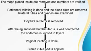
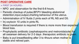
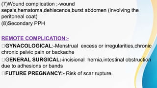
MITTAL COLLEGE OF NURSING
AJMER
CLASS PRESENTATION ON
LOWER SEGMENT CESAREAN SECTION
(LSCS)
SUBMITTED TO SUBMITTED BY
MRS. SNEHLATA PARASHAR MR. MAHIPAL LAMROR
( LECTURER ) BSC.NURSING 4th YEAR
• INTRODUCTION-
It is an operative procedure where by the fetus after the end of
28th weeks are delivered through an incision on the abdominal
and uterine walls.
• -The word cesarean is derived from the latin verb”cedere” which
means “to cut”.
• -The first operation performed on a patient is reffered to as a
primary cesarean section.
• -when the operational is performed in subsequent pregnancies,it
is called repeat cesarean section.
• -Kronig in 1912,introduced lower segment vertical incision and it
was popularized by De Lee (1922).
• INCIDENCE-”Steadily rising”
• Identification of at risk fetuses before term(IUGR)
• Identification of at risk mothers
• Wider use of repeat CS in cases with previous cesarean
delivery.
• Decrease in vaginal breach delivery.
• Increased no of women with age more than 30 years at
conception and association medical complication.
• Wider use of electronic fetal monitoring and increased
diagnosis of fetal distress.
• Cesarean delivery on demand
• INDICATION-
• The indication are broadly divided into two categories.
• (1)ABSOLUTE-(Vaginal delivery is not possible)
• Central placenta previa
• Contracted pelvis or cephalopelvic disproportion.
• Pelvic mass causing obstruction(cervical or broad ligament
fibroid).
• Advanced carcinoma cervix.
• Vaginal obstruction (atresia stenosis).
• (2)RELATIVE INDICATION-(Vaginal delivery is possible but high risk to mother & baby)
• Previous cesarean delivery
• Non reassuring FHR(fetal distress)
• Dystocia
• Antepartum hemorrhage (placenta previa & abruptio placenta)
• Malpresentation
• Failed surgical lnduction of labour, failure to progress in labor.
• Bad obsteric history with recurrent fetal wastage.
• Hypertensive disorder of pregnancy (severe pre eclampsia, eclampsia uncontrolled
fits even with antiseizure therapy.
• Medical gynecological disorder
• a)Uncontrolled diabetes
• b)Heart disease
• c)Mechanical obstruction due to benign or malignant pelvic tumors (carcinoma
cervix)or following repair of VVF.
• TIME OF OPERATION
• 1.Elective lscs
• 2.Emergency lscs
• 1.ELECTIVE LSCS :- Operation done at per arranged time
during pregnancy to ensure the best quality of obstetrics,
anaesthesia, neonatal resuscitation &nursing services.
• a)Maturity Certain:-Operation done at 1 week prior to the
expected date of delivery.
• b)Maturity Uncertain :-Amniocentesis is done for L/S ratio to
ensure the fetal lung maturity, otherwise spontaneous onset of
labor is awaited, then CS is done.
2.EMERGENCY LSCS :- Operation is done due to acute obstetric
emergencies.
Category 1:- When there is immediate threat to the life of the
woman or the fetus. Decision delivery interval should be 30
minutes.
Category 2:- when there is maternal or fetal compromise which
is not immediately life threatening. CS should be done within 75
minutes of making decision.
Category 3:-There is no maternal or fetal compromise but needs
early delivery.
Category 4:-Delivery is planned to suit the woman,family
members and the hospital staff.
• TYPES OF OPERATION :-
a)Lower segment cesarean section
b)Classic cesarean section
a)LOWER SEGMENT CESAREAN SECTION :-
In this operation, the extraction of the baby is done through an
incision made in the lower segment through a transperitoneal
approach.
B)CLASSICAL CESAREAN SECTION :-
In this operation, the baby is extracted through an incision made
in the upper segment of the uterus.
• PECULIARITIES OF LOWER UTERINE SEGMENT:-
 Peritoneum is more loosely attached to the uterus.
 Contraction is less than in upper part of uterus.
 Lower segment is less vascular.
 Thin muscle layer.
 Healing is more efficient.
 Sutures are intact (less problem with suture loosening)
 PRE OPERATIVE PERPARATIONS:-
 Informed written high risk consent for the procedure,
anesthesia and arrangement of properly cross matched blood.
 Antacid (sodium citrate 30ml ) given orally before transferring
the patient to the OT.
 Ranitidine 150mg orally night before and is repeated by 50mg.
IV 1 hr before surgery.
 Metoclopramide (10mg/IV) is given to increase the tone of the
lower esophagel sphincter.
 Bladder is emptied by a foleys catheter.
 FSH should be checked once more at this stage.
 Neonatologist should be made available.
• ANAESTHESIA :- May be spinal, epidural or general.
• POSITION OF THE PATIENT:-
• Patient is kept in dorsal position. In susceptible cases, to
minimize any adverse effects of venacaval compression, a 15
degree tilt to her left using a wedge till delivery of the baby is
benificial.
• ANTISEPTIC PAINTING :-
• Abdomen is painted with 7.5% povidine iodine solution.
• INCISION ON THE ABDOMEN :-
• Either a vertical or transverse skin incision
• Vertical incision :- Modified pfannenstell incision is made 3cm
above the public symphysis .
Advantages of transverse /
Pfannenstell incision
• Post operative comfort is more.
• Fundus of the uterus can be better palpated during immediate
post operative period.
• Less chance of incisional hernia.
• Cosmetic value.
• STEPS (PROCEDUR )
Assessment of the presenting part
Identifying the incision line
Pfannenstell incision
Incision to the rectus sheath
Open the rectus muscle and retract laterally
Open the peritoneal cavity
Parietoperitonum of the bladder & uterus is seprated by fingers
Doyen’s retractor is introduced & bladder is
pushed downward
Recognition of the lower uterine segment is made
by the presence of loose peritoneum over it
The loose peritoneum is incised transverse
Lower uterine segment incision is made in the middle,
deepended till the membranes are reached and
then extended laterally.
Amniotic sac is ruptured and the doyen’s retactor is removed
The presenting part is part is hooked by the surgeon
and delivered while Assistant applies fundal pressure
Green armytage hemostatic forceps are applied to the
angles and margins of uterine incision to control bleeding.
Umblical cord is clamped at two places and cut
Doyen’s retactor is reintroduced and the placenta and
membranes are delivered
Uteurs is exteriorized and the inside of the uterus is insepected for
any abnormailties and completeness of removal of contents
blade
handle
lock
STRUCTURE OF THE UTERINE WOUND IN 3 LAYERS
(1)Suture is placed on the for side in lateral angle of uterine
incision.
Suture material is no. 0 chronic catgut suture
Continuous running suture taking deeper muscles.
(2) SECOND LAYER-a similar continuous suture is placed taking
the superficial muscles and adjacent fascia overlapping the first
layer of suture
(3)peritoneal flaps are opposed by continuous inverting. Suture but
it is not necessary to close the visceral and parital peritoneal
layer.
The maps placed inside are removed and numbers are verified
Peritoneal toileting is done and the blood clots are removed
bilateral tubes and ovaries are examined
Doyen’s retractor is removed
After being satisfied that the uterus is well contracted.
the abdomen is closed in layers
Vaginal toileting is done
Sterile vulva pad is applied
POST OPERATIVE CARE
 FIRST 24 HOURS:-
• NPO and observation for the first 6-8 hours.
• Periodic checkup of pulse,BP,PV bleeding abdominal
distention,input,output,charting behaviour of the uterus.
• Administration of IV fluids 2 pints each of RL NS and D5.
• Inj oxytocin 10 units in pints RL
• Blood transfusion is required if there is more more than expected
blood loss.
• Prophylactic antibiotic (cephalosporins and metronidaizole) for
all cesarean delivery for 2-3 days .theraputoic antibiotc is given.
• Baby is put breastfeeding after 3 to 4hours when mother is
stable and relived pain.
 1st post operative day;-oral feeding in the form of sips is
given.ambulation is done and patient shifted to ward and oral
antibiotic is given.
 2nd post operative :-soft diet and ambulation.
 3rd post operative day:-observation of wound for any soakage
and bleeding soft to normal diet.
 5th post operative day:-suture out is done and the patient is
discharged and contraceptive device is given.
COMPLICATION:-
Intraoperative complication:-
(1)Extension of the uterine incision:-may lead to bleeding from the
uterine vessels and formation of broad ligament hematoma
(2)Uterine laceration at lower uterine segment may extended
laterally and inferiorly into the vagina.
(3)Bladder injury
(4)Urethral injury
(5)GI tract injury
(6)Excessive hemorrhage
FETAL COMPLICAL:-
Latrogenic prematurity and developmental of RDS
POST OPERATIVE COMPLICATION:-
IMMEDIATE COMPLICATION:-
(1)Post partum haemorrhage:-due to uterine atony
(2)Shock
(3)Anaesthetic hazards:-aspiration of gastric contents ,may result
in aspiration atelectasis and aspiration pneumonitis.
OTHERS:- Hypotension and spinal headache
(4)infections:-endomyometritis ,UTI,wound infections,peritonitis
(5)Intestinal obstraction ;-due to formation of adhesions and bands
or paralyticileus following peritonitis.
(6)DVT and thromboembolic disorder
(7)Wound complication ;-wound
sepsis,hematoma,dehiscence,burst abdomen (involving the
peritoneal coat)
(8)Secondary PPH
REMOTE COMPLICATION:-
GYNACOLOGICAL:-Menstrual excess or irregularities,chronic
chronic pelvic pain or backache
GENERAL SURGICAL:-incisional hernia,intestinal obstruction
due to adhesions or bands
FUTURE PREGNANCY:- Risk of scar rupture.
REFERENCE:-
DC DUTT’S textbook of obstrectis 8th edition.
LCSC slide share
lscs ppt.pptx

lscs ppt.pptx

  • 1.
    MITTAL COLLEGE OFNURSING AJMER CLASS PRESENTATION ON LOWER SEGMENT CESAREAN SECTION (LSCS) SUBMITTED TO SUBMITTED BY MRS. SNEHLATA PARASHAR MR. MAHIPAL LAMROR ( LECTURER ) BSC.NURSING 4th YEAR
  • 2.
    • INTRODUCTION- It isan operative procedure where by the fetus after the end of 28th weeks are delivered through an incision on the abdominal and uterine walls. • -The word cesarean is derived from the latin verb”cedere” which means “to cut”. • -The first operation performed on a patient is reffered to as a primary cesarean section. • -when the operational is performed in subsequent pregnancies,it is called repeat cesarean section. • -Kronig in 1912,introduced lower segment vertical incision and it was popularized by De Lee (1922).
  • 4.
    • INCIDENCE-”Steadily rising” •Identification of at risk fetuses before term(IUGR) • Identification of at risk mothers • Wider use of repeat CS in cases with previous cesarean delivery. • Decrease in vaginal breach delivery. • Increased no of women with age more than 30 years at conception and association medical complication. • Wider use of electronic fetal monitoring and increased diagnosis of fetal distress. • Cesarean delivery on demand
  • 5.
    • INDICATION- • Theindication are broadly divided into two categories. • (1)ABSOLUTE-(Vaginal delivery is not possible) • Central placenta previa • Contracted pelvis or cephalopelvic disproportion. • Pelvic mass causing obstruction(cervical or broad ligament fibroid). • Advanced carcinoma cervix. • Vaginal obstruction (atresia stenosis).
  • 6.
    • (2)RELATIVE INDICATION-(Vaginaldelivery is possible but high risk to mother & baby) • Previous cesarean delivery • Non reassuring FHR(fetal distress) • Dystocia • Antepartum hemorrhage (placenta previa & abruptio placenta) • Malpresentation • Failed surgical lnduction of labour, failure to progress in labor. • Bad obsteric history with recurrent fetal wastage. • Hypertensive disorder of pregnancy (severe pre eclampsia, eclampsia uncontrolled fits even with antiseizure therapy. • Medical gynecological disorder • a)Uncontrolled diabetes • b)Heart disease • c)Mechanical obstruction due to benign or malignant pelvic tumors (carcinoma cervix)or following repair of VVF.
  • 7.
    • TIME OFOPERATION • 1.Elective lscs • 2.Emergency lscs • 1.ELECTIVE LSCS :- Operation done at per arranged time during pregnancy to ensure the best quality of obstetrics, anaesthesia, neonatal resuscitation &nursing services. • a)Maturity Certain:-Operation done at 1 week prior to the expected date of delivery. • b)Maturity Uncertain :-Amniocentesis is done for L/S ratio to ensure the fetal lung maturity, otherwise spontaneous onset of labor is awaited, then CS is done.
  • 8.
    2.EMERGENCY LSCS :-Operation is done due to acute obstetric emergencies. Category 1:- When there is immediate threat to the life of the woman or the fetus. Decision delivery interval should be 30 minutes. Category 2:- when there is maternal or fetal compromise which is not immediately life threatening. CS should be done within 75 minutes of making decision. Category 3:-There is no maternal or fetal compromise but needs early delivery. Category 4:-Delivery is planned to suit the woman,family members and the hospital staff.
  • 9.
    • TYPES OFOPERATION :- a)Lower segment cesarean section b)Classic cesarean section a)LOWER SEGMENT CESAREAN SECTION :- In this operation, the extraction of the baby is done through an incision made in the lower segment through a transperitoneal approach. B)CLASSICAL CESAREAN SECTION :- In this operation, the baby is extracted through an incision made in the upper segment of the uterus.
  • 10.
    • PECULIARITIES OFLOWER UTERINE SEGMENT:-  Peritoneum is more loosely attached to the uterus.  Contraction is less than in upper part of uterus.  Lower segment is less vascular.  Thin muscle layer.  Healing is more efficient.  Sutures are intact (less problem with suture loosening)
  • 11.
     PRE OPERATIVEPERPARATIONS:-  Informed written high risk consent for the procedure, anesthesia and arrangement of properly cross matched blood.  Antacid (sodium citrate 30ml ) given orally before transferring the patient to the OT.  Ranitidine 150mg orally night before and is repeated by 50mg. IV 1 hr before surgery.  Metoclopramide (10mg/IV) is given to increase the tone of the lower esophagel sphincter.  Bladder is emptied by a foleys catheter.  FSH should be checked once more at this stage.  Neonatologist should be made available.
  • 12.
    • ANAESTHESIA :-May be spinal, epidural or general. • POSITION OF THE PATIENT:- • Patient is kept in dorsal position. In susceptible cases, to minimize any adverse effects of venacaval compression, a 15 degree tilt to her left using a wedge till delivery of the baby is benificial. • ANTISEPTIC PAINTING :- • Abdomen is painted with 7.5% povidine iodine solution. • INCISION ON THE ABDOMEN :- • Either a vertical or transverse skin incision • Vertical incision :- Modified pfannenstell incision is made 3cm above the public symphysis .
  • 13.
    Advantages of transverse/ Pfannenstell incision • Post operative comfort is more. • Fundus of the uterus can be better palpated during immediate post operative period. • Less chance of incisional hernia. • Cosmetic value.
  • 14.
    • STEPS (PROCEDUR) Assessment of the presenting part Identifying the incision line Pfannenstell incision Incision to the rectus sheath Open the rectus muscle and retract laterally Open the peritoneal cavity Parietoperitonum of the bladder & uterus is seprated by fingers
  • 15.
    Doyen’s retractor isintroduced & bladder is pushed downward Recognition of the lower uterine segment is made by the presence of loose peritoneum over it The loose peritoneum is incised transverse Lower uterine segment incision is made in the middle, deepended till the membranes are reached and then extended laterally.
  • 16.
    Amniotic sac isruptured and the doyen’s retactor is removed The presenting part is part is hooked by the surgeon and delivered while Assistant applies fundal pressure Green armytage hemostatic forceps are applied to the angles and margins of uterine incision to control bleeding. Umblical cord is clamped at two places and cut Doyen’s retactor is reintroduced and the placenta and membranes are delivered Uteurs is exteriorized and the inside of the uterus is insepected for any abnormailties and completeness of removal of contents
  • 17.
  • 18.
    STRUCTURE OF THEUTERINE WOUND IN 3 LAYERS (1)Suture is placed on the for side in lateral angle of uterine incision. Suture material is no. 0 chronic catgut suture Continuous running suture taking deeper muscles. (2) SECOND LAYER-a similar continuous suture is placed taking the superficial muscles and adjacent fascia overlapping the first layer of suture (3)peritoneal flaps are opposed by continuous inverting. Suture but it is not necessary to close the visceral and parital peritoneal layer.
  • 19.
    The maps placedinside are removed and numbers are verified Peritoneal toileting is done and the blood clots are removed bilateral tubes and ovaries are examined Doyen’s retractor is removed After being satisfied that the uterus is well contracted. the abdomen is closed in layers Vaginal toileting is done Sterile vulva pad is applied
  • 20.
    POST OPERATIVE CARE FIRST 24 HOURS:- • NPO and observation for the first 6-8 hours. • Periodic checkup of pulse,BP,PV bleeding abdominal distention,input,output,charting behaviour of the uterus. • Administration of IV fluids 2 pints each of RL NS and D5. • Inj oxytocin 10 units in pints RL • Blood transfusion is required if there is more more than expected blood loss. • Prophylactic antibiotic (cephalosporins and metronidaizole) for all cesarean delivery for 2-3 days .theraputoic antibiotc is given. • Baby is put breastfeeding after 3 to 4hours when mother is stable and relived pain.
  • 21.
     1st postoperative day;-oral feeding in the form of sips is given.ambulation is done and patient shifted to ward and oral antibiotic is given.  2nd post operative :-soft diet and ambulation.  3rd post operative day:-observation of wound for any soakage and bleeding soft to normal diet.  5th post operative day:-suture out is done and the patient is discharged and contraceptive device is given.
  • 22.
    COMPLICATION:- Intraoperative complication:- (1)Extension ofthe uterine incision:-may lead to bleeding from the uterine vessels and formation of broad ligament hematoma (2)Uterine laceration at lower uterine segment may extended laterally and inferiorly into the vagina. (3)Bladder injury (4)Urethral injury (5)GI tract injury (6)Excessive hemorrhage FETAL COMPLICAL:- Latrogenic prematurity and developmental of RDS
  • 23.
    POST OPERATIVE COMPLICATION:- IMMEDIATECOMPLICATION:- (1)Post partum haemorrhage:-due to uterine atony (2)Shock (3)Anaesthetic hazards:-aspiration of gastric contents ,may result in aspiration atelectasis and aspiration pneumonitis. OTHERS:- Hypotension and spinal headache (4)infections:-endomyometritis ,UTI,wound infections,peritonitis (5)Intestinal obstraction ;-due to formation of adhesions and bands or paralyticileus following peritonitis. (6)DVT and thromboembolic disorder
  • 24.
    (7)Wound complication ;-wound sepsis,hematoma,dehiscence,burstabdomen (involving the peritoneal coat) (8)Secondary PPH REMOTE COMPLICATION:- GYNACOLOGICAL:-Menstrual excess or irregularities,chronic chronic pelvic pain or backache GENERAL SURGICAL:-incisional hernia,intestinal obstruction due to adhesions or bands FUTURE PREGNANCY:- Risk of scar rupture.
  • 25.
    REFERENCE:- DC DUTT’S textbookof obstrectis 8th edition. LCSC slide share