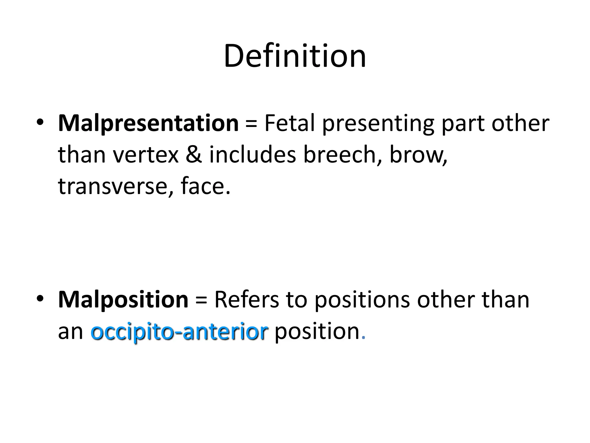
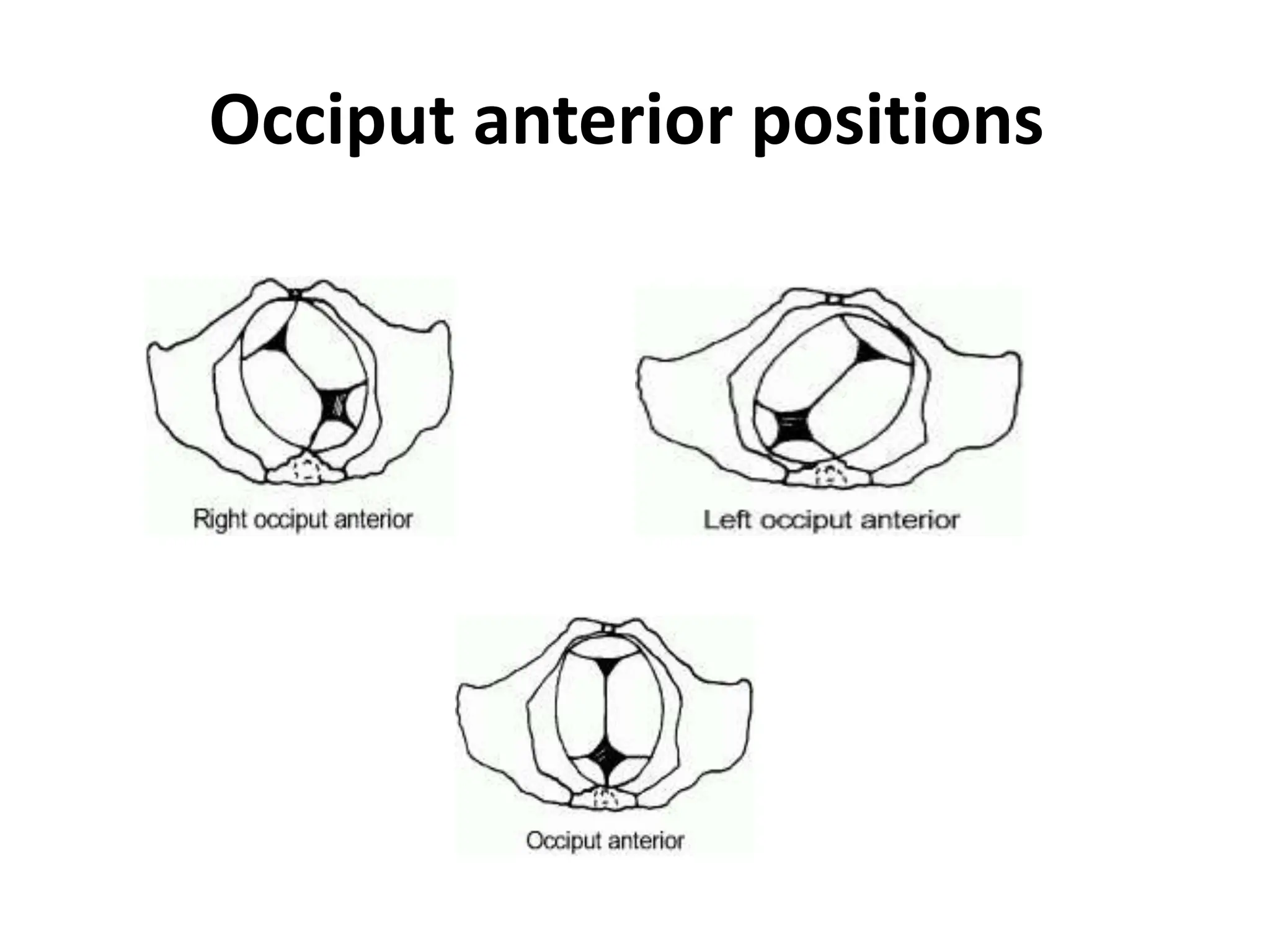
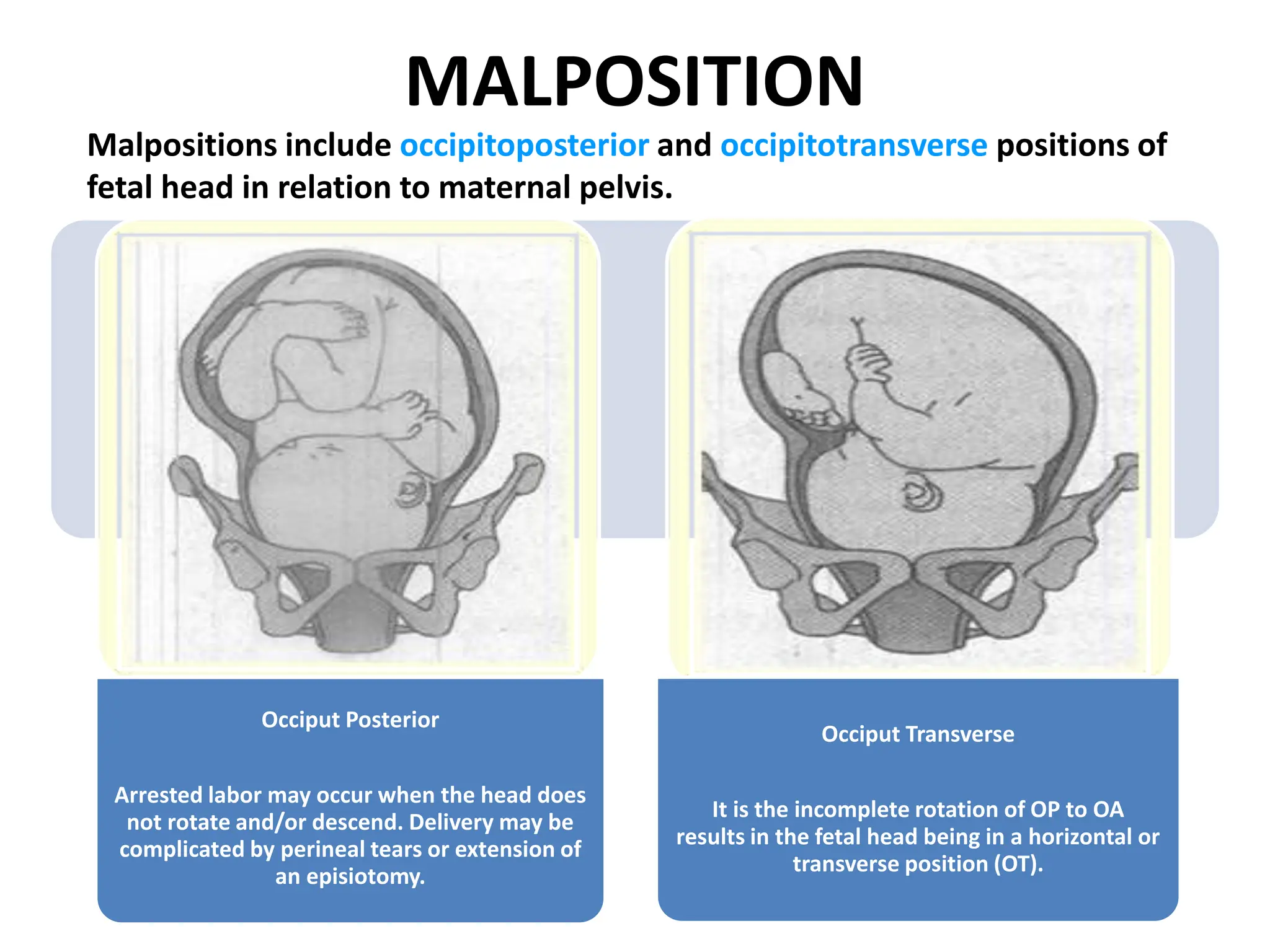
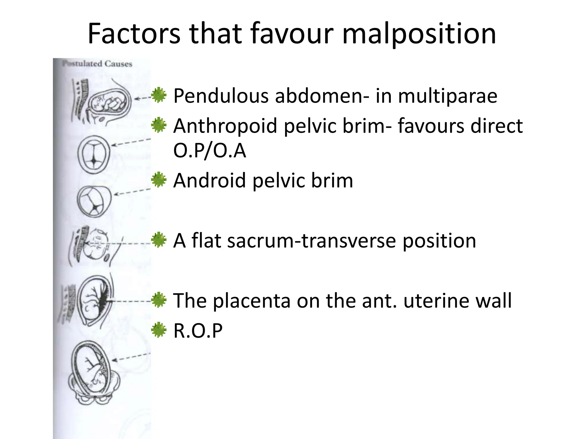
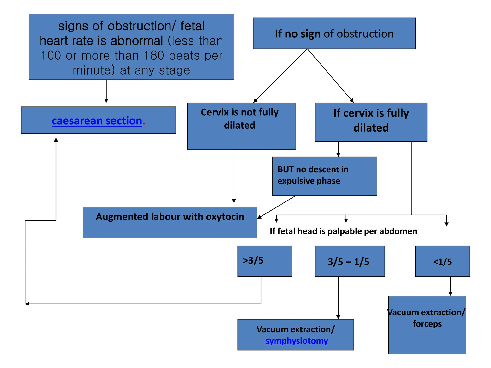
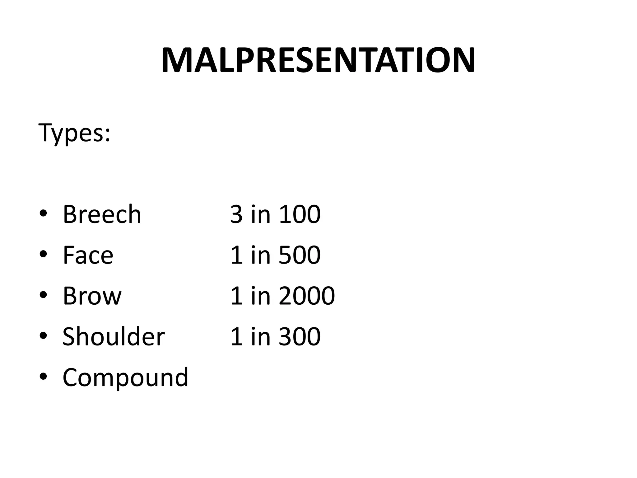
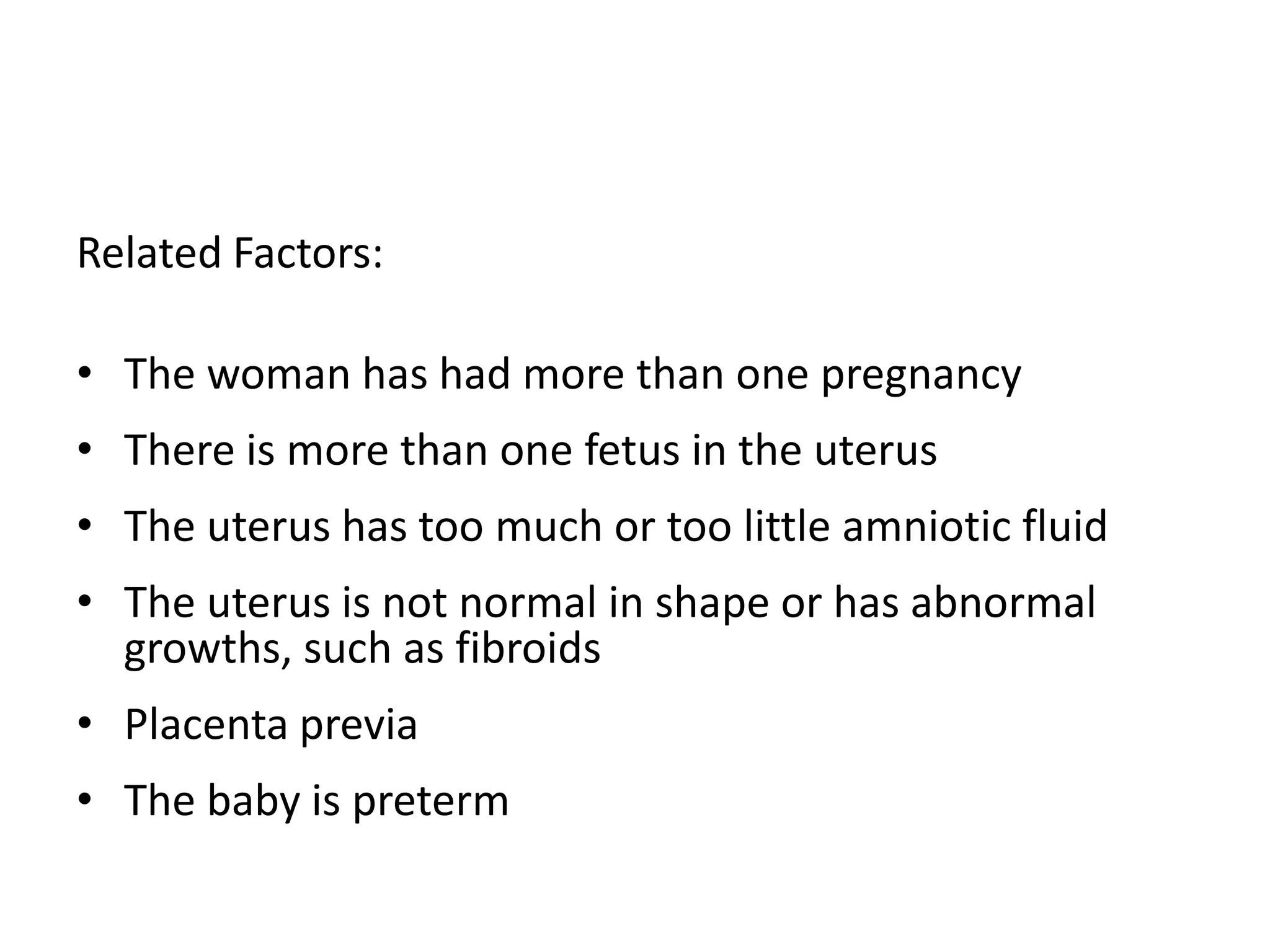
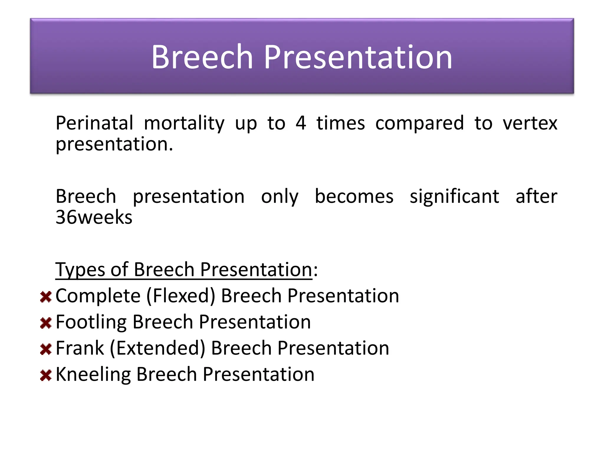
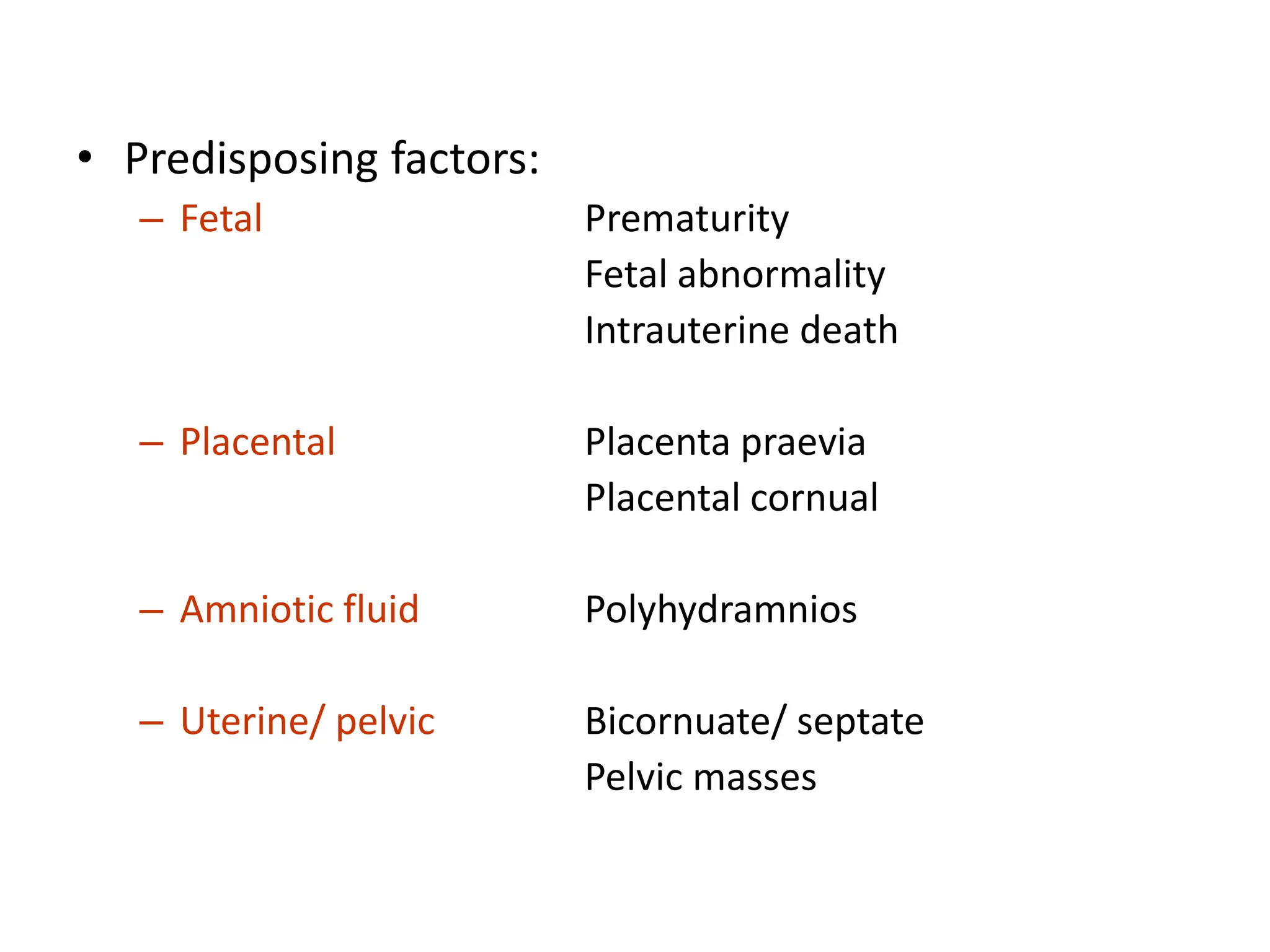
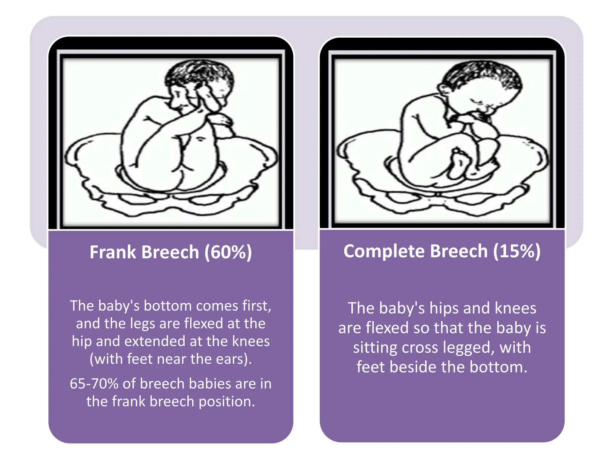
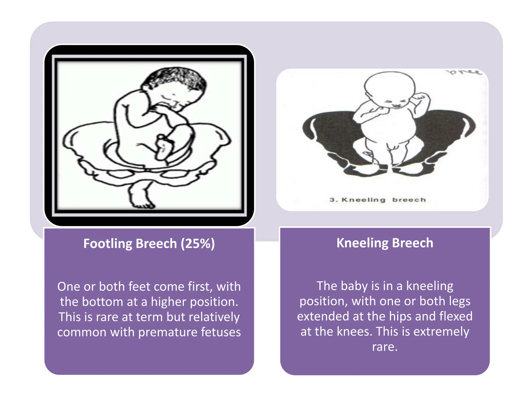
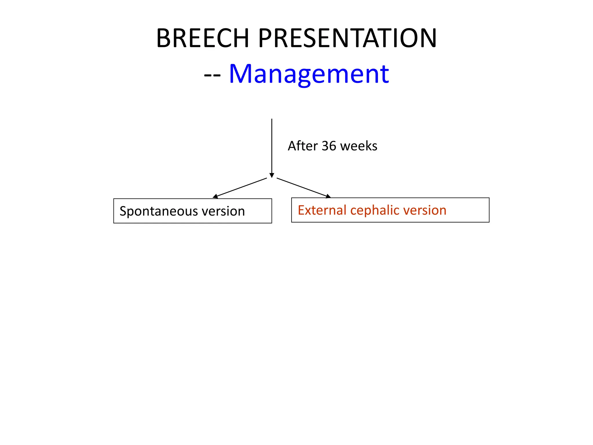
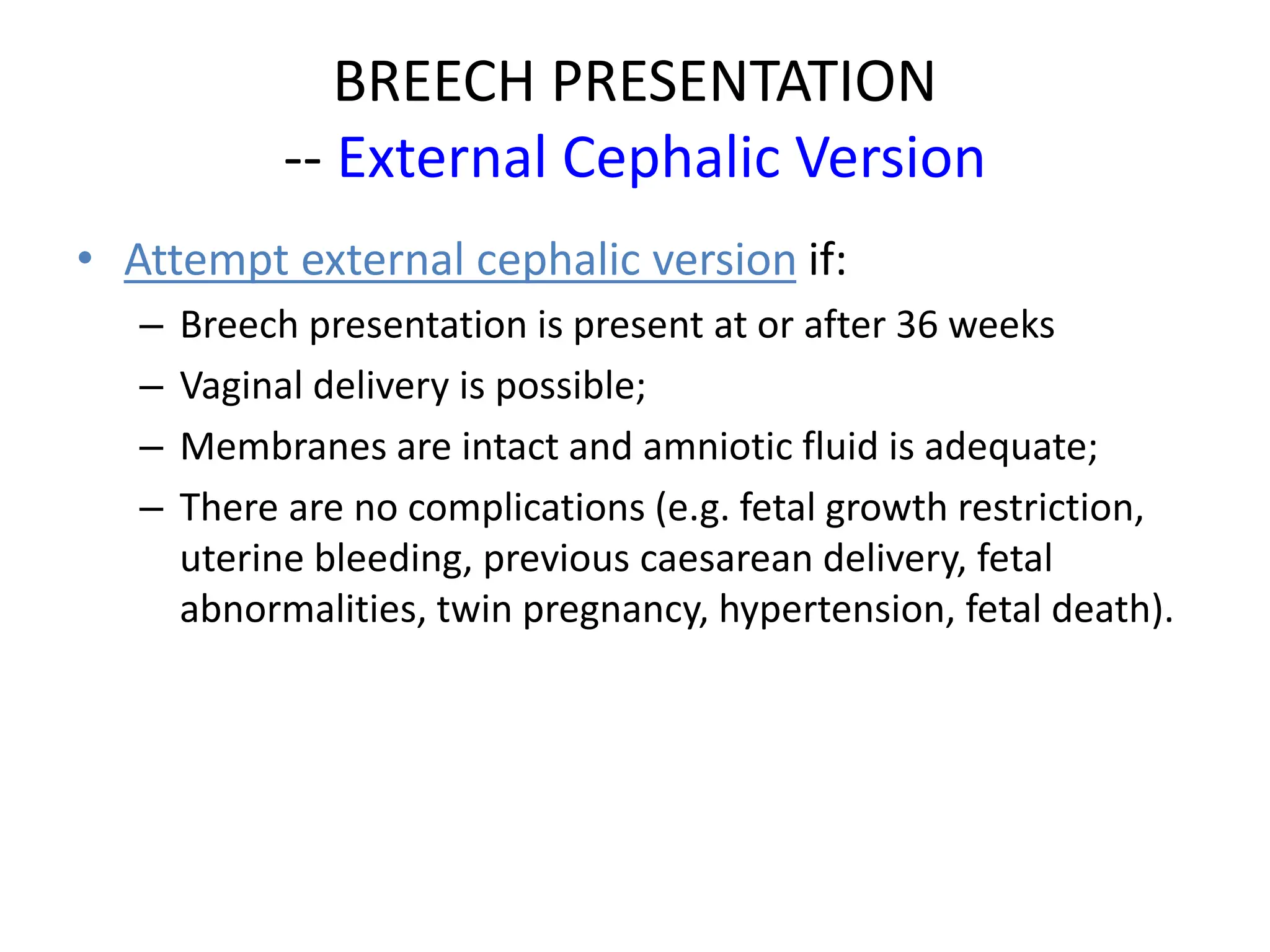
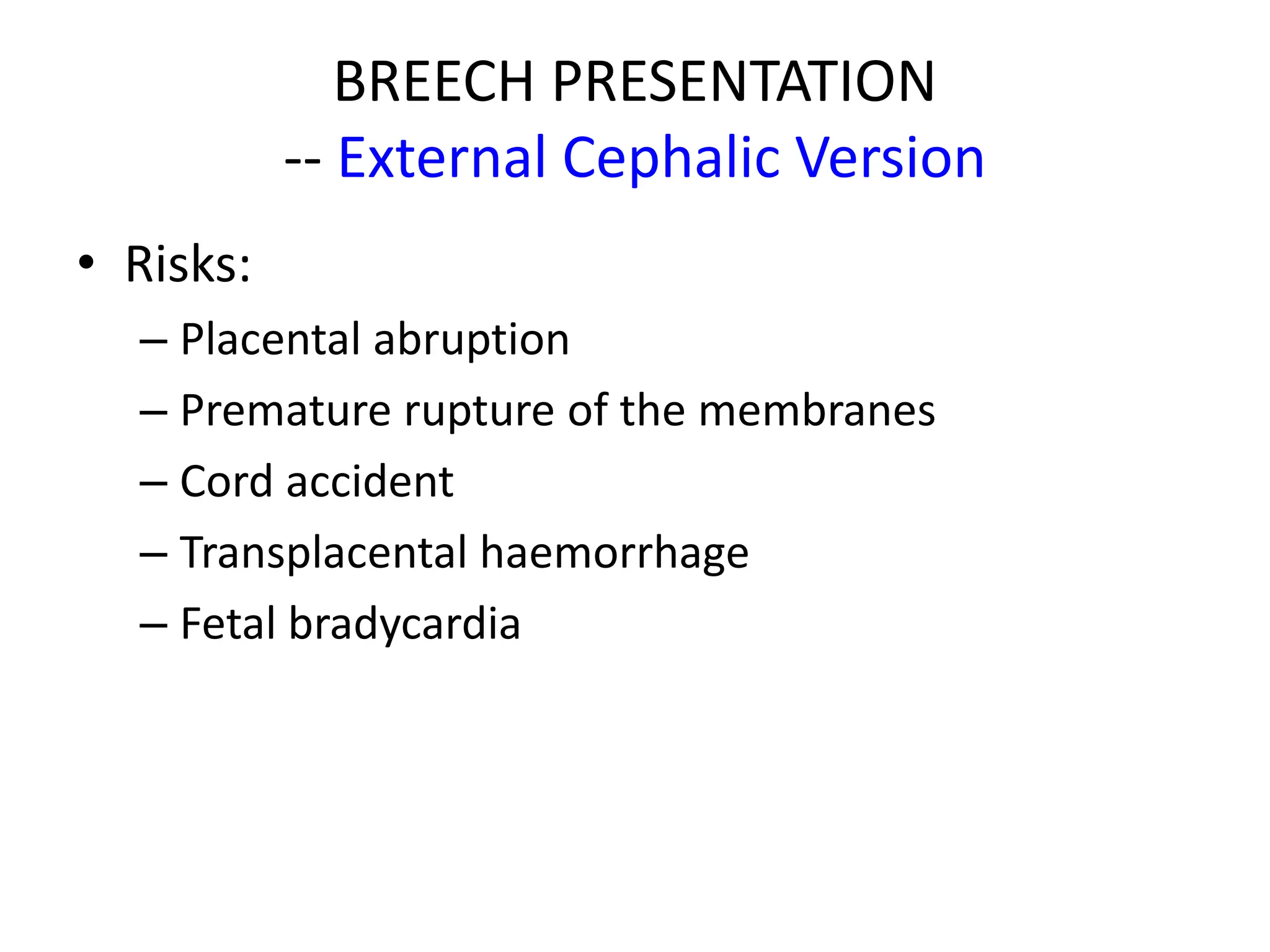
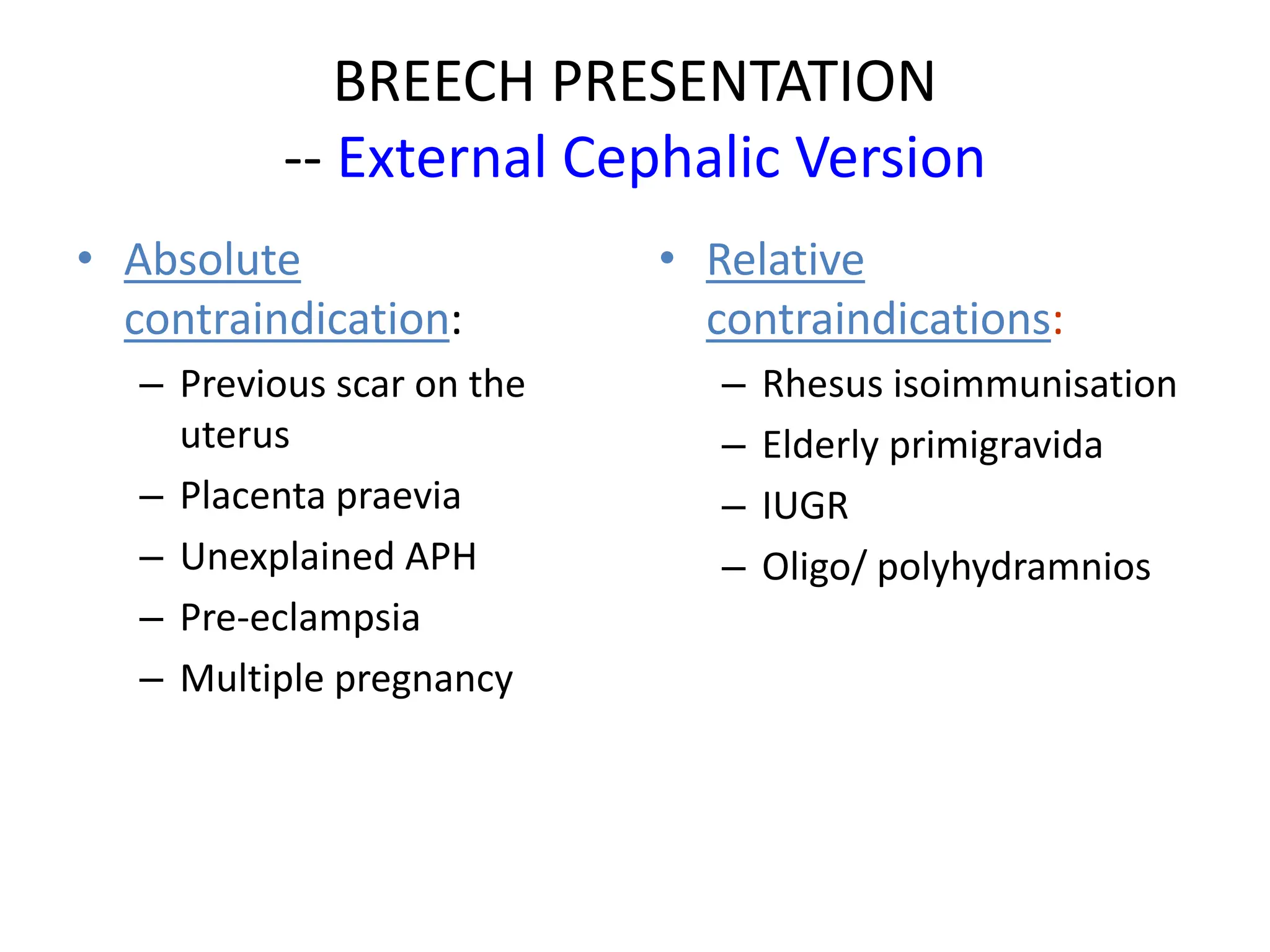
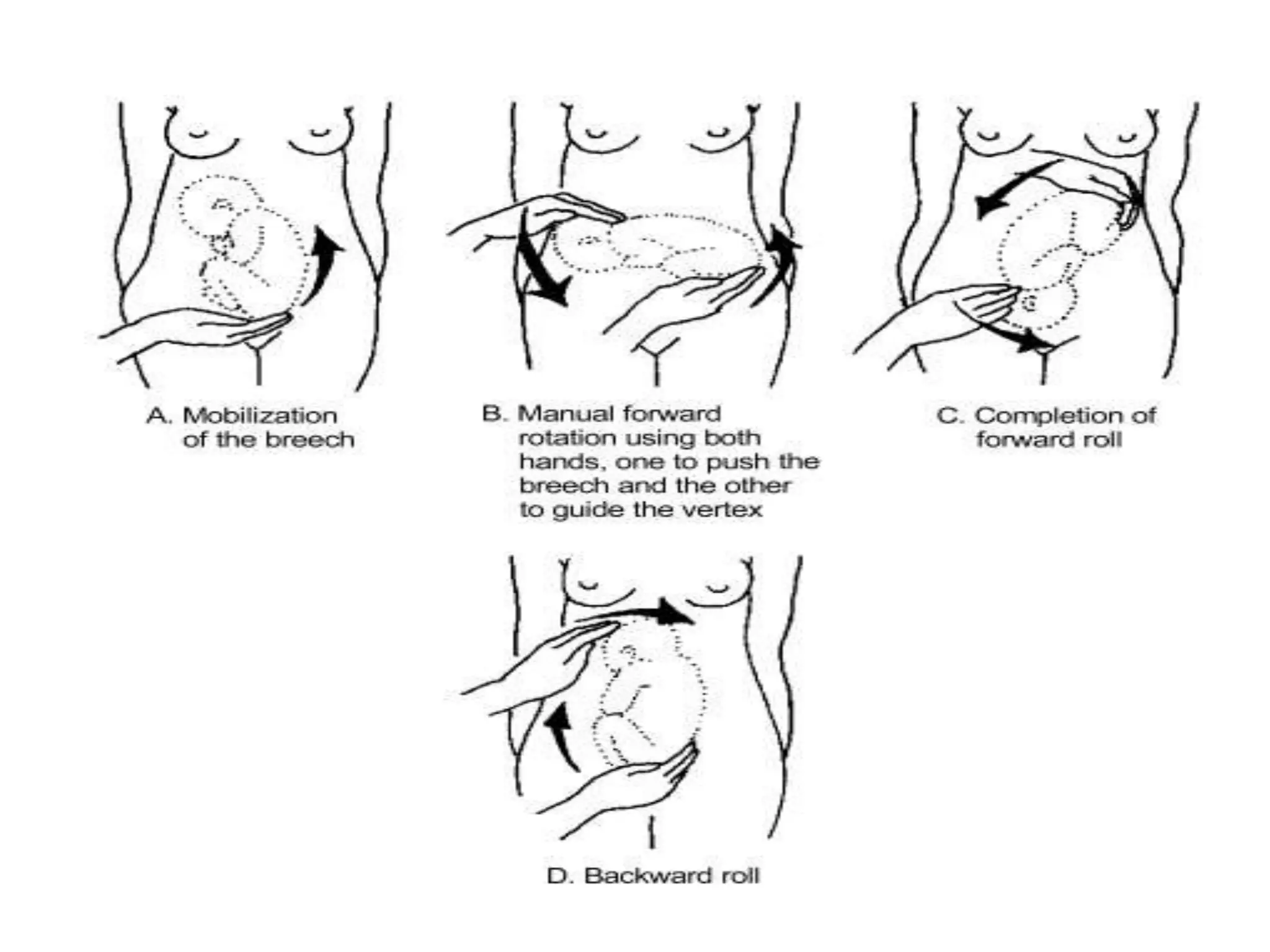
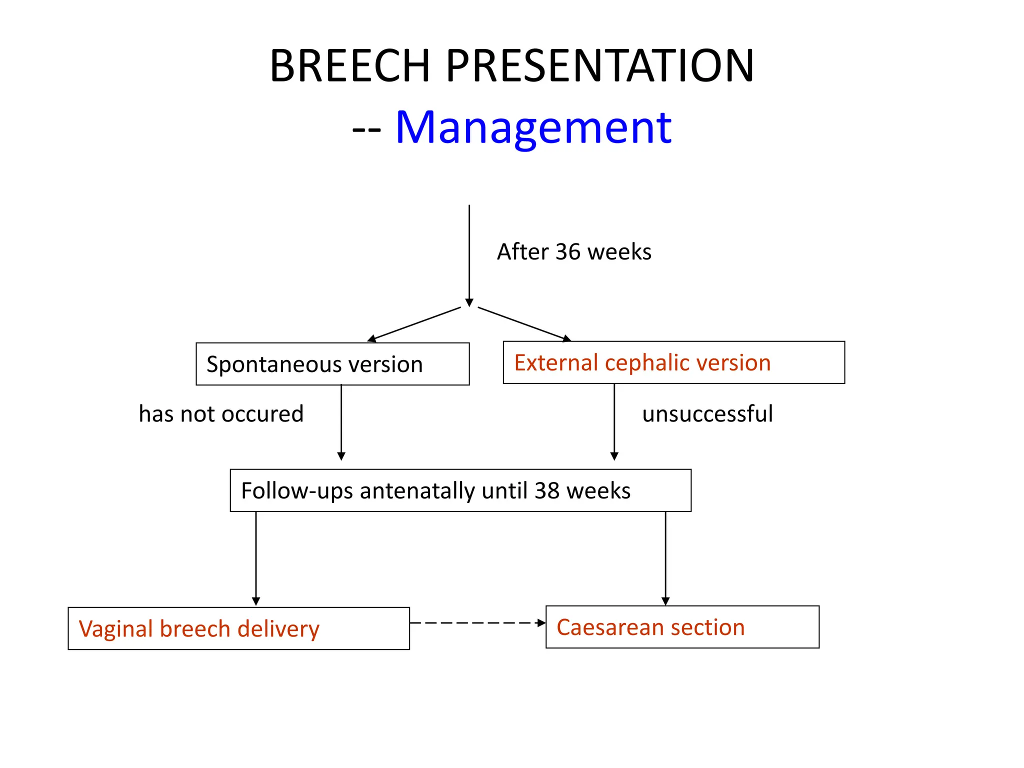
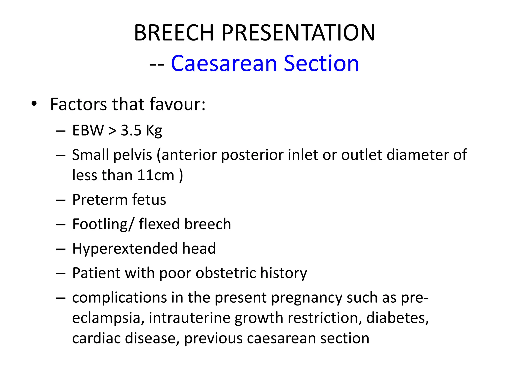
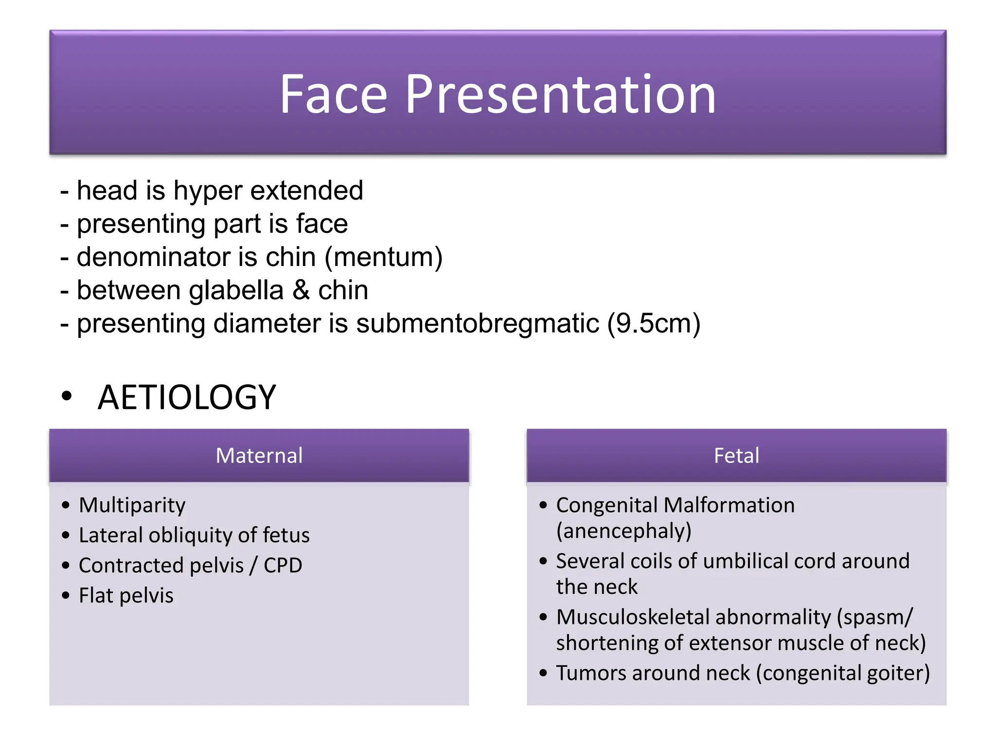
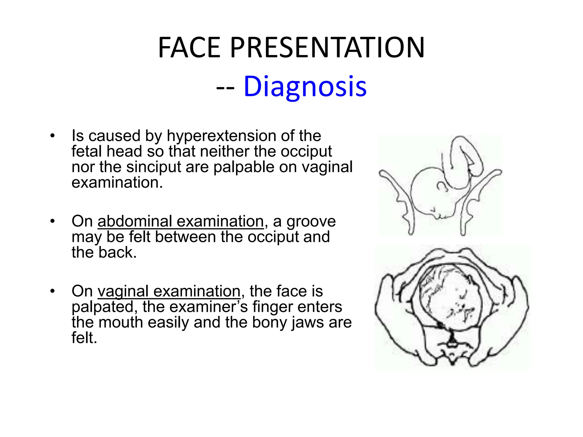
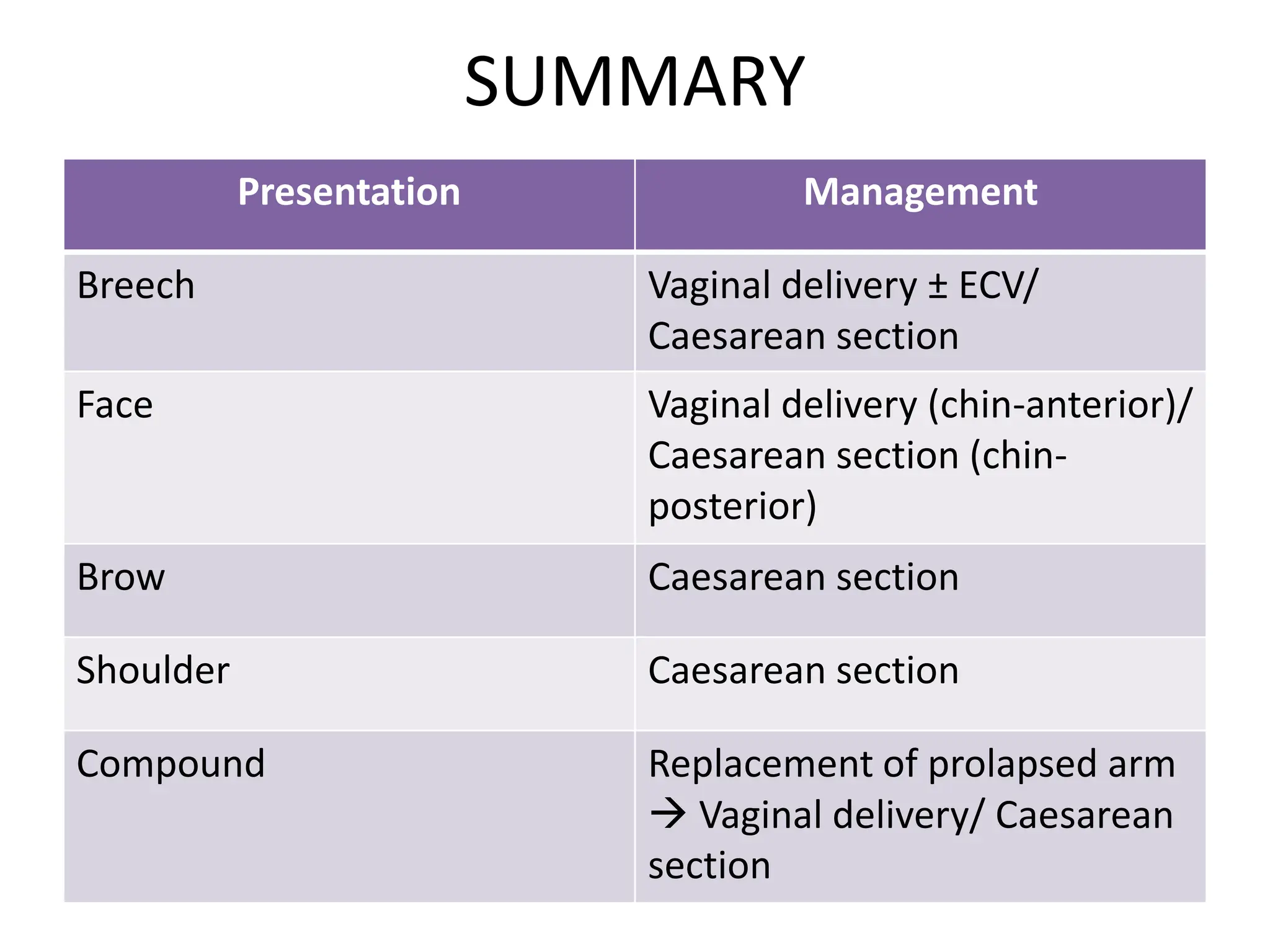
2. Definition
• Malpresentation = Fetal presenting part other
than vertex & includes breech, brow,
transverse, face.
• Malposition = Refers to positions other than
an occipito-anterior position.
3.
4. Commonest Presentation & Position
Suboccipito-Bregmatic
Extends from center of neck to bregma.
Presentation: Vertex
Attitude: Complete Flexion
D = 9.5cm
6. MALPOSITION
Occiput Posterior
Arrested labor may occur when the head does
not rotate and/or descend. Delivery may be
complicated by perineal tears or extension of
an episiotomy.
Occiput Transverse
It is the incomplete rotation of OP to OA
results in the fetal head being in a horizontal or
transverse position (OT).
Malpositions include occipitoposterior and occipitotransverse positions of
fetal head in relation to maternal pelvis.
7. How to diagnose?
• Palpation
– Fetal back is found to one side or may be difficult to identified.
– The fetal head is posterolateral and will be free above the brim.
• Auscultation
– The fetal heart best heard in the flank but descends to just above the
pubis as the head rotates and descends.
• VE
– the membrane tend to ruptured early before the labour is establish if
the membrane is intact they may protrude through the cervix giving
finger-like forewaters.
8. Factors that favour malposition
Pendulous abdomen- in multiparae
Anthropoid pelvic brim- favours direct
O.P/O.A
Android pelvic brim
A flat sacrum-transverse position
The placenta on the ant. uterine wall
R.O.P
9. Problems
Occiput Posterior - the baby's head faces the front
of the mother's pelvis instead of turning toward the
mother's back. The baby would then be delivered
with the head facing the ceiling, which is often a
more difficult way to deliver. A large episiotomy may
be required.This position occurs more often in
women who are having their first baby and women
who have a narrow midpelvis.
OP- may lead to dysfunctional labour (in
primigravida). Contraction may be painful and
accompanied by backache
11. signs of obstruction/ fetal
heart rate is abnormal (less than
100 or more than 180 beats per
minute) at any stage
caesarean section.
If no sign of obstruction
Cervix is not fully
dilated
If cervix is fully
dilated
Augmented labour with oxytocin
BUT no descent in
expulsive phase
3/5 – 1/5 <1/5
If fetal head is palpable per abdomen
>3/5
Vacuum extraction/
symphysiotomy
Vacuum extraction/
forceps
14. Related Factors:
• The woman has had more than one pregnancy
• There is more than one fetus in the uterus
• The uterus has too much or too little amniotic fluid
• The uterus is not normal in shape or has abnormal
growths, such as fibroids
• Placenta previa
• The baby is preterm
15. Breech Presentation
Perinatal mortality up to 4 times compared to vertex
presentation.
Breech presentation only becomes significant after
36weeks
Types of Breech Presentation:
Complete (Flexed) Breech Presentation
Footling Breech Presentation
Frank (Extended) Breech Presentation
Kneeling Breech Presentation
17. Frank Breech (60%)
The baby's bottom comes first,
and the legs are flexed at the
hip and extended at the knees
(with feet near the ears).
65-70% of breech babies are in
the frank breech position.
Complete Breech (15%)
The baby's hips and knees
are flexed so that the baby is
sitting cross legged, with
feet beside the bottom.
18. Footling Breech (25%)
One or both feet come first, with
the bottom at a higher position.
This is rare at term but relatively
common with premature fetuses
Kneeling Breech
The baby is in a kneeling
position, with one or both legs
extended at the hips and flexed
at the knees. This is extremely
rare.
20. BREECH PRESENTATION
-- External Cephalic Version
• Attempt external cephalic version if:
– Breech presentation is present at or after 36 weeks
– Vaginal delivery is possible;
– Membranes are intact and amniotic fluid is adequate;
– There are no complications (e.g. fetal growth restriction,
uterine bleeding, previous caesarean delivery, fetal
abnormalities, twin pregnancy, hypertension, fetal death).
21. BREECH PRESENTATION
-- External Cephalic Version
• Risks:
– Placental abruption
– Premature rupture of the membranes
– Cord accident
– Transplacental haemorrhage
– Fetal bradycardia
24. Principle: ‘Masterly inactivity
• The following points are important for the safe
conduct
of a breech delivery:
• Don’t be in hurry.
• Never pull from below and let the mother expel the
fetus by her own effort with uterine contractions
• Always keep the fetus with its back anterior
• Keep a pair of obstetrics forceps ready should it
become necessary to assist the aftercoming head
• Anesthetist and pediatrician should attend the
delivery
• Inform the operation theater, if C/S is needed.
25. BREECH PRESENTATION
-- Vaginal Breech Delivery
• Await for spontaneous labour
• A vaginal examination is done not only to assess the progress
of labour
• If the membranes rupture, do a vaginal examination
immediately to exclude uterine cord prolapse.
• If the membranes not rupture, examine for cord
presentation.
• Do not rupture the membranes
• Examine and monitor the woman regularly and adhere strictly
to the partogram.
• Poor progress may occur if sacrum is posterior/ bigger baby
than expected
• If there is any delay, the fetus is best delivered by an
emergency caesarean section.
26. BREECH PRESENTATION
-- Vaginal Breech Delivery
• Delivery of the buttocks
– Occur naturally
• Delivery of the legs and lower body
– Legs flexed : spontaneous delivery
– Legs extended : ‘Pinard’s manoeuvre’
• Delivery of the shoulders
– Loveset’s manoeuvre
• Delivery of the head
– Burns Marshall method
– Mariceau-Smellie-Veit manoeuvre
– Forceps delicery of the aftercoming head
27. Pinard’s manoeuvre
• In breech with extended legs
• once the groin is visible gentle pressure can be
applied to abduct the thigh and reach the
knee.
• The knee can be flexed with pressure in the
popliteal fossa and the leg delivered.
• Anterior leg is always delivered first.
28. BREECH PRESENTATION
-- Vaginal Breech Delivery
Loveset’s
manoeuvre
This procedure automatically corrects
any upward displacement of arms.
In Lovset’s maneuver baby’s trunk is
made to rotate with downward
traction holding the baby at the iliac
crest so that posterior shoulder comes
below symphysis pubis and the arm is
delivered by flexing the shoulder
followed by hooking at the elbow and
flexing it followed by bringing down
the forearm ‘like a hand shake’.
The same procedure is repeated by
reverse rotation of 180 degree so that
anterior shoulder comes below the
symphysis pubis.
29. Burns Marshall method
• For delivery of the aftercoming head
• It is commonly practice where the baby is allowed to hang for
a minute or so,
• The assistant gives a suprapubic downward and pressure
(Kristellar’s maneuver) to promote the head.
• Once the nape of the neck is visible, identified by the hairline,
the baby’s trunk is gently lifted and swung toward mother’s
abdomen holding the baby just above the ankle through an
arc of 180 degree.
• Left hand guards and slips the perineum over fetal mouth.
• As the mouth is born air passage is cleared of mucus and now
depressing the trunk the head is allowed to born.
31. Mariceau-Smellie-Veit
Manoeuvre
Jaw flexion and shoulder traction—JFST(Mariceau-
Smellie-Veit
Manoeuvre)
Here the baby is allowed to rest on the left
supinated forearm of the obstetrition, with the
limbs hanging on either side.
Left index and middle finger is placed on the
malar bones, while the right index and ring
fingers are placed on the respective shoulders
and the middle finger on the sub-occipital region.
To achieve flexion, traction is now given in
downward and backward direction and
simultaneous suprapubic pressure is maintained
by the assitant until the nape of the neck is
visible.
Thereafter, the baby is pulled in upward and
forward direction so that the face is born and by
depressing the trunk the head is born.
32. Forceps delicery of the aftercoming head
• The long Das/Simpson’s obstetric forceps can
be used instead of Piper’s forceps.
• The important prerequisite is that head must
be in the pelvic cavity and the occiput is
directly anterior, i.e. the face is facing the
posterior pelvic wall.
• Baby is lifted up by the assistant without
deviating the trunk to any side and forceps is
applied from ventral side.
33. BREECH PRESENTATION
-- Management
After 36 weeks
Spontaneous version External cephalic version
Follow-ups antenatally until 38 weeks
has not occured unsuccessful
Vaginal breech delivery Caesarean section
34. BREECH PRESENTATION
-- Caesarean Section
• Factors that favour:
– EBW > 3.5 Kg
– Small pelvis (anterior posterior inlet or outlet diameter of
less than 11cm )
– Preterm fetus
– Footling/ flexed breech
– Hyperextended head
– Patient with poor obstetric history
– complications in the present pregnancy such as pre-
eclampsia, intrauterine growth restriction, diabetes,
cardiac disease, previous caesarean section
35. BREECH PRESENTATION
-- Caesarean Section
• However in 2000 the result of the Canadian
Term Breech Trial were published. It came out
overwhelmingly with the conclusion that
singleton breech presentations at term should
preferably be delivered by caesarean section.
• Not to do so would invite unacceptable fetal
morbidity or mortality.
• There is therefore now a trend to deliver all
breeches at term by caesarean section.
36. • be remembered however, the results of the
study do not apply to
1)twin pregnancy with breech presentations,
2)preterm breech deliveries
3)breech presentations that arrive late
4)in advanced labour.
• In those situations there still appears to be a
role for delivering the baby vaginally.
37. Face Presentation
- head is hyper extended
- presenting part is face
- denominator is chin (mentum)
- between glabella & chin
- presenting diameter is submentobregmatic (9.5cm)
• AETIOLOGY
Maternal
• Multiparity
• Lateral obliquity of fetus
• Contracted pelvis / CPD
• Flat pelvis
Fetal
• Congenital Malformation
(anencephaly)
• Several coils of umbilical cord around
the neck
• Musculoskeletal abnormality (spasm/
shortening of extensor muscle of neck)
• Tumors around neck (congenital goiter)
38. FACE PRESENTATION
-- Diagnosis
• Is caused by hyperextension of the
fetal head so that neither the occiput
nor the sinciput are palpable on vaginal
examination.
• On abdominal examination, a groove
may be felt between the occiput and
the back.
• On vaginal examination, the face is
palpated, the examiner’s finger enters
the mouth easily and the bony jaws are
felt.
39. FACE PRESENTATION
-- Diagnosis
• The chin serves as the
reference point in
describing the position of
the head.
• It is necessary to
distinguish only chin-
anterior positions in which
the chin is anterior in
relation to the maternal
pelvis from chin-posterior
positions.
40. FACE PRESENTATION
-- Management
• Prolonged labour is common.
• Descent and delivery of the head by flexion
may occur in the chin-anterior position.
• In the chin-posterior position, however, the
fully extended head is blocked by the sacrum.
This prevents descent and labour is
arrested→ caesarean section
41. FACE PRESENTATION
-- Management of Chin-anterior
Cervix fully dilated Cervix not fully dilated
Allow normal child birth
Slow progress
with no signs
of obstruction
Descent
unsatisfactory
Augmentation
of labour
Forceps
delivery
Augmentation of labour
42. Brow Presentation
• The brow presentation is caused by partial extension of the
fetal head so that the occiput is higher than the sinciput.
• MGT: If the fetus is alive or
dead, deliver by caesarean
section.
*Do NOT deliver brow presentation
by vacuum extraction, outlet
forceps or symphysiotomy.
Mentovertical D = 14cm
Attitude = Partial Extension
43. Shoulder Presentation
• Occurs as a result of
transverse lie or oblique lie
• Predisposing factors = breech
presentation
• On abdominal examination,
neither the head nor the
buttocks can be felt at the
symphysis pubis and the head
is usually felt in the flank.
• On vaginal examination, a
shoulder may be felt, but not
always. An arm may prolapse
and the elbow, arm or hand
may be felt in the vagina.
• Ultrasound examination
44. Management
• Monitor for signs of cord prolapse. If the cord
prolapses and delivery is not imminent,
deliver by caesarean section.
• In modern practice, persistent transverse lie in
labour is delivered by caesarean section
whether the fetus is alive or dead.
45. Compound Presentation
• Occurs when an arm
prolapses alongside
the presenting part.
Both the prolapsed
arm and the fetal
head present in the
pelvis
simultaneously.
46. Management
• Replacement of the prolapsed
arm
• Assist the woman to
assume the knee-chest
position
• Push the arm above the
pelvic brim and hold it
there until a contraction
pushes the head into the
pelvis.
• Proceed with
management for normal
childbirth
• If the procedure fails or if the
cord prolapses, deliver by
caesarean section
It is usually seen in multipara or those with lax abdominal wall. Fetal malpositions are assessed during labor.
*The perinatal mortality can be up to 4 times that of vertex presentation. Reasons:
Higher incidence of fetal abnormality
Higher incidence of cord prolapse
Difficulty in delivering the shoulders
Difficulty in delivering the head.
In inexperienced hands