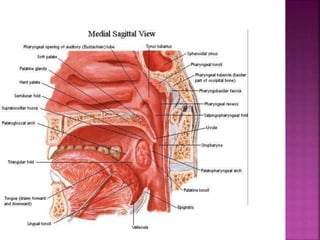
 As a conduit for passage of air and food.
 Respiratory function (Patent pathway
between nose & larynx).
 Reflex actions (sneezing, coughing &
vomiting).
 Speech resonance.
 Deglutition.
 Drainage of mucus from nasopharynx.
SOFT PALATE IS A THIN MOBILE MUSCLE COMPLEX SEPARATING
NASOPHARYNX FROM OROPHARYNX
 Annual World wide Incidence : 10%
 Annual Incidence in US :4.8/lac
 Annual Incidence in India :31735 (males)
6956 (females)
(Globocan 2012)
 Male : Female ratio = 4:1
 Tobacco
 Smoking
 Alcohol
 Viruses - HPV
 Genetic factors
 Lack of certain
vitamins
 Poor oral hygiene
 DS DNA virus with
>150 strains
 HPV types 6, 11, 16,
and 18 are high risk
(usually 16)
 Strongest association
with oropharyngeal
carcinoma, especially
tonsillar
Large tonsillar
lesion
Histology :
Basaloid pattern
HPV-E7 probe:
HPV DNA in nearly
All neoplastic cells
 >90% are squamous cell carcinoma or its variants – often
poorly differentiated
 Lymphomas – 10-15% tonsil
1-2% BOT
 Malignant melanomas – 6%
 Lymphoepithelioma (tonsil & BOT) - <1.5%
 Minor salivary gland tumors
 Sarcomas
 Plasmacytomas I to IV depending on degree of
differentiation
 Mostly asymptomatic
 Sore throat - most frequent
 Difficulty in swallowing
 Referred otalgia
 Neck swelling
 Change in voice – Hot potato voice
 Occasionally painless mass at BOT
 Trismus
 Impaired deglutition
 Poor articulation
 Deep ulceration & necrosis
 Tumor size and extension (stage);
 Presence of palpable lymph nodes; and
location, number, and size of involved lymph
nodes
 P53 and epidermal growth factor receptor
overexpression has been associated with
increased survival .
 Histologic differentiation
Earliest Lesion : Red
lesions with ill defined
borders
Tonsillar pillar and
hard palate
Superior constrictor
muscle
Skull Base
Lateral wall of
nasopharynx
 1st to be involved : Level II
 Followed by Level III and Level IV
 At presentation : 56% are node positive
diseases
 Bilateral disease : 16%
 Incidence of occult neck disease : 20%
 Incidence of clinically positive nodes
increases with T stage
 The Primary tumor usually
remains in tongue unless it
begins at peripheral margin
 Primary at lateral base of
tongue
Glossopharyngeal
sulcus
Neck
Vallecula
Lingual surface of epiglottis
Pharyngoepiglottic fold
Ant wall of pyriform sinus
 Vallecular lesions frequently penetrate
through hyoepiglottic ligament to enter pre
epiglottic space
 Most common : Level II followed by Level III
and Level IV
 Level Ib may be involved if tumor extends
into oral tongue or if massive upper neck
disease in present
 At presentation : 75% are node positive
diseases
 Bilateral disease : 30%
 Incidence of occult neck disease : 40% - 50%
 Intially superficial lesions which progresses
to develop a central ulcer with a rolled
margin and infiltrate the palatoglossus.
 Superior : Soft palate and Hard Plate
 Antero lateral : Retromolar trigone and
buccal mucosa
 Inferomedial : Tongue
 Progression leads to involvement of bone,
skull base, nasopharynx and medial pterygoid
muscle.
 Initial lesion :
Exophytic with
central ulceration
 Posterior tonsillar
pillar, oropharyngeal
wall
 Base of tongue
 Parapharyngeal space
 Skull base
 Mostly spreads inferiorly along
Palatopharyngeal muscle to
 Middle pharyngeal constrictor
 Pharyngoepiglottic fold
 Posterior border of thyroid cartilage
 At presentation : 45% are node positive cases
 Ipsilateral spread : Jugular and Level Ib
nodes.
 Contralateral spread : Uncommon (5% cases).
Confined to Jugular Chain
 Incidence of occult neck disease : 10% - 15%
 Incidence of clinically positive nodes
increases with T stage
 At presentation : 76% are node positive cases
 Ipsilateral spread : Jugular, Junctional, Level
V, Level Ib
 Contralateral spread : Seen in 11% cases.
Confined to Jugular Chain
 Incidence of occult neck disease : 50% - 60%
 Routine: Blood counts , KFT and LFT.
 Diagnostic
 Detailed history and physical examination
including inspection and palpation of the lesion .
 Laryngoscopy.
 CT scan of Head and neck .
 Biopsy from the primary lesion and
 Aspiration cytology from involved lymph node if
present .
 Metastatic:
 Chest x ray/CT Chest
 USG whole abdomen
 Standard in delineating extent of tumor
 Evaluation of LN status
 Depth of tumor penetration
 Evaluating involvement of mandible or extension into
base of skull
 Extracapsular extension
 For treatment planning
 Primary Tumors : Contrast enhancing masses,
distorting normal anatomic relationships.
 Pathological Lymph Nodes : Enlarged,
enhanced with contrast, having a necrotic
centre.
 Extracapsular extension : Irregular nodal
margin without clear distinction with
surrounding fat or when there is thickening of
surrounding fibroadipose tissue or muscle.
 Superior soft tissue contrast
 Sensitive in detecting tumor extension and LN
distribution
 MRI is useful in patients with compromised
renal function, who are not able to receive
contrast for CT
 T1Tumor ≤2 cm in greatest dimension
 T2Tumor >2 cm but not >4 cm in greatest
dimension
 T3Tumor >4 cm in greatest dimension
 T4aTumor invades the larynx, deep/extrinsic
muscle of the tongue, medial pterygoid, hard
palate, or mandible
 T4bTumor invades lateral pterygoid muscle,
pterygoid plates, lateral nasopharynx, or
skull base or encases carotid artery
 N0: No regional Lymph node metastasis
 N1: Metastasis in single ipsilateral node
<=3cm
 N2
 N2a: Metastasis in single ipsilateral node
>3cm but < 6cm
 N2b: Metastasis in multiple ipsilateral nodes
>3cm but < 6 cm
 N2c: Metstasis in bilateral or contralateral
lymph nodes, none > 6cm
 N3: Metastasis in a lymph node >6 cm.
 MX: Distant metastasis cannot be evaluated.
 M0: Cancer has not spread to other parts of
the body.
 M1: Cancer has spread to other parts of the
body
premanagement ca oropharynx

premanagement ca oropharynx

  • 7.
     As aconduit for passage of air and food.  Respiratory function (Patent pathway between nose & larynx).  Reflex actions (sneezing, coughing & vomiting).  Speech resonance.  Deglutition.  Drainage of mucus from nasopharynx.
  • 9.
    SOFT PALATE ISA THIN MOBILE MUSCLE COMPLEX SEPARATING NASOPHARYNX FROM OROPHARYNX
  • 14.
     Annual Worldwide Incidence : 10%  Annual Incidence in US :4.8/lac  Annual Incidence in India :31735 (males) 6956 (females) (Globocan 2012)  Male : Female ratio = 4:1
  • 15.
     Tobacco  Smoking Alcohol  Viruses - HPV  Genetic factors  Lack of certain vitamins  Poor oral hygiene
  • 16.
     DS DNAvirus with >150 strains  HPV types 6, 11, 16, and 18 are high risk (usually 16)  Strongest association with oropharyngeal carcinoma, especially tonsillar Large tonsillar lesion Histology : Basaloid pattern HPV-E7 probe: HPV DNA in nearly All neoplastic cells
  • 18.
     >90% aresquamous cell carcinoma or its variants – often poorly differentiated  Lymphomas – 10-15% tonsil 1-2% BOT  Malignant melanomas – 6%  Lymphoepithelioma (tonsil & BOT) - <1.5%  Minor salivary gland tumors  Sarcomas  Plasmacytomas I to IV depending on degree of differentiation
  • 19.
     Mostly asymptomatic Sore throat - most frequent  Difficulty in swallowing  Referred otalgia  Neck swelling  Change in voice – Hot potato voice  Occasionally painless mass at BOT
  • 20.
     Trismus  Impaireddeglutition  Poor articulation  Deep ulceration & necrosis
  • 21.
     Tumor sizeand extension (stage);  Presence of palpable lymph nodes; and location, number, and size of involved lymph nodes  P53 and epidermal growth factor receptor overexpression has been associated with increased survival .  Histologic differentiation
  • 23.
    Earliest Lesion :Red lesions with ill defined borders Tonsillar pillar and hard palate Superior constrictor muscle Skull Base Lateral wall of nasopharynx
  • 24.
     1st tobe involved : Level II  Followed by Level III and Level IV  At presentation : 56% are node positive diseases  Bilateral disease : 16%  Incidence of occult neck disease : 20%  Incidence of clinically positive nodes increases with T stage
  • 25.
     The Primarytumor usually remains in tongue unless it begins at peripheral margin  Primary at lateral base of tongue Glossopharyngeal sulcus Neck
  • 26.
    Vallecula Lingual surface ofepiglottis Pharyngoepiglottic fold Ant wall of pyriform sinus  Vallecular lesions frequently penetrate through hyoepiglottic ligament to enter pre epiglottic space
  • 27.
     Most common: Level II followed by Level III and Level IV  Level Ib may be involved if tumor extends into oral tongue or if massive upper neck disease in present  At presentation : 75% are node positive diseases  Bilateral disease : 30%  Incidence of occult neck disease : 40% - 50%
  • 28.
     Intially superficiallesions which progresses to develop a central ulcer with a rolled margin and infiltrate the palatoglossus.  Superior : Soft palate and Hard Plate  Antero lateral : Retromolar trigone and buccal mucosa  Inferomedial : Tongue  Progression leads to involvement of bone, skull base, nasopharynx and medial pterygoid muscle.
  • 29.
     Initial lesion: Exophytic with central ulceration  Posterior tonsillar pillar, oropharyngeal wall  Base of tongue  Parapharyngeal space  Skull base
  • 30.
     Mostly spreadsinferiorly along Palatopharyngeal muscle to  Middle pharyngeal constrictor  Pharyngoepiglottic fold  Posterior border of thyroid cartilage
  • 31.
     At presentation: 45% are node positive cases  Ipsilateral spread : Jugular and Level Ib nodes.  Contralateral spread : Uncommon (5% cases). Confined to Jugular Chain  Incidence of occult neck disease : 10% - 15%  Incidence of clinically positive nodes increases with T stage
  • 32.
     At presentation: 76% are node positive cases  Ipsilateral spread : Jugular, Junctional, Level V, Level Ib  Contralateral spread : Seen in 11% cases. Confined to Jugular Chain  Incidence of occult neck disease : 50% - 60%
  • 34.
     Routine: Bloodcounts , KFT and LFT.  Diagnostic  Detailed history and physical examination including inspection and palpation of the lesion .  Laryngoscopy.  CT scan of Head and neck .  Biopsy from the primary lesion and  Aspiration cytology from involved lymph node if present .  Metastatic:  Chest x ray/CT Chest  USG whole abdomen
  • 40.
     Standard indelineating extent of tumor  Evaluation of LN status  Depth of tumor penetration  Evaluating involvement of mandible or extension into base of skull  Extracapsular extension  For treatment planning
  • 41.
     Primary Tumors: Contrast enhancing masses, distorting normal anatomic relationships.  Pathological Lymph Nodes : Enlarged, enhanced with contrast, having a necrotic centre.  Extracapsular extension : Irregular nodal margin without clear distinction with surrounding fat or when there is thickening of surrounding fibroadipose tissue or muscle.
  • 43.
     Superior softtissue contrast  Sensitive in detecting tumor extension and LN distribution  MRI is useful in patients with compromised renal function, who are not able to receive contrast for CT
  • 46.
     T1Tumor ≤2cm in greatest dimension  T2Tumor >2 cm but not >4 cm in greatest dimension  T3Tumor >4 cm in greatest dimension  T4aTumor invades the larynx, deep/extrinsic muscle of the tongue, medial pterygoid, hard palate, or mandible  T4bTumor invades lateral pterygoid muscle, pterygoid plates, lateral nasopharynx, or skull base or encases carotid artery
  • 48.
     N0: Noregional Lymph node metastasis  N1: Metastasis in single ipsilateral node <=3cm  N2  N2a: Metastasis in single ipsilateral node >3cm but < 6cm  N2b: Metastasis in multiple ipsilateral nodes >3cm but < 6 cm  N2c: Metstasis in bilateral or contralateral lymph nodes, none > 6cm  N3: Metastasis in a lymph node >6 cm.
  • 50.
     MX: Distantmetastasis cannot be evaluated.  M0: Cancer has not spread to other parts of the body.  M1: Cancer has spread to other parts of the body

Editor's Notes

  • #6 Superiorly: It communicates with the nasopharynx through pharyngeal isthmus. Inferiorly: It opens into the laryngopharynx at the upper border of epiglottis. In front: It communicates with the oral cavity through oropharyngeal isthmus. Behind: supported by the body of axis vertebra and upper part of C-3. Lateral wall: There lies the palatine tonsils which lies in the tonsillar fossa .