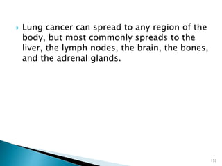
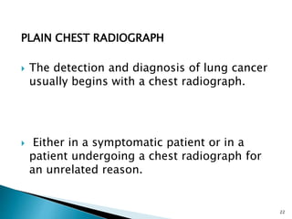
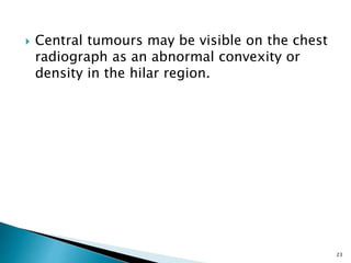
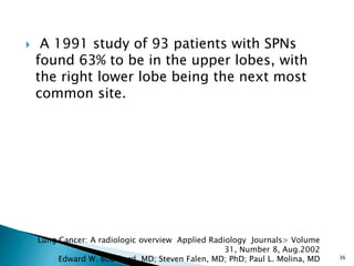
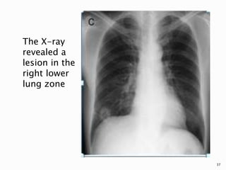
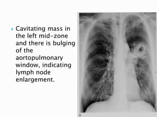
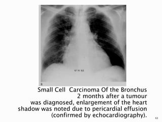
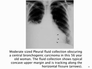
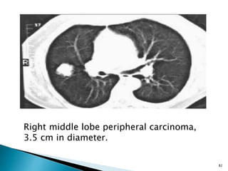
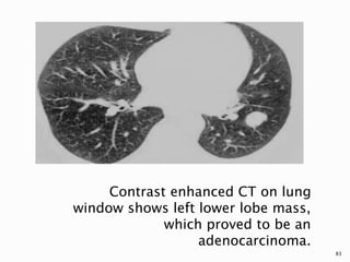
The discovery of malignant cells in pleural fluid
and/or parietal pleura signifies disseminated or
advanced disease and a reduced life expectancy in
patients with cancer.Median survival following
diagnosis ranges from 3 to 12 months and is
dependent on the stage and type of the underlying
malignancy. The shortest survival time is observed
in malignant effusions secondary to lung cancer
and the longest in ovarian cancer, while malignant
effusions due to an unknown primary have an
intermediate survival time.Historically, studies
showed that median survival times in effusions due
to carcinoma of the breast are 5-6 months.
However, more recent studies have suggested
longer survival times of up to 15 months. A
comparison of survival times in breast cancer
effusions in published studies to 1994 calculated
a median survival of 11 months.9
Currently, lung cancer is the most common
metastatic tumour to the pleura in men and breast
cancer in women.Together, both malignancies
account for 50- 65% of all malignant effusions. Lymphomas, tumours of the genitourinary
tract and gastrointestinal tract account for
a further 25% Pleural effusions from an
unknown primary are responsible for 15% of all
malignant pleural effusions.Few studies have
estimated the proportion of pleural effusions due to
mesothelioma: studies from 1975, 1985 and 1987
identified mesothelioma in 1/271, 3/472 and 22/592
patients, respectively, but there are no more recent
data to update this in light of the increasing incidence
of mesothelioma.
In this presentation our agenda is
Brief introduction
Radiological Modalities
Radiological Features
Radiological Imaging Of Complications of lung cancer.
I followed Dahnert and try to describe all findings in lung cancer.
Hope it will prove an atlas in Lung cancer imaging.
Imaging plays an important role in diagnosis and formulating differential diagnosis in case of Solitary pulmonary nodule. It helps in differentiating and predicting benign and malignant nodules.
The basics of Chest Radiology explained for the undergraduate students. The technical aspects including the various views, exposure, rotation and breath described.
The inside out approach of interpretation explained. The ABCDEFGH description includes Airway, Bones & soft tissue, Cardiac shadow, Diaphragm, Effusion (pleura), Fields (lungs), Gastric bubble and Hila & mediastinum.
The basic cardiac and lung pathologies discussed.
PowerPoint presentation on the topic HRCT Chest. This presentation is divided into 5 different parts. 1)Introduction to HRCT chest 2)Technichal aspects of HRCT 3) Relevant anatomy for HRCT interpretation 4)Pattern of lung disease in HRCT 5)HRCT pattern in various ILD’s
The solitary lung nodule. A diagnostic dilemma. hazem youssef
Incidentally discovered pulmonary nodule are a diagnostic challenge. This presentation is focused on the different features of lung nodules and their management.
HOT NEW PRODUCT! BIG SALES FAST SHIPPING NOW FROM CHINA!! EU KU DB BK substit...GL Anaacs
Contact us if you are interested:
Email / Skype : kefaya1771@gmail.com
Threema: PXHY5PDH
New BATCH Ku !!! MUCH IN DEMAND FAST SALE EVERY BATCH HAPPY GOOD EFFECT BIG BATCH !
Contact me on Threema or skype to start big business!!
Hot-sale products:
NEW HOT EUTYLONE WHITE CRYSTAL!!
5cl-adba precursor (semi finished )
5cl-adba raw materials
ADBB precursor (semi finished )
ADBB raw materials
APVP powder
5fadb/4f-adb
Jwh018 / Jwh210
Eutylone crystal
Protonitazene (hydrochloride) CAS: 119276-01-6
Flubrotizolam CAS: 57801-95-3
Metonitazene CAS: 14680-51-4
Payment terms: Western Union,MoneyGram,Bitcoin or USDT.
Deliver Time: Usually 7-15days
Shipping method: FedEx, TNT, DHL,UPS etc.Our deliveries are 100% safe, fast, reliable and discreet.
Samples will be sent for your evaluation!If you are interested in, please contact me, let's talk details.
We specializes in exporting high quality Research chemical, medical intermediate, Pharmaceutical chemicals and so on. Products are exported to USA, Canada, France, Korea, Japan,Russia, Southeast Asia and other countries.
Explore natural remedies for syphilis treatment in Singapore. Discover alternative therapies, herbal remedies, and lifestyle changes that may complement conventional treatments. Learn about holistic approaches to managing syphilis symptoms and supporting overall health.
micro teaching on communication m.sc nursing.pdfAnurag Sharma
Microteaching is a unique model of practice teaching. It is a viable instrument for the. desired change in the teaching behavior or the behavior potential which, in specified types of real. classroom situations, tends to facilitate the achievement of specified types of objectives.
Tom Selleck Health: A Comprehensive Look at the Iconic Actor’s Wellness Journeygreendigital
Tom Selleck, an enduring figure in Hollywood. has captivated audiences for decades with his rugged charm, iconic moustache. and memorable roles in television and film. From his breakout role as Thomas Magnum in Magnum P.I. to his current portrayal of Frank Reagan in Blue Bloods. Selleck's career has spanned over 50 years. But beyond his professional achievements. fans have often been curious about Tom Selleck Health. especially as he has aged in the public eye.
Follow us on: Pinterest
Introduction
Many have been interested in Tom Selleck health. not only because of his enduring presence on screen but also because of the challenges. and lifestyle choices he has faced and made over the years. This article delves into the various aspects of Tom Selleck health. exploring his fitness regimen, diet, mental health. and the challenges he has encountered as he ages. We'll look at how he maintains his well-being. the health issues he has faced, and his approach to ageing .
Early Life and Career
Childhood and Athletic Beginnings
Tom Selleck was born on January 29, 1945, in Detroit, Michigan, and grew up in Sherman Oaks, California. From an early age, he was involved in sports, particularly basketball. which played a significant role in his physical development. His athletic pursuits continued into college. where he attended the University of Southern California (USC) on a basketball scholarship. This early involvement in sports laid a strong foundation for his physical health and disciplined lifestyle.
Transition to Acting
Selleck's transition from an athlete to an actor came with its physical demands. His first significant role in "Magnum P.I." required him to perform various stunts and maintain a fit appearance. This role, which he played from 1980 to 1988. necessitated a rigorous fitness routine to meet the show's demands. setting the stage for his long-term commitment to health and wellness.
Fitness Regimen
Workout Routine
Tom Selleck health and fitness regimen has evolved. adapting to his changing roles and age. During his "Magnum, P.I." days. Selleck's workouts were intense and focused on building and maintaining muscle mass. His routine included weightlifting, cardiovascular exercises. and specific training for the stunts he performed on the show.
Selleck adjusted his fitness routine as he aged to suit his body's needs. Today, his workouts focus on maintaining flexibility, strength, and cardiovascular health. He incorporates low-impact exercises such as swimming, walking, and light weightlifting. This balanced approach helps him stay fit without putting undue strain on his joints and muscles.
Importance of Flexibility and Mobility
In recent years, Selleck has emphasized the importance of flexibility and mobility in his fitness regimen. Understanding the natural decline in muscle mass and joint flexibility with age. he includes stretching and yoga in his routine. These practices help prevent injuries, improve posture, and maintain mobilit
ARTIFICIAL INTELLIGENCE IN HEALTHCARE.pdfAnujkumaranit
Artificial intelligence (AI) refers to the simulation of human intelligence processes by machines, especially computer systems. It encompasses tasks such as learning, reasoning, problem-solving, perception, and language understanding. AI technologies are revolutionizing various fields, from healthcare to finance, by enabling machines to perform tasks that typically require human intelligence.
Lung Cancer: Artificial Intelligence, Synergetics, Complex System Analysis, S...Oleg Kshivets
RESULTS: Overall life span (LS) was 2252.1±1742.5 days and cumulative 5-year survival (5YS) reached 73.2%, 10 years – 64.8%, 20 years – 42.5%. 513 LCP lived more than 5 years (LS=3124.6±1525.6 days), 148 LCP – more than 10 years (LS=5054.4±1504.1 days).199 LCP died because of LC (LS=562.7±374.5 days). 5YS of LCP after bi/lobectomies was significantly superior in comparison with LCP after pneumonectomies (78.1% vs.63.7%, P=0.00001 by log-rank test). AT significantly improved 5YS (66.3% vs. 34.8%) (P=0.00000 by log-rank test) only for LCP with N1-2. Cox modeling displayed that 5YS of LCP significantly depended on: phase transition (PT) early-invasive LC in terms of synergetics, PT N0—N12, cell ratio factors (ratio between cancer cells- CC and blood cells subpopulations), G1-3, histology, glucose, AT, blood cell circuit, prothrombin index, heparin tolerance, recalcification time (P=0.000-0.038). Neural networks, genetic algorithm selection and bootstrap simulation revealed relationships between 5YS and PT early-invasive LC (rank=1), PT N0—N12 (rank=2), thrombocytes/CC (3), erythrocytes/CC (4), eosinophils/CC (5), healthy cells/CC (6), lymphocytes/CC (7), segmented neutrophils/CC (8), stick neutrophils/CC (9), monocytes/CC (10); leucocytes/CC (11). Correct prediction of 5YS was 100% by neural networks computing (area under ROC curve=1.0; error=0.0).
CONCLUSIONS: 5YS of LCP after radical procedures significantly depended on: 1) PT early-invasive cancer; 2) PT N0--N12; 3) cell ratio factors; 4) blood cell circuit; 5) biochemical factors; 6) hemostasis system; 7) AT; 8) LC characteristics; 9) LC cell dynamics; 10) surgery type: lobectomy/pneumonectomy; 11) anthropometric data. Optimal diagnosis and treatment strategies for LC are: 1) screening and early detection of LC; 2) availability of experienced thoracic surgeons because of complexity of radical procedures; 3) aggressive en block surgery and adequate lymph node dissection for completeness; 4) precise prediction; 5) adjuvant chemoimmunoradiotherapy for LCP with unfavorable prognosis.
New Drug Discovery and Development .....NEHA GUPTA
The "New Drug Discovery and Development" process involves the identification, design, testing, and manufacturing of novel pharmaceutical compounds with the aim of introducing new and improved treatments for various medical conditions. This comprehensive endeavor encompasses various stages, including target identification, preclinical studies, clinical trials, regulatory approval, and post-market surveillance. It involves multidisciplinary collaboration among scientists, researchers, clinicians, regulatory experts, and pharmaceutical companies to bring innovative therapies to market and address unmet medical needs.
Recomendações da OMS sobre cuidados maternos e neonatais para uma experiência pós-natal positiva.
Em consonância com os ODS – Objetivos do Desenvolvimento Sustentável e a Estratégia Global para a Saúde das Mulheres, Crianças e Adolescentes, e aplicando uma abordagem baseada nos direitos humanos, os esforços de cuidados pós-natais devem expandir-se para além da cobertura e da simples sobrevivência, de modo a incluir cuidados de qualidade.
Estas diretrizes visam melhorar a qualidade dos cuidados pós-natais essenciais e de rotina prestados às mulheres e aos recém-nascidos, com o objetivo final de melhorar a saúde e o bem-estar materno e neonatal.
Uma “experiência pós-natal positiva” é um resultado importante para todas as mulheres que dão à luz e para os seus recém-nascidos, estabelecendo as bases para a melhoria da saúde e do bem-estar a curto e longo prazo. Uma experiência pós-natal positiva é definida como aquela em que as mulheres, pessoas que gestam, os recém-nascidos, os casais, os pais, os cuidadores e as famílias recebem informação consistente, garantia e apoio de profissionais de saúde motivados; e onde um sistema de saúde flexível e com recursos reconheça as necessidades das mulheres e dos bebês e respeite o seu contexto cultural.
Estas diretrizes consolidadas apresentam algumas recomendações novas e já bem fundamentadas sobre cuidados pós-natais de rotina para mulheres e neonatos que recebem cuidados no pós-parto em unidades de saúde ou na comunidade, independentemente dos recursos disponíveis.
É fornecido um conjunto abrangente de recomendações para cuidados durante o período puerperal, com ênfase nos cuidados essenciais que todas as mulheres e recém-nascidos devem receber, e com a devida atenção à qualidade dos cuidados; isto é, a entrega e a experiência do cuidado recebido. Estas diretrizes atualizam e ampliam as recomendações da OMS de 2014 sobre cuidados pós-natais da mãe e do recém-nascido e complementam as atuais diretrizes da OMS sobre a gestão de complicações pós-natais.
O estabelecimento da amamentação e o manejo das principais intercorrências é contemplada.
Recomendamos muito.
Vamos discutir essas recomendações no nosso curso de pós-graduação em Aleitamento no Instituto Ciclos.
Esta publicação só está disponível em inglês até o momento.
Prof. Marcus Renato de Carvalho
www.agostodourado.com
Pulmonary Thromboembolism - etilogy, types, medical- Surgical and nursing man...VarunMahajani
Disruption of blood supply to lung alveoli due to blockage of one or more pulmonary blood vessels is called as Pulmonary thromboembolism. In this presentation we will discuss its causes, types and its management in depth.
Ozempic: Preoperative Management of Patients on GLP-1 Receptor Agonists Saeid Safari
Preoperative Management of Patients on GLP-1 Receptor Agonists like Ozempic and Semiglutide
ASA GUIDELINE
NYSORA Guideline
2 Case Reports of Gastric Ultrasound
TEST BANK for Operations Management, 14th Edition by William J. Stevenson, Ve...kevinkariuki227
TEST BANK for Operations Management, 14th Edition by William J. Stevenson, Verified Chapters 1 - 19, Complete Newest Version.pdf
Prix Galien International 2024 Forum ProgramLevi Shapiro
June 20, 2024, Prix Galien International and Jerusalem Ethics Forum in ROME. Detailed agenda including panels:
- ADVANCES IN CARDIOLOGY: A NEW PARADIGM IS COMING
- WOMEN’S HEALTH: FERTILITY PRESERVATION
- WHAT’S NEW IN THE TREATMENT OF INFECTIOUS,
ONCOLOGICAL AND INFLAMMATORY SKIN DISEASES?
- ARTIFICIAL INTELLIGENCE AND ETHICS
- GENE THERAPY
- BEYOND BORDERS: GLOBAL INITIATIVES FOR DEMOCRATIZING LIFE SCIENCE TECHNOLOGIES AND PROMOTING ACCESS TO HEALTHCARE
- ETHICAL CHALLENGES IN LIFE SCIENCES
- Prix Galien International Awards Ceremony
Ethanol (CH3CH2OH), or beverage alcohol, is a two-carbon alcohol
that is rapidly distributed in the body and brain. Ethanol alters many
neurochemical systems and has rewarding and addictive properties. It
is the oldest recreational drug and likely contributes to more morbidity,
mortality, and public health costs than all illicit drugs combined. The
5th edition of the Diagnostic and Statistical Manual of Mental Disorders
(DSM-5) integrates alcohol abuse and alcohol dependence into a single
disorder called alcohol use disorder (AUD), with mild, moderate,
and severe subclassifications (American Psychiatric Association, 2013).
In the DSM-5, all types of substance abuse and dependence have been
combined into a single substance use disorder (SUD) on a continuum
from mild to severe. A diagnosis of AUD requires that at least two of
the 11 DSM-5 behaviors be present within a 12-month period (mild
AUD: 2–3 criteria; moderate AUD: 4–5 criteria; severe AUD: 6–11 criteria).
The four main behavioral effects of AUD are impaired control over
drinking, negative social consequences, risky use, and altered physiological
effects (tolerance, withdrawal). This chapter presents an overview
of the prevalence and harmful consequences of AUD in the U.S.,
the systemic nature of the disease, neurocircuitry and stages of AUD,
comorbidities, fetal alcohol spectrum disorders, genetic risk factors, and
pharmacotherapies for AUD.
Couples presenting to the infertility clinic- Do they really have infertility...Sujoy Dasgupta
Dr Sujoy Dasgupta presented the study on "Couples presenting to the infertility clinic- Do they really have infertility? – The unexplored stories of non-consummation" in the 13th Congress of the Asia Pacific Initiative on Reproduction (ASPIRE 2024) at Manila on 24 May, 2024.
4. Epidemiology:
Carcinoma of the bronchus is the
commonest fatal malignancy in adult males
in the western world (35% of all cancer
deaths).
Commoner in males but incidence in
women is rising(21% of all cancer deaths).
Most cases occur between 40-70 years of
age and peaks in the 50-60 age range; it is
unusual below the age of 30 years.
4
5. Epidemiology:
The country with the highest incidence of
lung cancer among males is the United
Kingdom .
In general, the incidence of lung cancer in
industrialized western countries is
increased compared to third world
countries
5
6. Epidemiology:
In a 30 month prospective study conducted by
N. Ezemba et al at the University of Nigeria
Teaching Hospital, Enugu from Jan 2003-June
2005; 51 new cases were identified during the
study period.
The ages ranged from 30-81 years, mean of 56
± 21years with a male: female ratio of 2:1.
In 42% of the males there was a history of
cigarette smoking. No history of smoking
found among the females.
6
Challenges of lung cancer in a developing country by Ndubueze Ezemba, Eyo
Ekpe & John Eze Nigerian Journal of Medicine 2012 Apr-Jun;21(2):214-7.
7. Risk factors:
Cigarette Smoking: The single most
important aetiological factor is cigarette
smoking.This is dose related, the risk being
proportional to number of cigarettes smoked.
Second hand smoke is also bad. Non-
smoking women married to smokers had a
1.2x risk of developing cancer.
7
8. Risk Factors:
Concomitant Disease: It is also reported that
lung scarring ( tuberculosis, scleroderma,
infarction, bronchiectasis ) is associated with
the incidence of lung cancer especially
adenocarcinoma.
8
9. Risk Factors:
Industrial Exposure: radiation exposure,
asbestos, workers exposed to nickel/
chromate/ arsenic/ and newspaper industry
workers.
Air pollution: both indoor and outdoor
especially radon gas which may be the second
leading cause for lung cancer with up to
20,000 deaths per year.
Combined risk factors approach 100% risk.
9
10. Pathology/ Classification:
Bronchogenic carcinomas begin as a small
focus of atypical epithelial cells within the
bronchial mucosa. As the lesion progresses,
the atypia becomes frankly malignant and
the neoplasm grows in size.
10
11. According to anatomy:
(1)Central lung cancer,mostly is squamous
cell carcinoma and small cell carcinoma.
(2) peripheral lung cancer, mostly is
adenocarcinoma and large cell carcinoma.
11
12. According to histologic classification:
(1) Small cell lung cancer(SCLC) 20%
(2 ) Non-small cell lung cancer(NSCLC)
includes ;
Adenocarcinoma 30-40%
Squamous cell carcinoma 30-40%
Large cell Undifferentiated carcinoma 10%
12
13. Pathology / Classification:
Squamous cell carcinoma: It is the most
common subtype. It arises from altered
bronchial epithelium and growth in situ. It
is related to cigarette smoking. Cavitation
can occur. Stronly associated with smoking.
Adenocarcinoma: It arises from the
submucosal glands, located in peripheral
airways and alveoli. Commonest subtype in
women & non-smokers.
13
14. Pathology / Classification:
Large-cell carcinoma: are usually located
peripherally. They can be quite large.
Strongly associated with smoking.
14
15. Small Cell Lung Cancer belongs in a group of
tumors derived from neuroendocrine cells
that are responsible for the production and
secretion of specific peptide products. They
may be related to paraneoplastic syndromes
such as syndrome of inappropriate ADH
secretion, Cushing’s syndrome etc.
15
16. Clinical Features:
Respiratory symptoms such as cough,
wheeze, dyspnoea, chest discomfort and
hemoptysis are the most common.
About 20% of patients are asymptomatic at
presrentation usually for an unrelated
complaint.
16
17. Clinical Features:
Other presentations include superior vena
caval obstruction, Horner’s syndrome,
dysphagia and signs of pericardial
tamponade.
Pneumonia particularly if it does not
respond to treatment may be due to an
underlying neoplasm.
17
18. Clinical Features:
A small number of patients present with
paraneoplastic syndromes such as
hypertrophic osteoarthropathy, endocrine
disturbances e.g Cushing’s syndrome,
syndrome of inappropriate ADH secretion,
hypercalcaemia.
18
19. RADIOLOGICAL FEATURES
The radiological features of bronchogenic
carcinoma are to be discussed under the
different imaging modalities.
19
22. PLAIN CHEST RADIOGRAPH
The detection and diagnosis of lung cancer
usually begins with a chest radiograph.
Either in a symptomatic patient or in a
patient undergoing a chest radiograph for
an unrelated reason.
22
23. Central tumours may be visible on the chest
radiograph as an abnormal convexity or
density in the hilar region.
23
26. In many cases, however, the major
radiographic abnormality is abnormal
parenchymal opacification due to atelectasis
or postobstructive pneumonitis, which may
obscure the central tumour.
The distribution of parenchymal findings
depends on the tumour location, and can
range from subsegmental atelectasis to the
collapse of an entire lobe or lung.
26
29. Occasionally, the cancer remains
identifiable as a central contour bulge, and
if it obstructs the right upper lobe
bronchus, it may result in the S-sign of
Golden.
29
31. Other, less frequently seen manifestations
of a central tumour include mucoid
impaction, air trapping, and pulmonary
vascular occlusion or reflex vasoconstriction
leading to oligemia or infarction
31
33. Often, the first indication that a cancer
exists is the finding of a solitary pulmonary
nodule (SPN) on a chest radiograph.
This is the commonest presentation of
peripheral tumours on a chest radiograph.
The SPN is usually defined as a single
round or oval opacity in the pulmonary
parenchyma, measuring <3 cm in diameter.
33
34. With studies of good quality, a SPN larger
than 1 to 2 cm is usually not difficult to
detect, but can be overlooked easily in certain
locations, i.e. the hidden areas of the lung.
Bronchogenic carcinoma is most often
located in the upper lobes, particularly the
right upper lobe, and most missed cancers
are in the right upper lobe.
34
35. A large, round
soft-tissue mass
is present at the
right apex.
Blunting of the
right
costophrenic
angle is due to
a small pleural
effusion.
35
36. A 1991 study of 93 patients with SPNs
found 63% to be in the upper lobes, with
the right lower lobe being the next most
common site.
36
Lung Cancer: A radiologic overview Applied Radiology Journals> Volume
31, Number 8, Aug.2002
Edward W. Bouchard, MD; Steven Falen, MD; PhD; Paul L. Molina, MD
38. Once discovered, certain characteristics of
an SPN, such as size, calcification, shape,
edge characteristics, cavitation, and growth
rate can help differentiate between a benign
and malignant lesion.
Once a nodule reaches a size >3 cm, it is
more likely to be malignant
38
39. However, the incidence of primary
malignancy in smaller lesions, even in those
<1.5 cm, is substantial enough that size
alone is insufficient for differentiation.
39
40. Certain types of radiographically visible
calcification, such as lamellated or central
calcification in granulomas, and the
popcorn pattern in hamartomas, are highly
specific for benignity.
Caution must be exercised, however, as a
growing lung cancer may surround a
calcified granuloma.
40
41. The margin of a lesion can also provide
useful information.
Lobulation of a nodule is a worrisome
feature that suggests uneven growth, and
supports malignancy.
41
42. Cavitation is seen in a minority of lung
cancer, mostly squamous cell carcinoma,
but also occasionally in adenocarcinoma or
large cell types.
Usually, the cavity wall is thick (>5 mm)
and may demonstrate a nodular internal
margin.
42
43. A maximum wall thickness <4 mm is
unlikely to be malignant, but rare cases do
exist with thin walls simulating bullae.
43
44. Cavitating mass in
the left mid-zone
and there is bulging
of the
aortopulmonary
window, indicating
lymph node
enlargement.
44
45. Irregular opacity in
left mid-zone with
central air density
due to cavitation
and inferior
horizontal margin
due to air-fluid
level.
45
46. Spiculations, defined as linear strands
extending from the nodule into the lung
parenchyma, are of even greater concern, and
are thought to represent a desmoplastic
response to local tumor extension.
This is called the ‘Corona Radiata’ Sign.
46
48. Cancers arising in the lung apex, known as
superior sulcus or Pancoast tumors (usually
squamous cell carcinomas), are a distinct
subgroup because of their characteristic
location and constellation of symptoms.
Radiographic findings can be quite subtle
and are frequently obscured by, or
misinterpreted as, overlying
musculoskeletal structures, brachiocephalic
vessels, or benign pleural thickening.
48
49. Findings suggestive of malignancy include
an apical cap >5 mm, asymmetry of apical
caps >5 mm, an apical mass, and adjacent
bone destruction.
Clinical symptoms of arm pain and a
Horner's syndrome are classically
associated with a Pancoast tumor.
49
52. Lung cancer occasionally takes the form of
focal or multifocal consolidation, typically
with bronchioalveolar carcinoma (BAC).
Although the most common appearance of
BAC is as a SPN (43%), consolidation is the
second most common radiographic pattern
(30%).
52
54. This pattern is caused by tumour growth
along the framework of peripheral airways
and alveoli, combined with mucoid
secretions.
Air bronchograms and air alveolograms are
characteristic, but not specific, features.
54
55. A pattern of focal or multifocal nodularity can
result from involvement of one or more acini,
and when confluent, can resemble non-
neoplastic conditions, such as pneumonia,
aspiration, or edema.
The consolidative pattern has a poorer
prognosis than the solitary nodular pattern.
55
56. Alveolar cell carcinoma. (A) Chest X-ray
shows solitary right upper zone mass
suggesting focal disease
56
57. Alveolar Cell Carcinoma(B) Eight months
later, the disease has rapidly progressed
to the diffuse pattern with widespread
nodules and consolidation 57
58. Hilar and/or mediastinal adenopathy is
sometimes the sole manifestation of lung
cancer.
Small-cell carcinoma tends to have bulky,
central adenopathy with a relatively
inconspicuous separate primary lung
parenchymal site, but all cell types can have
metastatic spread centrally.
58
59. Careful inspection of the normal contours,
lines, and stripes that classically define the
mediastinum may reveal enlargement of the
aortopulmonary window, right paratracheal
thickening, a double density adjacent to the
aortic knob, all of which are frequent
findings of mediastinal metastasis of lung
carcinoma.
59
60. Small cell carcinoma of bronchus.
(A) Chest X-ray shows right upper lobe
masses and extensive right paratracheal
and right hilar lymphadenopathy.
60
61. The lateral film can be especially helpful in
the evaluation of suspicious increased hilar
or mediastinal density.
Intrathoracic spread of lung carcinoma is
not limited solely to mediastinal and hilar
adenopathy.
61
62. The pleura, chest wall, heart, great vessels,
diaphragm, and nerves are additional
structures that can be involved secondarily.
Such involvement significantly impacts
tumour staging, treatment, and prognosis.
62
63. Small Cell Carcinoma Of the Bronchus
2 months after a tumour
was diagnosed, enlargement of the heart
shadow was noted due to pericardial effusion
(confirmed by echocardiography). 63
64. Pleural involvement usually manifests as a
pleural effusion, with or without pleural
masses.
Pleural effusion (either free-flowing or
loculated) implies seeding by tumour, but a
non-malignant effusion can result from
central lymphatic obstruction, or a
coincidental benign cause, such as
pneumonia, congestive heart failure, or
pulmonary embolus.
64
65. Moderate sized Pleural fluid collection obscuring
a central bronchogenic carcinoma in this 56 year
old woman. The fluid collection shows typical
concave upper margin and is tracking along the
horizontal fissure (arrows). 65
66. Bone involvement is common, and
can be due to direct extension or
metastatic spread.
Pancoast tumours are typically associated
with direct extension to ribs or vertebral
bodies, but this can also occur with other
peripheral cancers.
66
67. Metastatic disease may also involve other
bones on the chest radiograph, as
evidenced by bony destruction or lytic
lesions in the humerus, sternum, clavicle,
and scapula.
67
68. Elevation of the diaphragm may indicate
phrenic nerve involvement by tumour, or be
mimicked by a subpulmonic effusion.
68
69. Complete collapse of the left upper lobe,
and the left hemidiaphragm is elevated
due to phrenic nerve involvement.
69
70. However, once the suspected tumour is
identified, additional important information
is often necessary that cannot be provided
by the chest radiograph.
Therefore, the next step in the diagnostic
work-up of lung cancer is computed
tomography.
70
71. BRONCHOGRAPHY
This is now an obsolete investigation in the
diagnosis of bronchogenic carcinoma.
It has being replaced by CT which is now
the imaging modality of choice.
71
72. Bronchial alterations which are found in
pulmonary malignancy include abrupt
bronchial obstruction, localized bronchial
displacement, concentric bronchial
narrowing, "thumb-print" impression and
abrupt bronchial narrowing without
termination.
72
74. COMPUTED TOMOGRAPHY
Thoracic CT scanning plays several vital
roles in the evaluation of patients with
known or suspected lung cancer.
One is to further characterize a suspicious
abnormality seen on a chest radiograph,
and to provide a more complete evaluation
of a primary neoplasm.
74
75. A second and indispensable role is that of
pre-treatment or pre- operative staging, for
which CT is the primary imaging modality.
Additionally, chest CT helps provide a
roadmap for other staging procedures such
as bronchoscopy, mediastinoscopy,
transthoracic needle biopsy, and video-
assisted thorocoscopy.
75
76. Most, if not all, of the various manifestations
of lung cancer described for chest
radiography can be better evaluated with CT.
Cross-sectional imaging can help further
clarify a tumuor's location, whether in a
central or peripheral location, and delineate
its relationship to pleura, chest wall, and
mediastinal structures.
76
77. The level and degree of obstruction by central
tumours leading to atelectasis and
postobstructive pneumonitis can be
visualized easily with cross-sectional
imaging.
Trapped secretions distal to an obstructing
lesion can produce the so-called mucous
bronchogram.
77
78. Contrast-enhanced CT on lung window shows
collapsed left lung and demonstrates tumour
extending into the left main bronchus.78
79. Bronchocele due to carcinoma of the
bronchus. CT shows dilated, fluid-filled
bronchi in the lingula, secondary to
carcinoma at the left hilum. 79
80. Imaging features used to characterize an SPN
on a chest radiograph are equally as useful
on CT, including size and growth rate,
calcification, shape and margins, and
cavitation, along with the additional
characteristics of density and contrast
enhancement.
80
81. As with chest radiography, increasing size,
especially >3 cm, correlates with an
increasing chance of malignancy.
81
83. Contrast enhanced CT on lung
window shows left lower lobe mass,
which proved to be an
adenocarcinoma.
83
84. CT can better detect and evaluate
calcifications within a nodule.
The distribution of calcium, rather than its
presence alone, is a more important
diagnostic consideration.
84
85. Thin layers of calcium in a lamellar pattern
are indicative of a granuloma, and popcorn
calcifications with associated fat density,
are associated with a benign hamartoma.
85
87. A smooth peripheral margin on CT is
associated more frequently with benign
lesions.
As with chest radiographs, lobulations and
spiculations are worrisome findings.
87
90. The wall thickness of a cavitary lesion can be
measured more accurately with CT.
One study found that the majority (94%) of
cavitary solitary pulmonary nodules with a
wall thickness ≤4 mm were benign, and the
majority (95%) with a wall thickness ≥15 mm
were malignant.
90
Lung Cancer: A radiologic overview Applied Radiology Journals> Volume 31,
Number 8, Aug.2002
Edward W. Bouchard, MD; Steven Falen, MD; PhD; Paul L. Molina, MD
91. Lesions with wall thicknesses between 5
and 15 mm were almost equally divided
between benign and malignant.
CT also has the added advantage of better
evaluating the contour of a cavity's wall. A
smooth inner wall is more commonly
associated with a benign aetiology, while a
nodular internal margin reflects focal
tumour nodules.
91
94. Several other characteristics of an SPN can
be evaluated with CT, such as attenuation
and contrast enhancement.
Homogeneous attenuation has been found
to be associated more often with a benign,
rather than a malignant lesion.
94
95. A newer technique for the assessment of the
SPN is based on differential nodule
enhancement with IV contrast material, as
measured with thin-slice CT.
It relies on qualitative and quantitative
differences in the blood supply to benign and
malignant nodules.
95
96. Results from a 1992 study suggest that
malignant nodules tend to enhance
significantly more (20 HU increase) than
benign nodules, with the most diagnostically
important measurement made at 2 minutes
post-injection.
96
Lung Cancer: A radiologic overview Applied Radiology Journals> Volume 31,
Number 8, Aug.2002
Edward W. Bouchard, MD; Steven Falen, MD; PhD; Paul L. Molina, MD
97. One additional CT finding that may be helpful
in the evaluation of lobar consolidation and
the clinical suspicion of bronchioalveolar cell
carcinoma is the CT angiogram sign.
CT angiogram sign :This is defined as
branching pulmonary vessels extending >3
cm into completely consolidated pulmonary
parenchyma that is of diffusely homogeneous
lower attenuation than that of muscle.
97
98. Although initially thought to be specific for
bronchoalveolar cell carcinoma, it has now
been recognised as a generic appearance
provided the density of consolidation is
relatively low.
This sign has been associated with :
pulmonary lymphoma and infectious/post
obstructive pneumonia.
98
101. CT is the preferred imaging technique for
evaluating adenopathy.
The accurate localization of abnormal
lymph nodes, whether peribronchial, hilar,
mediastinal, scalene, or supraclavicular, is
important.
101
102. Lymph node involvement is usually florid with
small cell carcinomas.
Mediastinal invasion may involve the phrenic
nerve causing elevation of a hemidiaphragm
or the recurrent laryngeal nerve leading to
hoarseness of the voice.
102
105. Approximately 5% of all lung cancers invade
the parietal pleura and chest wall.
CT has demonstrated a wide range of
results when assessing for chest wall
invasion by tumour. Sensitivity ranges from
38% to 87%, and specificity ranges from
40% to 90%, depending on the study.
105
106. The best criterion for diagnosing chest-wall
invasion with CT is bony destruction, with
or without tumour extension into the chest
wall.
Other, less reliable signs of chest-wall
invasion include pleural thickening, loss of
the extra-pleural fat plane, and an obtuse
angle between the mass and the chest wall.
106
108. CT is also very useful in accurately identifying
the involvement of adjacent structures such
as the pleura (pleural effusion) and the heart
(pericardial effusion).
108
110. MAGNETIC RESONANCE IMAGING
MRI plays a complementary role to CT
because of its superior soft tissue contrast,
multiplanar imaging capability, and superb
delineation of thoracic vessels.
There are areas of the chest where the
geometry of the structures of interest are
better imaged with MRI.
110
111. Perhaps the best example is the evaluation
of Pancoast tumours, in which direct
coronal and sagittal imaging with MRI
facilitates assessment of invasion of the
chest wall, brachial plexus, subclavian
vessels, vertebral bodies, and neural
foramina.
111
113. MRI has been shown to be superior to CT in
detecting mediastinal extension when there
is associated vessel involvement.
MRI is also believed to be more accurate in
establishing superior vena caval patency or
obstruction, which may be due to
thrombus, compression by soft-tissue
mass, or direct invasion.
113
115. A significant disadvantage of MRI is its
poorer spatial resolution, which can lead to
adjacent nodes on CT appearing as an
enlarged mass on MRI, resulting in the
mistaken diagnosis of abnormal nodal
enlargement.
115
116. BARIUM STUDIES
Enlarged mediastinal lymph nodes may
compress or invade the esophagus.
Barium swallow may therefore be used to
evaluate the mediastinum, and is essential in
patients with dysphagia.
In these patients, esophageal compression or
invasion may be demonstrated.
116
118. ULTRASONOGRAPHY
Ultrasound scanning provides a radiation-
free access to certain types of
bronchogenic carcinoma.
This is particularly true for Pancoast
tumours which occur at the lung apex and
can be viewed from the supraclavicular
fossa.
Ultrasonography can also be used to
evaluate large pleural effusions in cases
where a mass is suspected.
118
121. POSITRON EMISSION TOMOGRAPHY
CT and MR imaging of the chest provides
valuable information about the morphology
of a lesion.
However, morphologic information alone
may not offer all the information necessary
to direct proper clinical management.
121
122. Many lesions are indeterminate as to
whether they are benign or malignant by
morphologic imaging techniques, such as
CT and MRI, and further investigation is
warranted.
122
123. One of the more recent advances in
oncologic imaging that has generated a
renewed interest in diagnosis, staging, and
response to therapy is positron emission
tomography (PET).
PET imaging with [2-18F]fluoro-2-deoxy-
D-glucose (F-18 FDG) allows for the
evaluation of the relative level of metabolic
activity of a lesion compared with other
tissues.
123
124. F-18 FDG PET imaging has been shown to
be an accurate, non-invasive imaging test
for the assessment of pulmonary nodules
and larger mass lesions
124
125. A comprehensive meta-analysis by Gould
et al of 40 eligible studies, including 1,474
focal pulmonary lesions of any size, found
the mean sensitivity and specificity for
detecting malignancy were 96.0% and
73.5%, respectively.
However, in this analysis, there was little
data for nodules <1 cm in diameter.
125
Lung Cancer: A radiologic overview Applied Radiology Journals> Volume 31,
Number 8, Aug.2002
Edward W. Bouchard, MD; Steven Falen, MD; PhD; Paul L. Molina, MD
126. When a lung mass is shown to be malignant,
it is important to stage the extent of disease
accurately.
Several studies have shown that PET is more
accurate than CT for the staging of NSCLC.
PET appears to be more accurate than CT in
detecting metastatic mediastinal
lymphadenopathy.
126
127. Valk et al conducted a prospective study in
76 patients of PET imaging for staging of
NSCLC in which mediastinal PET and CT
findings were compared with the results of
surgical staging.
They reported the sensitivity and specificity
for the diagnosis of mediastinal nodal
disease were 83% and 94% for PET and 63%
and 73% for CT, respectively.
127
Lung Cancer: A radiologic overview Applied Radiology Journals> Volume 31,
Number 8, Aug.2002
Edward W. Bouchard, MD; Steven Falen, MD; PhD; Paul L. Molina, MD
128. Detection of unsuspected metastatic disease
by PET may permit reduction in the number
of thoracotomies performed for non-
resectable disease.
128
133. ANGIOGRAPHY
This is mainly carried out to assess the
vascularity of a diagnosed tumour and also
for pre-operative embolisation to reduce
tumour bulk/ reduce intra-op bleeding.
133
135. STAGING OF BRONCHOGENIC CARCINOMA
Staging is done using the lung cancer TNM
staging system.
T= Tumour size
N= Level of nodal involvement
M= Presence or absence of metastases.
13
5
136. 136
TNM STAGING
T1 <3cm in diameter, sorrounded by lung/visceral
pleura
T2 >3cm in diameter/invasion of visceral
pleura/lobar atelectasis/obstructive
pneumonitis/at least 2cm from the carina.
T3 Tumour of any size; less than 2cm from the
carina/ invasion of parietal pleura, chest wall,
diaphragm, mediastinal pleura, pericardium.
T4 Invasion of the heart, great vessels, trachea,
esophagus, vertebral body, carina/ malignant
effusion
N1 Peribronchial / ipsilateral hilar nodes
N2 Ipsilateral mediastinal nodes.
N3 Contralateral hilar/ mediastinal nodes
M0 No metastases
M1 Distant metastases present.
138. 1.SVC SYNDROME
SVC (Superior Vena Cava) Syndrome is a set
of symptoms that result when blood flow
through the superior vena cava is
obstructed by extrinsic compression or by
tumour invasion.
138
139. Lung cancer is the leading malignant cause
of SVC syndrome, with non–small cell lung
cancer accounting for about 50% of the
cases and SCLC accounting for about 25% of
cases occurring in malignancy.
This syndrome is a complication that occurs
in 2% to 4% of people living with lung
cancer, and in some cases is the first
symptom that leads to the diagnosis.
139
140. Clinical features include:
Swelling of the face, arms, or chest wall
Difficulty breathing (dyspnoea)
Widening of the veins in the neck and chest
140
144. 2.INTRACTABLE HEMOPTYSIS
Bronchial artery angiography with
embolization has become a mainstay in the
treatment of intractable hemoptysis in
some patients with lung cancer.
Major complications are rare and immediate
clinical success defined as cessation of
hemorrhage ranges in most series from 85%
to 100%, although recurrence of
hemorrhage ranges from 10% to 33%.
144
145. Reports of neurological damage following
bronchial angiography indicate care in
avoiding obstruction of the artery of
Adamkiewicz.
145
146. Angiographic image
showing blood ejecting
from a ruptured bronchial
artery branch (arrow)
Selective embolization of
the feeding artery
obtained with gel foam.
146
147. 3.HYPERTROPHIC OSTEOARTHROPATHY
A.k.a Bamberger-Marie syndrome
Hypertrophic osteoarthropathy is a
paraneoplastic syndrome most often found
in non-small cell lung cancer.
147
148. It is a medical condition combining
clubbing and periostitis of the long bones
of the upper and lower extremities.
Distal expansion of the long bones as well
as painful, swollen joints and synovial
villous proliferation are often seen.
148
149. Diagnosis is confirmed by the characteristic
bone changes on plain radiograph and
periostitis on bone scintigram.
The syndrome generally resolves
dramatically with treatment of the
underlying malignancy.
149
152. 4.DISTANT METASTASES
Small cell> Adeno > Large> Squamous
Lung cancer spread (metastatases) is sadly too
common.
Nearly 40% of people with lung cancer have
metastases to a distant region of the body at
the time of diagnosis.
152
153. Lung cancer can spread to any region of the
body, but most commonly spreads to the
liver, the lymph nodes, the brain, the bones,
and the adrenal glands.
153
154. LIVER METASTASES
The staging CT scan of the thorax is usually
extended to include the liver and adrenal
glands.
CT scanning has a sensitivity of about 85%
in the detection of liver metastases. Similar
rates may be obtained with MRI and
ultrasonography performed by experienced
imagers.
154
157. ADRENAL METASTASES
Adrenal metastases are common and often
solitary.
They must be differentiated from adrenal
adenomas, which occur in 1% of the adult
population.
157
158. Lesions smaller than 1 cm are usually
benign.
Metastases are usually larger than 3 cm; on
non-enhanced CT scans, they have an
attenuation coefficient of 10 HU or higher.
Adenomas and metastases can also be
distinguished by using MRI and PET
scanning.
158
162. BONE METASTASES
Osteolytic (70%) Osteoblastic (30%)
Technetium-99m (99m Tc) radionuclide bone
scanning is indicated in patients with bone
pain or local tenderness.
The test has a 95% sensitivity for the
detection of metastases but a high false-
positive rate because of degenerative
disease and trauma.
162
163. The assessment of these metastases
requires comparison of the bone scans with
plain radiographs.
Vertebrae(70%), Pelvis(40%), Femora(25%)
Plain radiographs typically show destructive
lytic lesions ± pathological fractures.
Similar features are seen on CT scans.
163
168. BRAIN METASTASES
SCLC and adenocarcinoma are the most
common sources of cerebral metastases.
MRI is superior to CT, especially in the
depiction of the posterior fossa and the
area adjacent to the skull base.
168
169. However, the brain is not routinely imaged
in asymptomatic patients with NSCLC,
because the incidence of silent cerebral
metastases is only 2-4%.
Brain metastases are typically hemorrhagic
and occur at the grey-white mater junction
of the brain.
169
174. CONCLUSION / SUMMARY
Lung cancer is an extremely prevalent
disease that most radiologists will
encounter on a frequent basis.
Familiarity with the various manifestations
of lung cancer on the different imaging
modalities may help suggest the initial
diagnosis, especially in an older patient
with a history of cigarette smoking.
174
175. 175
CHEST RADIOGRAPHY 1st line investigation;
cheap and readily
available; can depict most
of the features of overt
lung cancer and its
complications.
COMPUTED TOMOGRAPHY The gold standard in
diagnosis and staging of
lung cancer; gives cross-
sectional imaging with
better representation of
anatomy; clearly depicts
mediastinal adenopathy
and involvement of
adjacent structures.
176. MAGNETIC RESONANCE
IMAGING
Excellent soft tissue
resolution; clearly depicts
vascular invasion better
than CT; imaging
modality of choice for
assessing Pancoast
tumours; of importance in
cases where CT findings
are indeterminate or
equivocal.
POSITRON EMISSION
TOMOGRAPHY
Provides excellent
depiction of functional
status of suspicious lung
masses; helps to sort out
status of nodal
enlargement coexisting
with lung cancer. 176