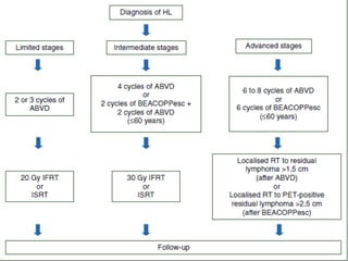
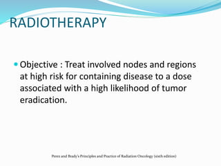
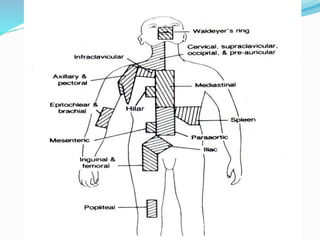
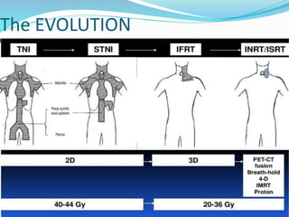
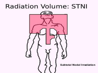
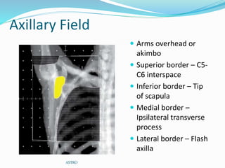
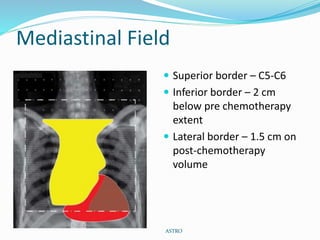
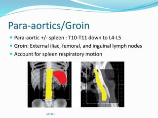
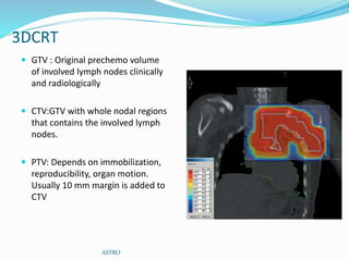
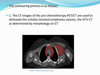
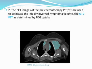
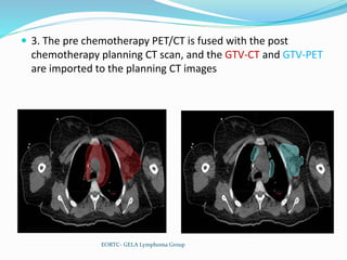
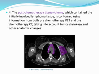
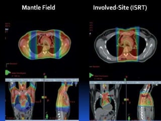
RADIOTHERAPY
 Objective : Treat involved nodes and regions
at high risk for containing disease to a dose
associated with a high likelihood of tumor
eradication.
Perez and Brady’s Principles and Practice of Radiation Oncology (sixth edition)
As Primary Treatment
i. Early-stage nodular lymphocyte-predominant Hodgkin
lymphoma (nLPHL)
ii. Selected cases of early-stage classic HL in patients who are
not candidates for primary chemotherapy
Radiation Therapy as Part of a
Combined Modality Approach
i. Early-stage classic HL
 After adequate systemic chemotherapy
ii. Advanced-stage disease
 Localized RT may be used for residual lymphoma after
full chemotherapy
 RT may be an integral part of some regimens for
advanced-stage disease
The EVOLUTION
MANTLE FIELD
 Involves all nodes from skull base to 10th thoracic level
 Includes :
 Bilateral Cervical
 Bilateral Supraclavicular
 Bilateral Infraclavicular
 Bilateral Axillary
 Bilateral Hilar
 Bilateral Mediastinal
Perez and Brady’s Principles and Practice of Radiation Oncology (sixth edition)
FIELD SIMULATION
• Supine
• Neck Extended
• Arms above the head, or at 90* angle towards the
side, or in ‘akimbo’ position
Perez and Brady’s Principles and Practice of Radiation Oncology (sixth edition)
FIELD DESIGN
 Superior : Bissects the mandible and passes through the
mastoid process
 Lateral : Both the axillae
 Inferior axillary margin: At the level of the inferior tips of
the scapulae.
 Inferior mediastinal border : T10-11 interspace.
Perez and Brady’s Principles and Practice of Radiation Oncology (sixth edition)
BLOCKS
• Head of Humerus is shielded both anteriorly and posteriorly.
• Larynx is shielded anteriorly.
• Heart is shielded below the hilar level without including the
mediastinal LN’s both anteriorly and posteriorly.
• Spinal cord shielding is done in midline for dosages >40 Gy.
• A small block is put at the inferior border of spinal cord
posteriorly.
• Oral cavity is shielded if the superior border includes the oral
cavity.
Perez and Brady’s Principles and Practice of Radiation Oncology (sixth edition)
MINI MANTLE & MODIFIED
MANTLE
Inverted-Y
 Target Volume:
 Para aortic
 Pelvis
 Inguinal nodes(b/l)
 Spleen
Perez and Brady’s Principles and Practice of Radiation Oncology (sixth edition)
Treatment Fields:
 For Paraaortic
 Superiorly:The T10-11 vertebrae
 Inferiorly:The lower limit of L4
 Laterally : width of transverse process
 Pelvis Field:
 Laterally : 1.5-2 cm lat to the widest point in pelvis
 Inferiorly : Lesser trochanter.
Perez and Brady’s Principles and Practice of Radiation Oncology (sixth edition)
Inverted “Y” Field
Para aortic fields pelvic field
BLOCKS:
 Central midline block for
 Bladder
 Small bowel
 Oophoropexy
 Testicular shielding
Perez and Brady’s Principles and Practice of Radiation Oncology (sixth edition)
Oophoropexy
 Medial or lateral transposition of the ovaries via laparoscopy
 The surgeon marks the ovaries with radiopaque sutures or clips and
relocates them medially and as low as possible behind the uterine body.
 A double-thickness midline block is then used; its location is guided by the
position of the opacified nodes and transposed ovaries.
 When the ovaries are at least 2 cm from the edge of this block, the dose is
decreased to 8% of that delivered to the iliac Nodes.
 Alternatively, one or both of the ovaries can be transposed laterally to a
position overlying the iliac wings
Perez and Brady’s Principles and Practice of Radiation Oncology (sixth edition)
RATIONALE
 Chemotherapy is effective notably for microscopic disease,
therefore large fields are no longer necessary.
 Consolidating radiation therapy to involved lymph nodes after
a limited number of chemotherapy cycles remains a necessity.
 Radiotherapy-induced complications are dependent on the
irradiated volume and the total radiation dose.
 It is therefore of utmost necessity to decrease the size of
radiation fields and to limit radiation doses.
Perez and Brady’s Principles and Practice of Radiation Oncology (sixth edition)
IFRT
 Involved field radiotherapy.
 IFRT is the most commonly used technique at present
 Targets a smaller area rather than a classical extended field.
 IFRT (ASTRO 2002) DEFINITION
 IFRT encompasses region and not an individual lymph node.
 Initially involved Pre chemo sites and volume are treated
 Exception to above rule is for transverse diameter of mediastinum and
paraaortic lymph nodes for which reduced post chemo volume is treated.
ASTRO
DEFINITIONS AND RULES
 All patients must have pre- and post- chemotherapy
cervical and thoracic CT scans (axillary lymph node areas
must be clearly visible on thoracic CT scans).
 Patients must be examined by the radiation oncologist
before chemotherapy.
 The remission status after chemotherapy should be
determined for each initially involved lymph node
exclusively using CT scans.
ASTRO
 Complete remission (CR) is defined as the complete
disappearance of clinically and/or radiologically
detectable disease.
 CRu is defined as at least a 75% decrease in tumor size.
 A partial response (PR) is at least a 50% decrease in
tumor size.
 Failure is less than a 50% decrease or any increase in
tumor size
ASTRO
Fields for IFRT
Cervical Chain
Arms position: Akimbo or at
sides
Upper Border: 1 to 2 cm
above the lower tip of the
mastoid process and
midpoint through the
chin.
Lower Border: 2 cm below
the bottom of the clavicle.
Lateral Border: To include
the medial two-thirds of
the clavicle.
ASTRO
Axillary Field
 Arms overhead or
akimbo
 Superior border – C5-
C6 interspace
 Inferior border – Tip
of scapula
 Medial border –
Ipsilateral transverse
process
 Lateral border – Flash
axilla
ASTRO
Mediastinal Field
 Superior border – C5-C6
 Inferior border – 2 cm
below pre chemotherapy
extent
 Lateral border – 1.5 cm on
post-chemotherapy
volume
ASTRO
Para-aortics/Groin
 Para-aortic +/- spleen : T10-T11 down to L4-L5
 Groin: External iliac, femoral, and inguinal lymph nodes
 Account for spleen respiratory motion
ASTRO
3DCRT
 GTV : Original prechemo volume
of involved lymph nodes clinically
and radiologically
 CTV:GTV with whole nodal regions
that contains the involved lymph
nodes.
 PTV: Depends on immobilization,
reproducibility, organ motion.
Usually 10 mm margin is added to
CTV
ASTRO
INVOLVED NODE RADIOTHERAPY
 Includes the originally involved nodes before
chemotherapy
 Requires FDG PET before and after chemotherapy for
accurate target delineation
 Concept : Recurrence usually occurred in the initial
involved nodes – Shahidi ( 2006 )
EORTC- GELA Lymphoma Group
VOLUME DEFINITION - EORTC
 CTV : Initial volume of LN with exclusion of normal
displaced structures (e.g. muscles, blood vessels)
 GTV : If CR, not applicable.
If PR : post chemotherapy volume
 PTV : If CR, 1 cm isotropic margin of CTV
If PR, PTV1: CTV + 1 cm isotropic margin
PTV2: GTV + 1 cm isotropic margin
EORTC- GELA Lymphoma Group
Techniques needed - EORTC
 Whenever feasible, pre chemotherapy PET-CT in RT
planning position
 3D, 4D, IMRT
 If conventional then field size need to be approximately
5x 5 cm
EORTC- GELA Lymphoma Group
 The contouring process is as follows:
 1. The CT images of the pre chemotherapy PET/CT are used to
delineate the initially involved lymphoma volume, the GTV-CT
as determined by morphology on CT
EORTC- GELA Lymphoma Group
 2. The PET images of the pre chemotherapy PET/CT are used
to delineate the initially involved lymphoma volume, the GTV-
PET as determined by FDG uptake
EORTC- GELA Lymphoma Group
 3. The pre chemotherapy PET/CT is fused with the post
chemotherapy planning CT scan, and the GTV-CT and GTV-PET
are imported to the planning CT images
EORTC- GELA Lymphoma Group
 4. The post chemotherapy tissue volume, which contained the
initially involved lymphoma tissue, is contoured using
information from both pre chemotherapy PET and pre
chemotherapy CT, taking into account tumor shrinkage and
other anatomic changes.
EORTC- GELA Lymphoma Group
 The CTV
 Encompasses all of the initial lymphoma volume
 Still respecting normal structures that were never involved by
lymphoma, such as lungs, chest wall, muscles, and mediastinal
normal structures
EORTC- GELA Lymphoma Group
 The concept of ISRT was developed on the basis of the INRT
 ISRT accommodates cases in which optimal pre chemotherapy
imaging is not available
 It is not possible to reduce the CTV to the same extent as with
INRT because the pre chemotherapy GTV information may not
be optimal
 In ISRT, clinical judgment in conjunction with the best available
imaging is used to contour a larger CTV that will accommodate
the uncertainties in defining the pre chemotherapy GTV
EORTC- GELA Lymphoma Group
 If pre chemotherapy imaging is available, but image fusion
with the post chemotherapy planning CT scan is not possible
 To contour the pre chemotherapy target volume on the
planning CT scan allowance should be made for the
uncertainty of the contouring and differences in positioning by
including a larger volume in the CTV
EORTC- GELA Lymphoma Group
 If no pre chemotherapy imaging is available
 To gather description of :
 The pre chemotherapy physical examination of the patient
 The location of scars and scar tissue on the post
chemotherapy planning CT scan
 The patient’s and the family’s recollections of the location
of the presenting lymph node(s)
 The CTV should be contoured taking into account all of this
information, making generous allowance for the many
uncertainties in the process
Radiotherapy lymphoma

Radiotherapy lymphoma

  • 3.
    RADIOTHERAPY  Objective :Treat involved nodes and regions at high risk for containing disease to a dose associated with a high likelihood of tumor eradication. Perez and Brady’s Principles and Practice of Radiation Oncology (sixth edition)
  • 5.
    As Primary Treatment i.Early-stage nodular lymphocyte-predominant Hodgkin lymphoma (nLPHL) ii. Selected cases of early-stage classic HL in patients who are not candidates for primary chemotherapy
  • 6.
    Radiation Therapy asPart of a Combined Modality Approach i. Early-stage classic HL  After adequate systemic chemotherapy ii. Advanced-stage disease  Localized RT may be used for residual lymphoma after full chemotherapy  RT may be an integral part of some regimens for advanced-stage disease
  • 8.
  • 11.
    MANTLE FIELD  Involvesall nodes from skull base to 10th thoracic level  Includes :  Bilateral Cervical  Bilateral Supraclavicular  Bilateral Infraclavicular  Bilateral Axillary  Bilateral Hilar  Bilateral Mediastinal Perez and Brady’s Principles and Practice of Radiation Oncology (sixth edition)
  • 12.
    FIELD SIMULATION • Supine •Neck Extended • Arms above the head, or at 90* angle towards the side, or in ‘akimbo’ position Perez and Brady’s Principles and Practice of Radiation Oncology (sixth edition)
  • 13.
    FIELD DESIGN  Superior: Bissects the mandible and passes through the mastoid process  Lateral : Both the axillae  Inferior axillary margin: At the level of the inferior tips of the scapulae.  Inferior mediastinal border : T10-11 interspace. Perez and Brady’s Principles and Practice of Radiation Oncology (sixth edition)
  • 14.
  • 15.
    • Head ofHumerus is shielded both anteriorly and posteriorly. • Larynx is shielded anteriorly. • Heart is shielded below the hilar level without including the mediastinal LN’s both anteriorly and posteriorly. • Spinal cord shielding is done in midline for dosages >40 Gy. • A small block is put at the inferior border of spinal cord posteriorly. • Oral cavity is shielded if the superior border includes the oral cavity. Perez and Brady’s Principles and Practice of Radiation Oncology (sixth edition)
  • 16.
    MINI MANTLE &MODIFIED MANTLE
  • 18.
    Inverted-Y  Target Volume: Para aortic  Pelvis  Inguinal nodes(b/l)  Spleen Perez and Brady’s Principles and Practice of Radiation Oncology (sixth edition)
  • 19.
    Treatment Fields:  ForParaaortic  Superiorly:The T10-11 vertebrae  Inferiorly:The lower limit of L4  Laterally : width of transverse process  Pelvis Field:  Laterally : 1.5-2 cm lat to the widest point in pelvis  Inferiorly : Lesser trochanter. Perez and Brady’s Principles and Practice of Radiation Oncology (sixth edition)
  • 20.
    Inverted “Y” Field Paraaortic fields pelvic field
  • 21.
    BLOCKS:  Central midlineblock for  Bladder  Small bowel  Oophoropexy  Testicular shielding Perez and Brady’s Principles and Practice of Radiation Oncology (sixth edition)
  • 22.
    Oophoropexy  Medial orlateral transposition of the ovaries via laparoscopy  The surgeon marks the ovaries with radiopaque sutures or clips and relocates them medially and as low as possible behind the uterine body.  A double-thickness midline block is then used; its location is guided by the position of the opacified nodes and transposed ovaries.  When the ovaries are at least 2 cm from the edge of this block, the dose is decreased to 8% of that delivered to the iliac Nodes.  Alternatively, one or both of the ovaries can be transposed laterally to a position overlying the iliac wings Perez and Brady’s Principles and Practice of Radiation Oncology (sixth edition)
  • 27.
    RATIONALE  Chemotherapy iseffective notably for microscopic disease, therefore large fields are no longer necessary.  Consolidating radiation therapy to involved lymph nodes after a limited number of chemotherapy cycles remains a necessity.  Radiotherapy-induced complications are dependent on the irradiated volume and the total radiation dose.  It is therefore of utmost necessity to decrease the size of radiation fields and to limit radiation doses. Perez and Brady’s Principles and Practice of Radiation Oncology (sixth edition)
  • 28.
    IFRT  Involved fieldradiotherapy.  IFRT is the most commonly used technique at present  Targets a smaller area rather than a classical extended field.  IFRT (ASTRO 2002) DEFINITION  IFRT encompasses region and not an individual lymph node.  Initially involved Pre chemo sites and volume are treated  Exception to above rule is for transverse diameter of mediastinum and paraaortic lymph nodes for which reduced post chemo volume is treated. ASTRO
  • 29.
    DEFINITIONS AND RULES All patients must have pre- and post- chemotherapy cervical and thoracic CT scans (axillary lymph node areas must be clearly visible on thoracic CT scans).  Patients must be examined by the radiation oncologist before chemotherapy.  The remission status after chemotherapy should be determined for each initially involved lymph node exclusively using CT scans. ASTRO
  • 30.
     Complete remission(CR) is defined as the complete disappearance of clinically and/or radiologically detectable disease.  CRu is defined as at least a 75% decrease in tumor size.  A partial response (PR) is at least a 50% decrease in tumor size.  Failure is less than a 50% decrease or any increase in tumor size ASTRO
  • 31.
  • 32.
    Cervical Chain Arms position:Akimbo or at sides Upper Border: 1 to 2 cm above the lower tip of the mastoid process and midpoint through the chin. Lower Border: 2 cm below the bottom of the clavicle. Lateral Border: To include the medial two-thirds of the clavicle. ASTRO
  • 33.
    Axillary Field  Armsoverhead or akimbo  Superior border – C5- C6 interspace  Inferior border – Tip of scapula  Medial border – Ipsilateral transverse process  Lateral border – Flash axilla ASTRO
  • 34.
    Mediastinal Field  Superiorborder – C5-C6  Inferior border – 2 cm below pre chemotherapy extent  Lateral border – 1.5 cm on post-chemotherapy volume ASTRO
  • 35.
    Para-aortics/Groin  Para-aortic +/-spleen : T10-T11 down to L4-L5  Groin: External iliac, femoral, and inguinal lymph nodes  Account for spleen respiratory motion ASTRO
  • 36.
    3DCRT  GTV :Original prechemo volume of involved lymph nodes clinically and radiologically  CTV:GTV with whole nodal regions that contains the involved lymph nodes.  PTV: Depends on immobilization, reproducibility, organ motion. Usually 10 mm margin is added to CTV ASTRO
  • 38.
    INVOLVED NODE RADIOTHERAPY Includes the originally involved nodes before chemotherapy  Requires FDG PET before and after chemotherapy for accurate target delineation  Concept : Recurrence usually occurred in the initial involved nodes – Shahidi ( 2006 ) EORTC- GELA Lymphoma Group
  • 39.
    VOLUME DEFINITION -EORTC  CTV : Initial volume of LN with exclusion of normal displaced structures (e.g. muscles, blood vessels)  GTV : If CR, not applicable. If PR : post chemotherapy volume  PTV : If CR, 1 cm isotropic margin of CTV If PR, PTV1: CTV + 1 cm isotropic margin PTV2: GTV + 1 cm isotropic margin EORTC- GELA Lymphoma Group
  • 40.
    Techniques needed -EORTC  Whenever feasible, pre chemotherapy PET-CT in RT planning position  3D, 4D, IMRT  If conventional then field size need to be approximately 5x 5 cm EORTC- GELA Lymphoma Group
  • 41.
     The contouringprocess is as follows:  1. The CT images of the pre chemotherapy PET/CT are used to delineate the initially involved lymphoma volume, the GTV-CT as determined by morphology on CT EORTC- GELA Lymphoma Group
  • 42.
     2. ThePET images of the pre chemotherapy PET/CT are used to delineate the initially involved lymphoma volume, the GTV- PET as determined by FDG uptake EORTC- GELA Lymphoma Group
  • 43.
     3. Thepre chemotherapy PET/CT is fused with the post chemotherapy planning CT scan, and the GTV-CT and GTV-PET are imported to the planning CT images EORTC- GELA Lymphoma Group
  • 44.
     4. Thepost chemotherapy tissue volume, which contained the initially involved lymphoma tissue, is contoured using information from both pre chemotherapy PET and pre chemotherapy CT, taking into account tumor shrinkage and other anatomic changes. EORTC- GELA Lymphoma Group
  • 45.
     The CTV Encompasses all of the initial lymphoma volume  Still respecting normal structures that were never involved by lymphoma, such as lungs, chest wall, muscles, and mediastinal normal structures EORTC- GELA Lymphoma Group
  • 49.
     The conceptof ISRT was developed on the basis of the INRT  ISRT accommodates cases in which optimal pre chemotherapy imaging is not available  It is not possible to reduce the CTV to the same extent as with INRT because the pre chemotherapy GTV information may not be optimal  In ISRT, clinical judgment in conjunction with the best available imaging is used to contour a larger CTV that will accommodate the uncertainties in defining the pre chemotherapy GTV EORTC- GELA Lymphoma Group
  • 50.
     If prechemotherapy imaging is available, but image fusion with the post chemotherapy planning CT scan is not possible  To contour the pre chemotherapy target volume on the planning CT scan allowance should be made for the uncertainty of the contouring and differences in positioning by including a larger volume in the CTV EORTC- GELA Lymphoma Group
  • 51.
     If nopre chemotherapy imaging is available  To gather description of :  The pre chemotherapy physical examination of the patient  The location of scars and scar tissue on the post chemotherapy planning CT scan  The patient’s and the family’s recollections of the location of the presenting lymph node(s)  The CTV should be contoured taking into account all of this information, making generous allowance for the many uncertainties in the process EORTC- GELA Lymphoma Group
  • 52.
     If nopre chemotherapy imaging is available  To gather description of :  The pre chemotherapy physical examination of the patient  The location of scars and scar tissue on the post chemotherapy planning CT scan  The patient’s and the family’s recollections of the location of the presenting lymph node(s)  The CTV should be contoured taking into account all of this information, making generous allowance for the many uncertainties in the process

Editor's Notes

  • #12 CLOAK
  • #15 Lung blocks : Made separate for anterior and posterior. Upper border in anterior : 2 cms below medial clavicle, and a thin lung band is left at the lateral clavicles. Upper border in posterior : a thin band is left under the clavicles as the infraclavicular LN’s are located anteriorly. Lateral Borders : a 1 cm band is left in costal curves which extends until the 5th or 6th costa and finishes horizontally in chest wall. Medial Border : bilateral mediastinal and hilar lymphatics are included (mediastinal enlargement should be included).