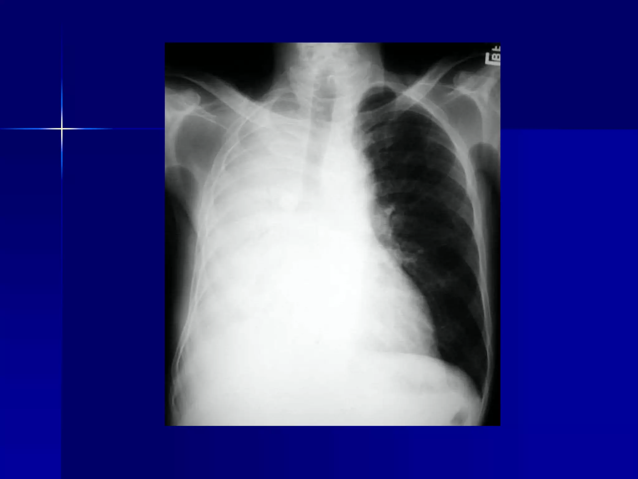
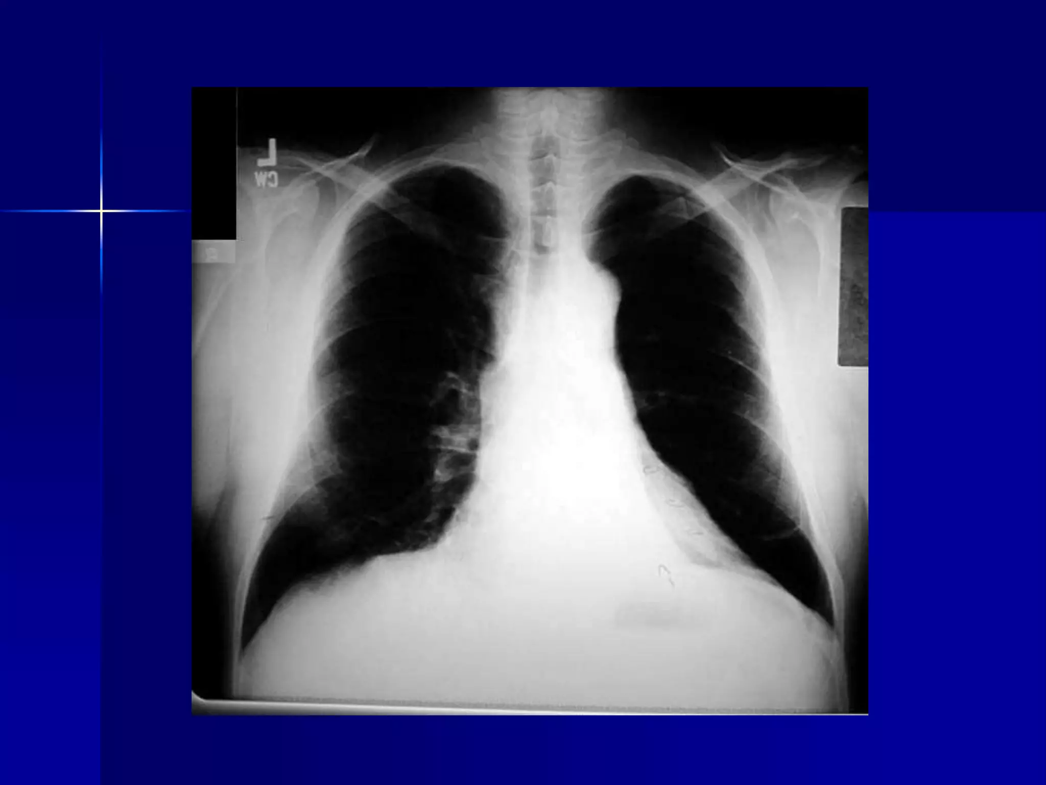
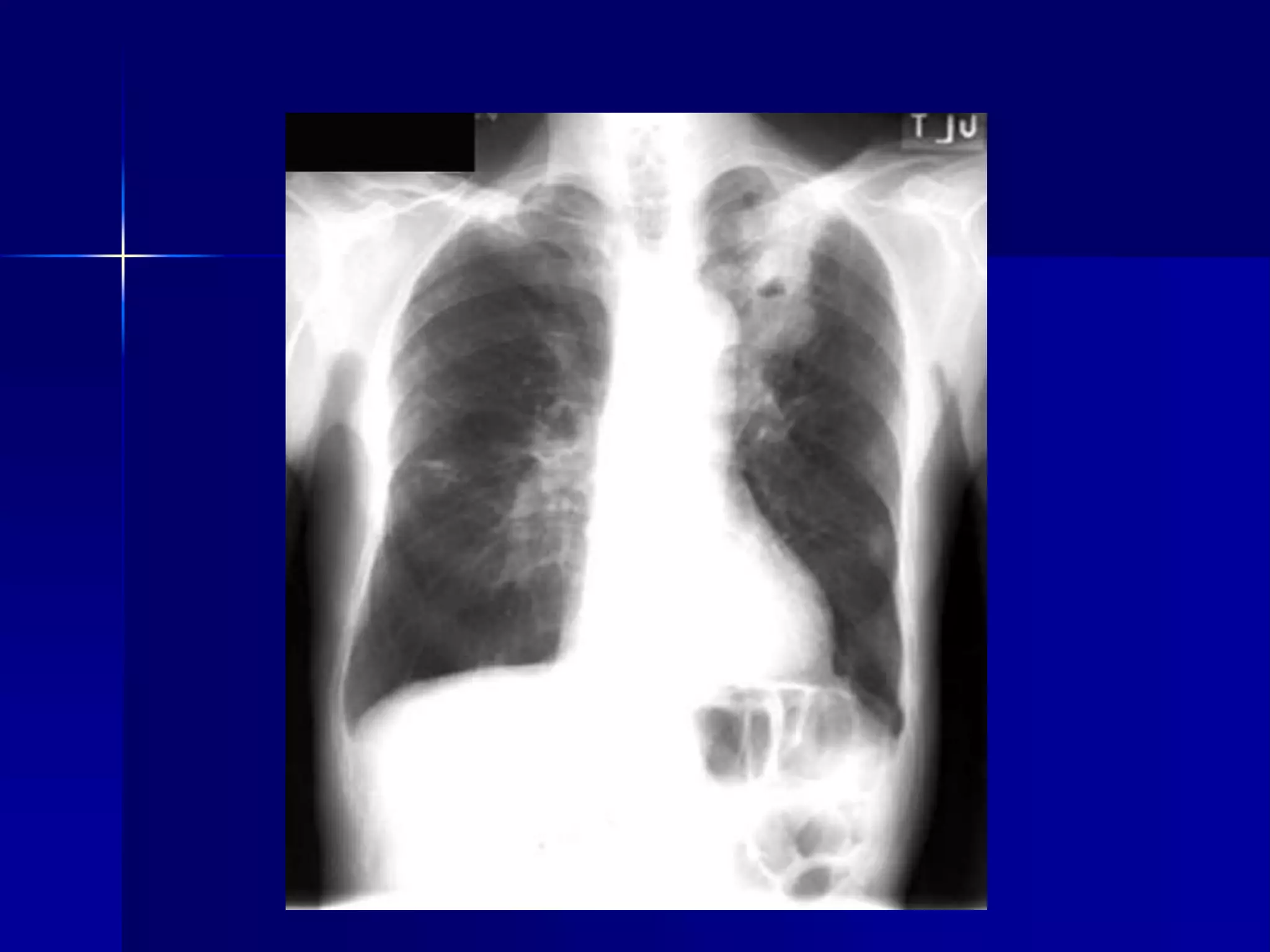
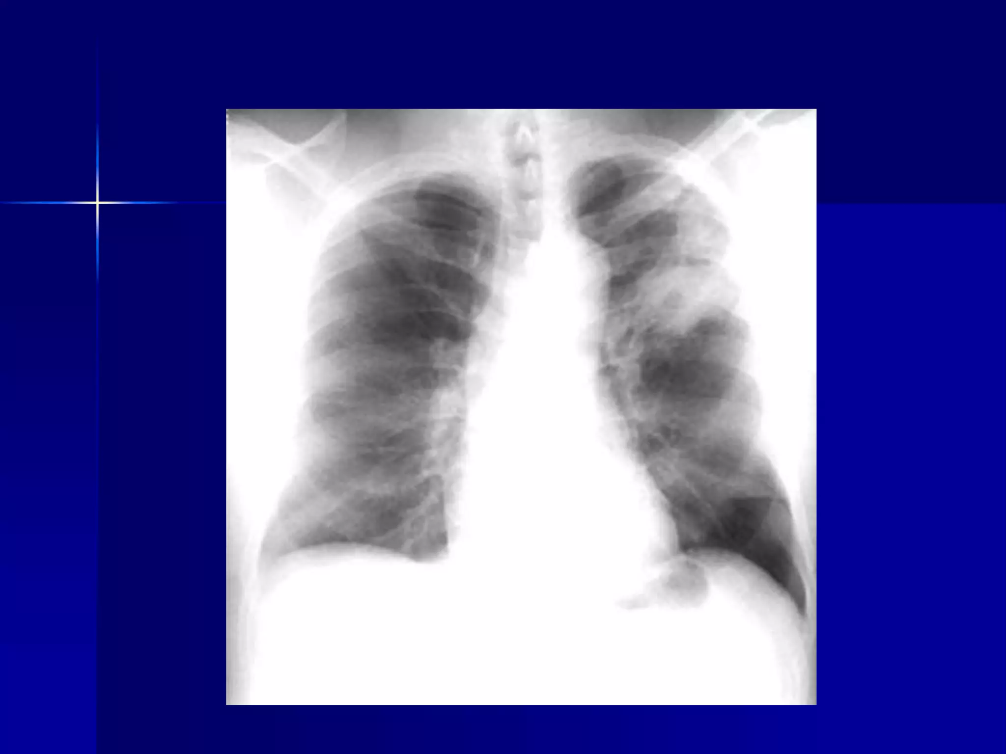
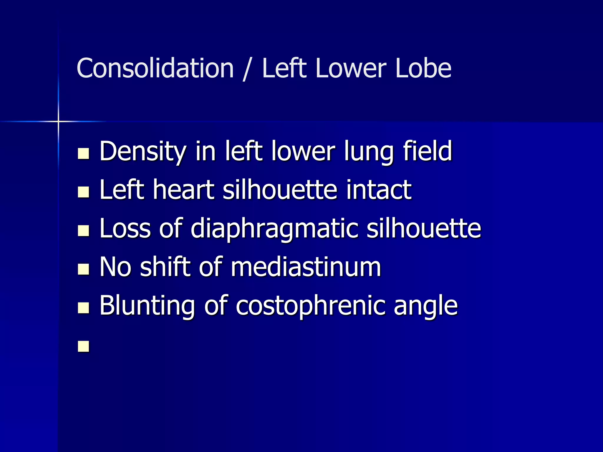
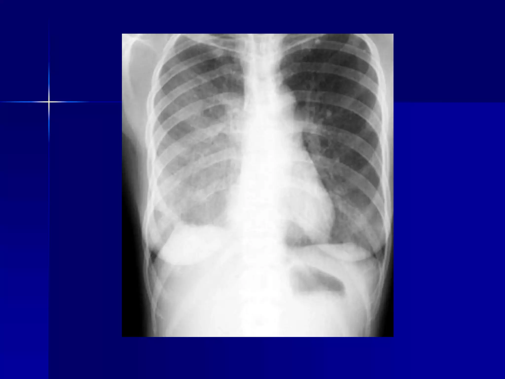
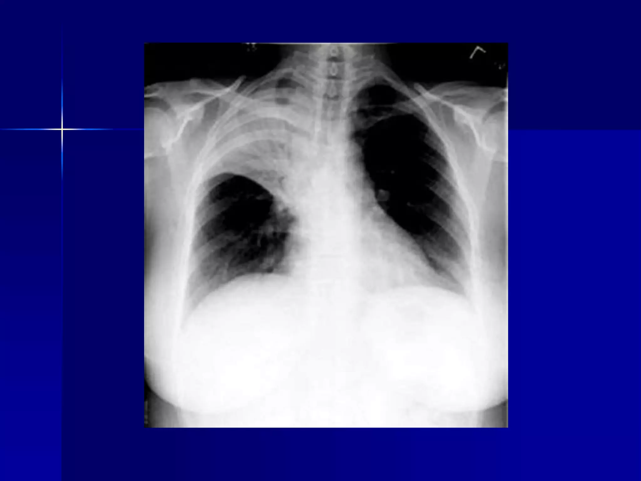
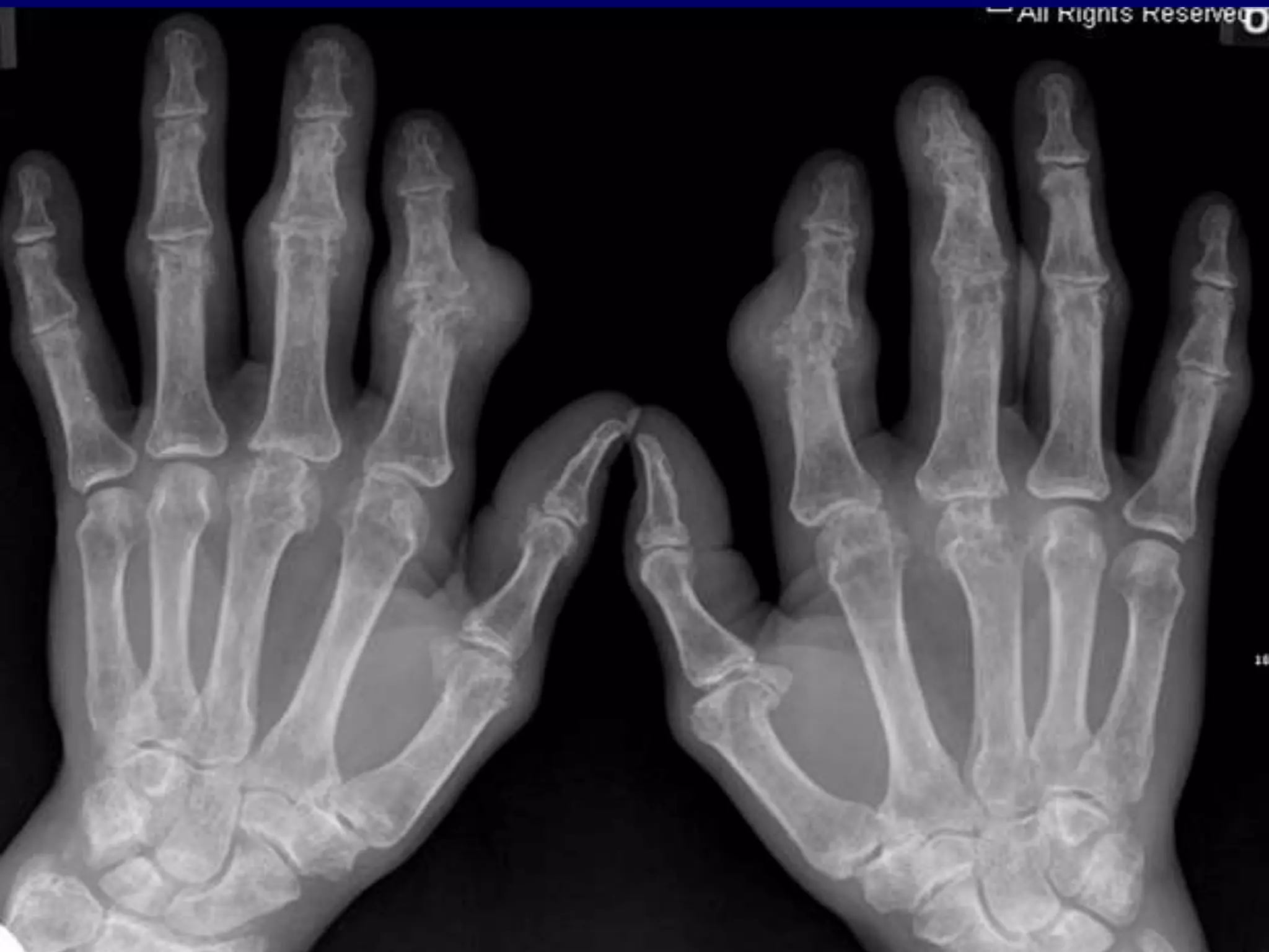
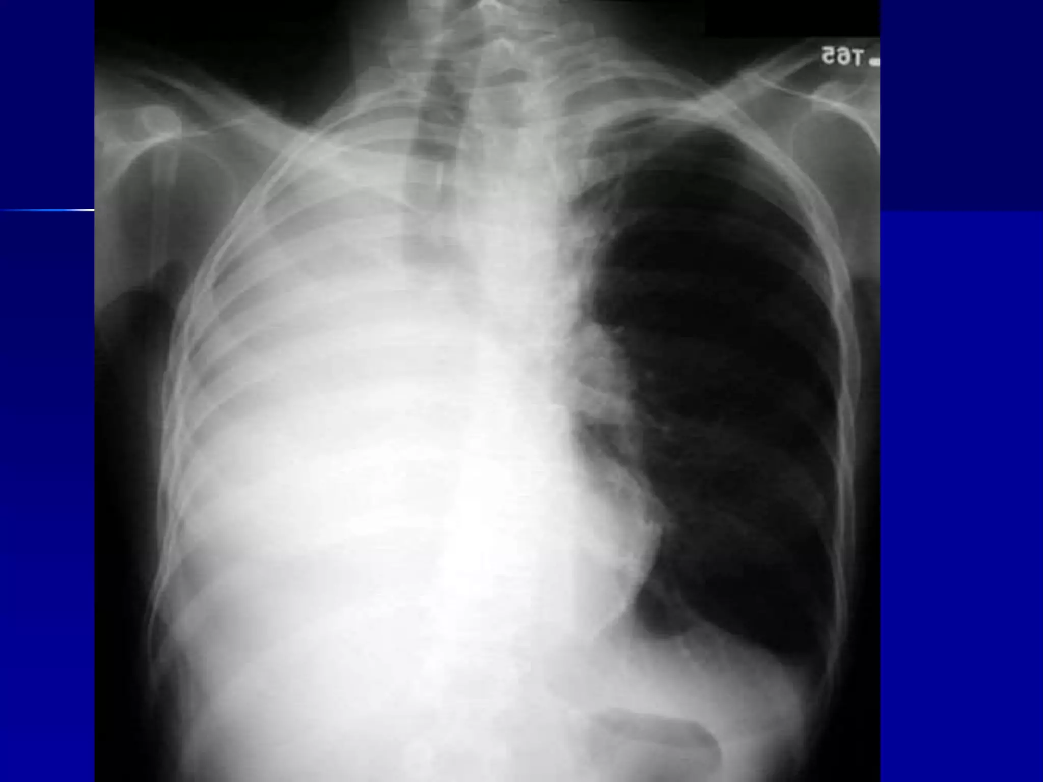
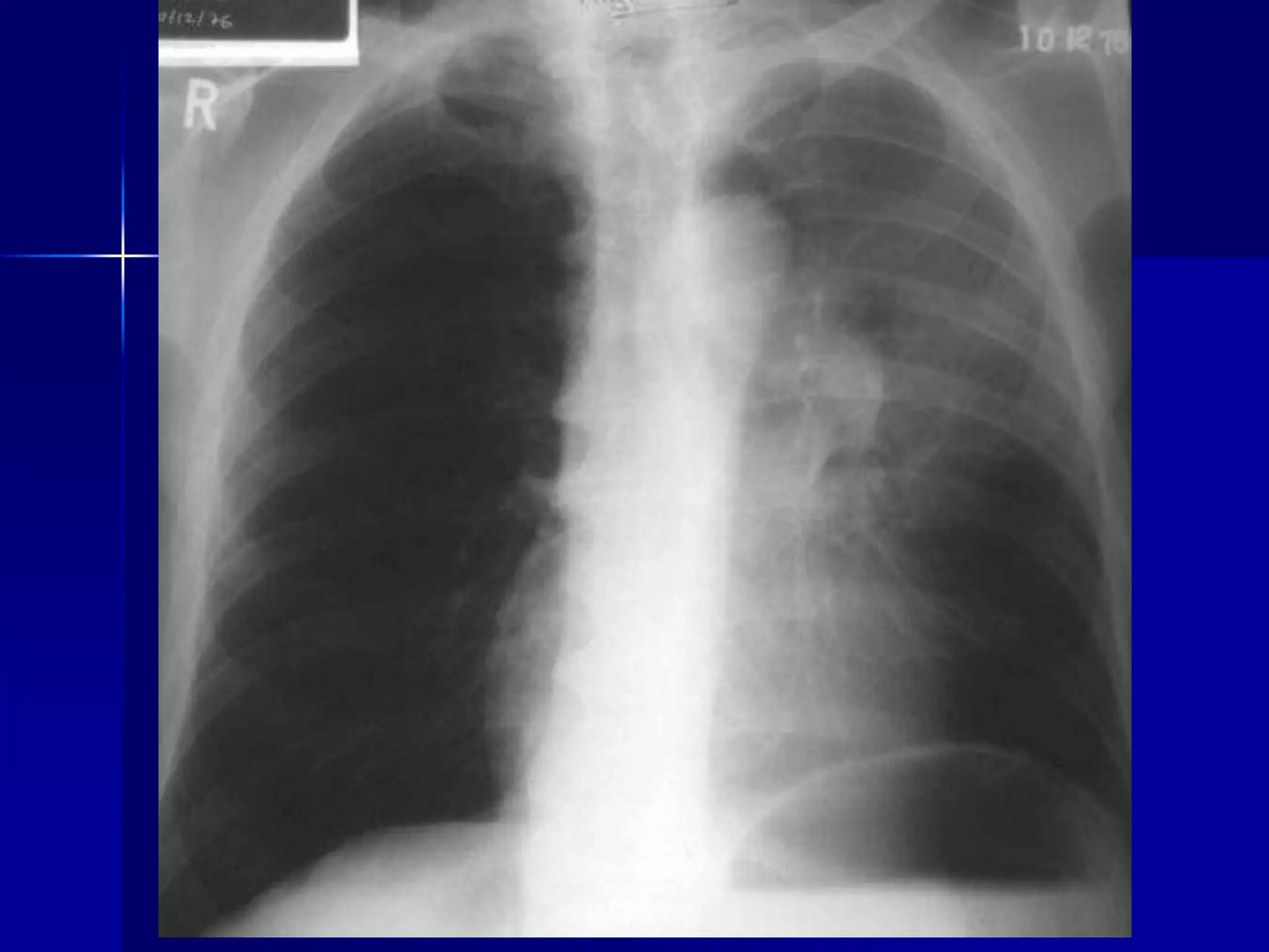
X- Ray Review 
By: Dr Wedad Bardisi
Atelectasis Right Lung 
 Homogenous density right hemithorax 
 Mediastinal shift to right 
 Right heart and diaphragmatic 
silhouette are not identifiable
 Atelectasis Right Lung 
 Open Bronchus Sign 
 Homogenous density right hemithorax 
 Mediastinal shift to right 
 Right heart and diaphragmatic silhouette 
are not identifiable 

Atelectasis Left Lung 
 Homogenous density left hemithorax 
 Mediastinal shift to the left 
 Diaphragmatic and heart silhouette 
are not identifiable
 Atelectasis Left Lower Lobe 
 Inhomogeneous cardiac density 
 Triangular retrocardiac density 
 Left hilum pulled down 

 Atelectasis Right Upper Lobe 
 Density in the right upper lung field 
 Transverse fissure pulled up 
 Right hilum pulled up 
 Smaller right lung 
 Smaller right hemithorax 

Cancer Breast 
 Inflammatory Carcinoma 
 Post-Radiation 
 Larger right breast 
 Inverted nipple 
 Radiation Fibrosis of Lung 
 Right lung smaller 
 Right hemithorax smaller 
 Paramediastinal fibrosis 

Cystic Fibrosis - Bronchiectasis 
 Bilateral diffuse 
 Multiple cavities / Bronchiectasis 
 Peribronchial fibrosis 
 Prominent hilum 
 Hyperinflated
Silicosis 
 Egg shell calcification of lymph nodes 
 Other findings include: 
 Diaphragmatic pleural calcification 
 Multiple cavities with fluid levels 

Histoplasmosis 
 Calcified nodes 
 Calcified nodules in lungs 

Visceral pleural calcification 
 Open drainage with air fluid levels in 
pleural space
Silicosis 
 Diaphragmatic pleural calcification 
 Other findings include: 
 Multiple cavities with fluid levels 
 Egg shell calcification of lymph nodes
Squamous Cell Carcinoma Lung 
 Cavity 
 LUL mass 
 Thick walled cavity 
 Eccentric location of cavity 
 Fluid level
Lung Cancer / Squamous Cell 
 Mass density 
 Anterior segment of LUL 
 Thick wall cavitation
 Rib Fracture / Hematoma
Consolidation / Lingula 
 Density in left lower lung field 
 Loss of left heart silhouette 
 Diaphragmatic silhouette intact 
 No shift of mediastinum 
 Blunting of costophrenic angle 

Consolidation / Left Lower Lobe 
 Density in left lower lung field 
 Left heart silhouette intact 
 Loss of diaphragmatic silhouette 
 No shift of mediastinum 
 Blunting of costophrenic angle 

Lobar Pneumonia Right Middle Lobe 
 Vague density right lower lung field 
 Indistinct right cardiac silhouette 
 Intact diaphragmatic silhouette 
 Lateral Density corresponding to RML 
 No loss of lung volume 
 No air bronchogram
Consolidation Right Middle Lobe 
 Density in right middle lung field 
 Loss of right cardiac silhouette 
 Pulmonary artery overlay sign 
 Air bronchogram not visible 
 Minor movement of fissure 

Consolidation Left Lower Lobe 
 Density in left lower lung field 
 Left heart silhouette intact 
 Loss of diaphragmatic silhouette 
 No shift of mediastinum 
 Pneumatocele
Pneumoperitoneum 
 
 Air under diaphragm
 Diaphragmatic paralysis
 Lung Metastasis/Alveolar Form 
Cancer Pancreas 
 Soft fluffy lesions 
 Air bronchogram 
 Coalesing lesions 

Pulmonary Edema 
Acute Diffuse Alveolar 
 Bilateral 
 Diffuse 
 Butterfly pattern 
 Soft fluffy lesions 
 Coalescing 
 Air bronchogram
Emphysema 
 Hyperlucent lung fields 
 Multiple blebs 
 Avascular zones 
 Prominent pulmonary arteries 
 Radiologic TLC
Milary Tuberculosis 
 Interstitial nodules 
– Uniform size 
– Sharper edges
Lymphangitic Metastasis 
 Cancer Breast 
 Kerley lines 
 Subpulmonic effusion on right
Tuberculosis Spine 
 Loss of intervertebral space 
 Vertebral collapse 
 Cold abscess is not present 
 PA view is not diagnostic.
 Mediastinal Lymph Nodes 
 Extrapleural 
 Polycyclic margin 
 Anterior mediastinum
 Air Fluid Level 
 Inhomogeneous cardiac density 
 Retrocardiac density 
 In mediastinum in PA view 
 Hiatal hernia 
 Other findings include: 
 Pleural fibrosis on right
 Sarcoidosis / Miliary Nodules / Hilar 
Nodes
Mass density 
 Mass density can be encountered in lung cancer, 
benign tumors, sarcoma, lymphoma, Wegener's 
and blastomycosis and tuberculoma. 
 Radiological criteria for a mass lesion are chest 
lateral and PA views. 
 Density 
 Round or oval 
 Sharp margins 
 Homogenous density (exception: air 
bronchogram in lymphoma and blastomycosis)
 No respect for anatomy (in cancer) 
 Can break down leading to thick walled cavity 
 May show calcification (histoplasmoma, 
tuberculoma, hamartoma) 
 Note in a gross cut section a mass which is well 
demarcated from the adjacent normal lung. 
Malignant tumors have infiltrating edges, while 
benign tumors are rounded and well circumscribed.
 Mass 
 Round or oval 
 Sharp margin 
 Homogenous 
 No respect for anatomy 
 Lung Cancer: Large cell 

 Round homogenous density 
 Sharp margins 
 Medial portion pleural based (acute 
angle) 
 This is a case of squamous cell lung 
cancer.
Hilar Nodes 
 bilateral symmetrical hilar nodes and 
para tracheal nodes. 
 A clear space between the nodes and 
heart, identifies the nodes as hilar. 

Sarcoidosis 
 Alveolar Form 
 Symmetrical hilar nodes 
 Mediastinal nodes 
 Multiple bilateral mass densities with 
alveolar features 
 Soft coalescing
 Lung Cancer 
 RUL primary lesion 
 Para tracheal nodes
Pleural Effusion 
 Fluid accumulates in the pleural space. 
 Irrespective of the nature of fluid, radiologically they will look 
similar. 
 Radiological criteria are: Density 
 In dependent portion 
– Costophrenic angle in PA view 
– Anterior and posterior portions of gutter in lateral view 
– Along sides in lateral decubitus position 
– Along posteriorly in supine position, giving diffuse haziness on 
the side of effusion 
 Silhouette of upper limit of density 
– Upper margin high in axilla in PA view 
– Upper margin high interiorly and posteriorly in lateral view 
 Blunting of costophrenic angle 
 Lack of identifiable diaphragm (silhouette sign principle).
 Massive 
 Unilateral VS bilateral 
 Sub pulmonic 
 Loculated 
 Supine position 
 Lateral decubitus position
 Pleural Effusion 
 Homogenous density 
 Meniscus maximum in axilla 
 Loss of cardiophrenic angle 
 Loss of diaphragmatic and right 
cardiac silhouette 

 Loculated Pleural Effusion 
 Empyema 
 Haziness of right hemithorax 
 Density not corresponding to lobar anatomy 
 Diaphragmatic and cardiac silhouettes intact 
 Lateral film below 
– Loculated fluid overlying vertebral column
 Pleural Fibrosis 
 Small right hemithorax 
 Diffuse haziness 
 Tracheal shift to right 
 Blunted costophrenic angle 
 Lines not corresponding to fissures 

 Consolidation Right Upper Lobe / 
 Air Bronchogram 
 Density in right upper lung field 
 Lobar density 
 Loss of ascending aorta silhouette 
 No shift of mediastinum 
 Transverse fissure not significantly 
shifted 
 Air bronchogram
 Pneumothorax 
 Atelectatic lung is dense implying that 
it is abnormal ("normal lung" will not 
be dense) 
 Bleb is easily recognized in the close-up 
below 

 Heart Failure/Rapid Resolution 
 Such rapid resolution as seen above is 
usually due to secondary cause such 
as fluid overload.
 Subcutaneous Emphysema 
 Diffuse interstitial fibrosis
Ankylosing Spondylitis 
 Spine film: Bamboo spine 
 Cervical spine 
 Bilateral non-specific upper lobe 
disease 
 .
 Aneurysm Arch of Aorta 
 Mediastinal mass 
 Extrapleural 

Bronchiectasis 
 Radiologic findings include: 
 Normal appearing CXR in most 
 Tubular shadows 
 Tram line 
 Gloved fingers 
 Mucocele 
 Ring shadows with thickened bronchial walls 
 Air fluid levels 
 Watch for dextrocardia 
– Immotile cilia syndrome 
 Diffuse lung fibrosis 
– Due to recurrent infections
Cystic Fibrosis - Bronchiectasis 
 Bilateral diffuse 
 Multiple cavities / Bronchiectasis 
 Peribronchial fibrosis 
 Prominent hilum 
 Hyperinflated 

 Bronchiectasis 
 Multiple bilateral basal air fluid levels
 Pleural Effusion 
 Rheumatoid Arthritis 
 Chest lateral and hand x-rays. 

 Diagnosis 
 Acute colonic pseudo-obstruction or 
Ogilvie syndrome 
 Differential 
 Mechanical colonic obstruction 
 Toxic megacolon 
 Mesenteric ischaemia
Case 
 This 19 year old male presented with a 
history of 3 stone weight loss (42 lbs to 
our US cousins, 19kg to the metric world) 
over a period of three months. He 
complained of malaise and was anaemic. 
Endoscopy of the upper gastrointestinal 
tract was normal. 
 A barium small bowel meal was 
performed.
 Findings 
 Changes of early/intermediate Crohn's 
disease, with thickened folds, tending to 
asymmetry and obliteration in places. There 
are small apthous ulcers, and nodules, with 
normal diameter bowel. There is a linear 
mesenteric ulcer (arrowed, lower image 
 Small bowel Crohn's disease
 Avascular necrosis of the femoral head 
 differential for the causes: 
 Toxic 
Steroids, Anti-inflammatory drugs, Alcohol, 
Immunosuppressives, Traumatic, Idiopathic, 
Fractures (femoral neck, talus, scaphoid), 
Radiotherapy, Heat (burns), Fat embolism
 Inflammatory 
Rheumatoid arthritis, SLE, 
Scleroderma, Infection - eg. pyogenic 
arthritis, Pancreatitis 
 
 Metabolic and endocrine 
Pregnancy, Diabetes, Cushing's 
syndrome, Hyperlipidaemias, Gout
 Haemopoetic disorders 
Haemoglobinopathies, Polycythaemia 
rubra vera, Haemophilia 
 Thrombotic and embolic 
Dysbaric osteonecrosis (divers), 
Arteritis
 Posterior dislocation of the shoulder 
 A painful shoulder after a fall 
 This patient complained of pain and 
restricted movement in the shoulder, 
having blacked out and fallen over. What 
abnormality is demonstrated? What other 
view would be useful? And if the patient 
was unable to abduct the arm, what 
other view can be performed?
 Findings 
 Posterior dislocation is much less common 
than anterior dislocation (approximately 4% 
of dislocations), and is frequently much less 
obvious on the AP view alone, requiring a 
further view for confirmation. This may be 
either an axial view of the shoulder, or a 
tangential view of the scapula if the patient 
can not raise their arm sufficiently for the 
former.
 An elderly patient with change in bowel habit and 
dysuria 
 This patient presented with a history of change of 
bowel habit and lower abdominal pain. There had 
been a brief episode of rectal bleeding a few weeks 
earlier, and some dysuria. 
 The General Practitioner had recently prescribed a 
course of antibiotics for a suspected urinary tract 
infecton, but this had not helped. 
 A barium enema was arranged to investigate the 
rectal bleeding
 Diagnosis: 
 Moderately differentiated 
adenoarcinoma of the colon with 
colovesical fistula
 Paget's disease of the humerus & 
pathological fracture
 Anterior mediastinal Seminoma Young 
adult man with chest discomfort and 
left shoulder pain
 Bone mets and R. pleural effusion
 Left upper lobe atelectasis
 Haemothorax 
 T tube
 Cardiomegaly bilateral
 Pulmonary artery enlargement
 Atlantoaxial instability (cervical 
rheumatoid arthritis). Flexion and 
Extension views show the distance 
between the atlas and the dens 
anteriorly in the extension view is 2.3 
mm while in the flexion view the 
distance is 7.5 mm.
 Adult woman with chronic left wrist 
pain. Avascular necrosis of the 
scaphoid bone. Age indeterminate 
scaphoid wrist fracture. Proximal 
scaphoid bone is sclerotic due to 
osteonecrosis.
 Gout : Marginal erosions with 
overhanging edges and sclerotic 
borders at numerous joints throughout 
the hands and feet. Interphalangeal 
joint space narrowing. Soft tissue 
swelling with associated soft tissue 
calcifications
 Bone metastases to the finger. 
Radiograph shows a destructive 
expanded osteolytic lesion in the 
metacarpal of the thumb in a 55-year-old 
man with lung carcinoma.
 Advanced RA. Radiograph of the hand 
shows severe destruction and 
mutilation of the radiocarpal, 
intercarpal, carpometacarpal, and 
metacarpophalangeal joints. 
Intercarpal ankylosis is noted. 
 There is also subluxation and deviation 
of the fourth and fifth fingers
 Skeletal sickle cell anemia. H 
vertebrae. Lateral view of the spine 
shows angular depression of the 
central portion of each upper and 
lower endplate.
 Ankylosing Spondylitis 35-year-old 
male presents with low back pain of 
many months duration that is not 
relieved by rest. In addition, significant 
neck stiffness.
 Findings: AP and lateral view of the 
lumbosacral spine demonstrate flowing 
osteophytes with bony bridges 
between the margins of adjacent 
vertebral bodies seen laterally at all 
the visualized levels.
 In addition on the lateral view there is 
anterior syndesmophyte formation 
with straightening of the normal 
curvature of the lumbar spine. 
Vertebral body height is maintained 
and there is squaring of the anterior 
vertebral margins. In addition, 
ankylosis of the posterior diarthrodial 
joints. The sacroiliac joints are fused 
bilaterally
 Lateral radiograph shows sclerotic 
metastasis of the L2 vertebra in a 54- 
year-old man with prostatic carcinoma
 Osteomalcia malabsorption
 Skeletal sickle cell anemia. 
Osteonecrosis
 Seventy-year-old with dysphagia 
Esophageal diverticula (probably pulsion 
variety), hiatal hernia, GE reflux Two wide-necked 
diverticula at the junction of the mid 
and distal esophagus that retain contrast 
after the esophagus empties. There were 
tertiary, nonpropulsive contractions, a large 
sliding hiatal hernia, and GE reflux to the 
thoracic inlet.
 Young man with hematuria IVP 
shows a dual renal pelvis on the left 
with partial duplication of the left 
ureter. Ultrasound shows a cortical 
bar, compatible with duplication of the 
collecting system.Diagnosis: Partial 
duplex, left kidney
 LLL col
 Massive 
 Massive Pleural Effusion
 Atelectasis Right Lung
 Left upper lobe collapse due to left 
hilar mass
Xray

Xray

  • 1.
    X- Ray Review By: Dr Wedad Bardisi
  • 3.
    Atelectasis Right Lung  Homogenous density right hemithorax  Mediastinal shift to right  Right heart and diaphragmatic silhouette are not identifiable
  • 5.
     Atelectasis RightLung  Open Bronchus Sign  Homogenous density right hemithorax  Mediastinal shift to right  Right heart and diaphragmatic silhouette are not identifiable 
  • 7.
    Atelectasis Left Lung  Homogenous density left hemithorax  Mediastinal shift to the left  Diaphragmatic and heart silhouette are not identifiable
  • 9.
     Atelectasis LeftLower Lobe  Inhomogeneous cardiac density  Triangular retrocardiac density  Left hilum pulled down 
  • 11.
     Atelectasis RightUpper Lobe  Density in the right upper lung field  Transverse fissure pulled up  Right hilum pulled up  Smaller right lung  Smaller right hemithorax 
  • 13.
    Cancer Breast Inflammatory Carcinoma  Post-Radiation  Larger right breast  Inverted nipple  Radiation Fibrosis of Lung  Right lung smaller  Right hemithorax smaller  Paramediastinal fibrosis 
  • 15.
    Cystic Fibrosis -Bronchiectasis  Bilateral diffuse  Multiple cavities / Bronchiectasis  Peribronchial fibrosis  Prominent hilum  Hyperinflated
  • 17.
    Silicosis  Eggshell calcification of lymph nodes  Other findings include:  Diaphragmatic pleural calcification  Multiple cavities with fluid levels 
  • 19.
    Histoplasmosis  Calcifiednodes  Calcified nodules in lungs 
  • 21.
    Visceral pleural calcification  Open drainage with air fluid levels in pleural space
  • 23.
    Silicosis  Diaphragmaticpleural calcification  Other findings include:  Multiple cavities with fluid levels  Egg shell calcification of lymph nodes
  • 25.
    Squamous Cell CarcinomaLung  Cavity  LUL mass  Thick walled cavity  Eccentric location of cavity  Fluid level
  • 27.
    Lung Cancer /Squamous Cell  Mass density  Anterior segment of LUL  Thick wall cavitation
  • 29.
     Rib Fracture/ Hematoma
  • 31.
    Consolidation / Lingula  Density in left lower lung field  Loss of left heart silhouette  Diaphragmatic silhouette intact  No shift of mediastinum  Blunting of costophrenic angle 
  • 33.
    Consolidation / LeftLower Lobe  Density in left lower lung field  Left heart silhouette intact  Loss of diaphragmatic silhouette  No shift of mediastinum  Blunting of costophrenic angle 
  • 35.
    Lobar Pneumonia RightMiddle Lobe  Vague density right lower lung field  Indistinct right cardiac silhouette  Intact diaphragmatic silhouette  Lateral Density corresponding to RML  No loss of lung volume  No air bronchogram
  • 37.
    Consolidation Right MiddleLobe  Density in right middle lung field  Loss of right cardiac silhouette  Pulmonary artery overlay sign  Air bronchogram not visible  Minor movement of fissure 
  • 39.
    Consolidation Left LowerLobe  Density in left lower lung field  Left heart silhouette intact  Loss of diaphragmatic silhouette  No shift of mediastinum  Pneumatocele
  • 41.
    Pneumoperitoneum  Air under diaphragm
  • 43.
  • 45.
     Lung Metastasis/AlveolarForm Cancer Pancreas  Soft fluffy lesions  Air bronchogram  Coalesing lesions 
  • 47.
    Pulmonary Edema AcuteDiffuse Alveolar  Bilateral  Diffuse  Butterfly pattern  Soft fluffy lesions  Coalescing  Air bronchogram
  • 49.
    Emphysema  Hyperlucentlung fields  Multiple blebs  Avascular zones  Prominent pulmonary arteries  Radiologic TLC
  • 51.
    Milary Tuberculosis Interstitial nodules – Uniform size – Sharper edges
  • 53.
    Lymphangitic Metastasis Cancer Breast  Kerley lines  Subpulmonic effusion on right
  • 55.
    Tuberculosis Spine Loss of intervertebral space  Vertebral collapse  Cold abscess is not present  PA view is not diagnostic.
  • 57.
     Mediastinal LymphNodes  Extrapleural  Polycyclic margin  Anterior mediastinum
  • 59.
     Air FluidLevel  Inhomogeneous cardiac density  Retrocardiac density  In mediastinum in PA view  Hiatal hernia  Other findings include:  Pleural fibrosis on right
  • 61.
     Sarcoidosis /Miliary Nodules / Hilar Nodes
  • 62.
    Mass density Mass density can be encountered in lung cancer, benign tumors, sarcoma, lymphoma, Wegener's and blastomycosis and tuberculoma.  Radiological criteria for a mass lesion are chest lateral and PA views.  Density  Round or oval  Sharp margins  Homogenous density (exception: air bronchogram in lymphoma and blastomycosis)
  • 63.
     No respectfor anatomy (in cancer)  Can break down leading to thick walled cavity  May show calcification (histoplasmoma, tuberculoma, hamartoma)  Note in a gross cut section a mass which is well demarcated from the adjacent normal lung. Malignant tumors have infiltrating edges, while benign tumors are rounded and well circumscribed.
  • 65.
     Mass Round or oval  Sharp margin  Homogenous  No respect for anatomy  Lung Cancer: Large cell 
  • 67.
     Round homogenousdensity  Sharp margins  Medial portion pleural based (acute angle)  This is a case of squamous cell lung cancer.
  • 69.
    Hilar Nodes bilateral symmetrical hilar nodes and para tracheal nodes.  A clear space between the nodes and heart, identifies the nodes as hilar. 
  • 71.
    Sarcoidosis  AlveolarForm  Symmetrical hilar nodes  Mediastinal nodes  Multiple bilateral mass densities with alveolar features  Soft coalescing
  • 73.
     Lung Cancer  RUL primary lesion  Para tracheal nodes
  • 74.
    Pleural Effusion Fluid accumulates in the pleural space.  Irrespective of the nature of fluid, radiologically they will look similar.  Radiological criteria are: Density  In dependent portion – Costophrenic angle in PA view – Anterior and posterior portions of gutter in lateral view – Along sides in lateral decubitus position – Along posteriorly in supine position, giving diffuse haziness on the side of effusion  Silhouette of upper limit of density – Upper margin high in axilla in PA view – Upper margin high interiorly and posteriorly in lateral view  Blunting of costophrenic angle  Lack of identifiable diaphragm (silhouette sign principle).
  • 75.
     Massive Unilateral VS bilateral  Sub pulmonic  Loculated  Supine position  Lateral decubitus position
  • 77.
     Pleural Effusion  Homogenous density  Meniscus maximum in axilla  Loss of cardiophrenic angle  Loss of diaphragmatic and right cardiac silhouette 
  • 79.
     Loculated PleuralEffusion  Empyema  Haziness of right hemithorax  Density not corresponding to lobar anatomy  Diaphragmatic and cardiac silhouettes intact  Lateral film below – Loculated fluid overlying vertebral column
  • 81.
     Pleural Fibrosis  Small right hemithorax  Diffuse haziness  Tracheal shift to right  Blunted costophrenic angle  Lines not corresponding to fissures 
  • 83.
     Consolidation RightUpper Lobe /  Air Bronchogram  Density in right upper lung field  Lobar density  Loss of ascending aorta silhouette  No shift of mediastinum  Transverse fissure not significantly shifted  Air bronchogram
  • 85.
     Pneumothorax Atelectatic lung is dense implying that it is abnormal ("normal lung" will not be dense)  Bleb is easily recognized in the close-up below 
  • 87.
     Heart Failure/RapidResolution  Such rapid resolution as seen above is usually due to secondary cause such as fluid overload.
  • 89.
     Subcutaneous Emphysema  Diffuse interstitial fibrosis
  • 91.
    Ankylosing Spondylitis Spine film: Bamboo spine  Cervical spine  Bilateral non-specific upper lobe disease  .
  • 93.
     Aneurysm Archof Aorta  Mediastinal mass  Extrapleural 
  • 94.
    Bronchiectasis  Radiologicfindings include:  Normal appearing CXR in most  Tubular shadows  Tram line  Gloved fingers  Mucocele  Ring shadows with thickened bronchial walls  Air fluid levels  Watch for dextrocardia – Immotile cilia syndrome  Diffuse lung fibrosis – Due to recurrent infections
  • 96.
    Cystic Fibrosis -Bronchiectasis  Bilateral diffuse  Multiple cavities / Bronchiectasis  Peribronchial fibrosis  Prominent hilum  Hyperinflated 
  • 98.
     Bronchiectasis Multiple bilateral basal air fluid levels
  • 100.
     Pleural Effusion  Rheumatoid Arthritis  Chest lateral and hand x-rays. 
  • 102.
     Diagnosis Acute colonic pseudo-obstruction or Ogilvie syndrome  Differential  Mechanical colonic obstruction  Toxic megacolon  Mesenteric ischaemia
  • 104.
    Case  This19 year old male presented with a history of 3 stone weight loss (42 lbs to our US cousins, 19kg to the metric world) over a period of three months. He complained of malaise and was anaemic. Endoscopy of the upper gastrointestinal tract was normal.  A barium small bowel meal was performed.
  • 105.
     Findings Changes of early/intermediate Crohn's disease, with thickened folds, tending to asymmetry and obliteration in places. There are small apthous ulcers, and nodules, with normal diameter bowel. There is a linear mesenteric ulcer (arrowed, lower image  Small bowel Crohn's disease
  • 107.
     Avascular necrosisof the femoral head  differential for the causes:  Toxic Steroids, Anti-inflammatory drugs, Alcohol, Immunosuppressives, Traumatic, Idiopathic, Fractures (femoral neck, talus, scaphoid), Radiotherapy, Heat (burns), Fat embolism
  • 108.
     Inflammatory Rheumatoidarthritis, SLE, Scleroderma, Infection - eg. pyogenic arthritis, Pancreatitis   Metabolic and endocrine Pregnancy, Diabetes, Cushing's syndrome, Hyperlipidaemias, Gout
  • 109.
     Haemopoetic disorders Haemoglobinopathies, Polycythaemia rubra vera, Haemophilia  Thrombotic and embolic Dysbaric osteonecrosis (divers), Arteritis
  • 111.
     Posterior dislocationof the shoulder  A painful shoulder after a fall  This patient complained of pain and restricted movement in the shoulder, having blacked out and fallen over. What abnormality is demonstrated? What other view would be useful? And if the patient was unable to abduct the arm, what other view can be performed?
  • 112.
     Findings Posterior dislocation is much less common than anterior dislocation (approximately 4% of dislocations), and is frequently much less obvious on the AP view alone, requiring a further view for confirmation. This may be either an axial view of the shoulder, or a tangential view of the scapula if the patient can not raise their arm sufficiently for the former.
  • 113.
     An elderlypatient with change in bowel habit and dysuria  This patient presented with a history of change of bowel habit and lower abdominal pain. There had been a brief episode of rectal bleeding a few weeks earlier, and some dysuria.  The General Practitioner had recently prescribed a course of antibiotics for a suspected urinary tract infecton, but this had not helped.  A barium enema was arranged to investigate the rectal bleeding
  • 115.
     Diagnosis: Moderately differentiated adenoarcinoma of the colon with colovesical fistula
  • 117.
     Paget's diseaseof the humerus & pathological fracture
  • 119.
     Anterior mediastinalSeminoma Young adult man with chest discomfort and left shoulder pain
  • 121.
     Bone metsand R. pleural effusion
  • 123.
     Left upperlobe atelectasis
  • 125.
  • 127.
  • 129.
  • 131.
     Atlantoaxial instability(cervical rheumatoid arthritis). Flexion and Extension views show the distance between the atlas and the dens anteriorly in the extension view is 2.3 mm while in the flexion view the distance is 7.5 mm.
  • 133.
     Adult womanwith chronic left wrist pain. Avascular necrosis of the scaphoid bone. Age indeterminate scaphoid wrist fracture. Proximal scaphoid bone is sclerotic due to osteonecrosis.
  • 135.
     Gout :Marginal erosions with overhanging edges and sclerotic borders at numerous joints throughout the hands and feet. Interphalangeal joint space narrowing. Soft tissue swelling with associated soft tissue calcifications
  • 137.
     Bone metastasesto the finger. Radiograph shows a destructive expanded osteolytic lesion in the metacarpal of the thumb in a 55-year-old man with lung carcinoma.
  • 139.
     Advanced RA.Radiograph of the hand shows severe destruction and mutilation of the radiocarpal, intercarpal, carpometacarpal, and metacarpophalangeal joints. Intercarpal ankylosis is noted.  There is also subluxation and deviation of the fourth and fifth fingers
  • 141.
     Skeletal sicklecell anemia. H vertebrae. Lateral view of the spine shows angular depression of the central portion of each upper and lower endplate.
  • 143.
     Ankylosing Spondylitis35-year-old male presents with low back pain of many months duration that is not relieved by rest. In addition, significant neck stiffness.
  • 144.
     Findings: APand lateral view of the lumbosacral spine demonstrate flowing osteophytes with bony bridges between the margins of adjacent vertebral bodies seen laterally at all the visualized levels.
  • 145.
     In additionon the lateral view there is anterior syndesmophyte formation with straightening of the normal curvature of the lumbar spine. Vertebral body height is maintained and there is squaring of the anterior vertebral margins. In addition, ankylosis of the posterior diarthrodial joints. The sacroiliac joints are fused bilaterally
  • 147.
     Lateral radiographshows sclerotic metastasis of the L2 vertebra in a 54- year-old man with prostatic carcinoma
  • 149.
  • 151.
     Skeletal sicklecell anemia. Osteonecrosis
  • 153.
     Seventy-year-old withdysphagia Esophageal diverticula (probably pulsion variety), hiatal hernia, GE reflux Two wide-necked diverticula at the junction of the mid and distal esophagus that retain contrast after the esophagus empties. There were tertiary, nonpropulsive contractions, a large sliding hiatal hernia, and GE reflux to the thoracic inlet.
  • 159.
     Young manwith hematuria IVP shows a dual renal pelvis on the left with partial duplication of the left ureter. Ultrasound shows a cortical bar, compatible with duplication of the collecting system.Diagnosis: Partial duplex, left kidney
  • 161.
  • 163.
     Massive Massive Pleural Effusion
  • 165.
  • 167.
     Left upperlobe collapse due to left hilar mass