This document describes the anatomy of various arteries and veins in the thorax as seen on different slices of a CT scan, including:
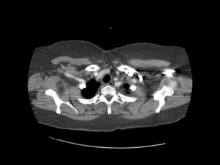
1) The brachiocephalic artery, left common carotid artery, left subclavian artery, and formation of the brachiocephalic vein by the left subclavian vein and internal jugular vein at the level of T2-T3.
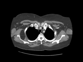
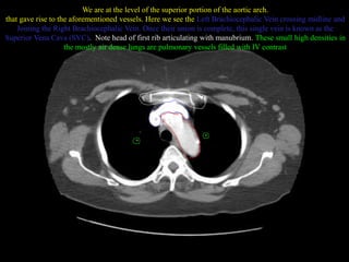
2) The aortic arch and origins of vessels like the brachiocephalic artery and left common carotid artery at a more superior level.
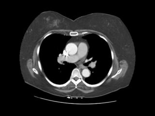
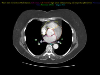
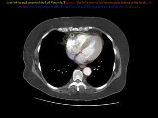
3) Various cardiac and vascular structures seen at different levels moving caudally, including the left atrium, ventricles, pulmonary veins and arteries, vena


![Segmental approach in congenital heart disease [autosaved].pptx 2.pptx final](https://cdn.slidesharecdn.com/ss_thumbnails/segmentalapproachincongenitalheartdiseaseautosaved-141103111700-conversion-gate01-thumbnail.jpg?width=640&height=640&fit=bounds)


