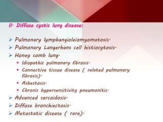
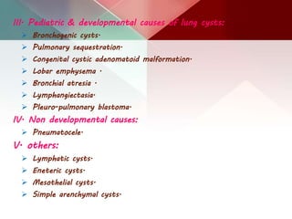
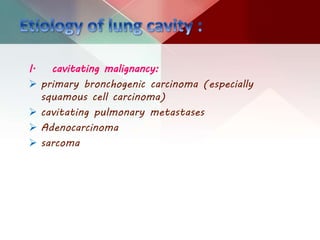
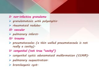
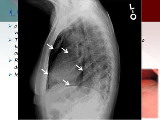
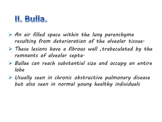
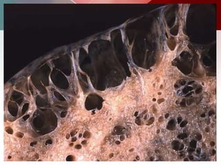
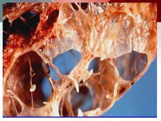
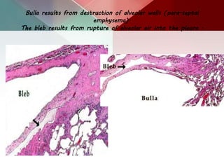
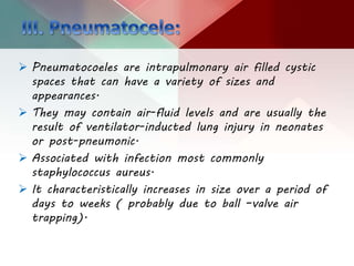
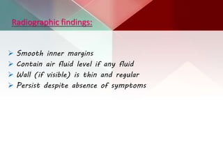
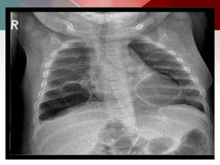
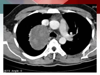
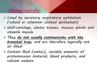
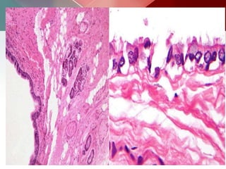
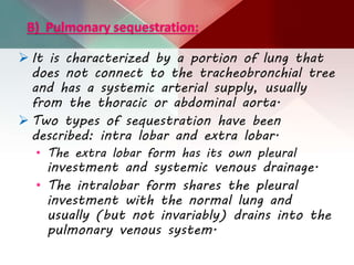
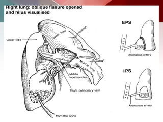
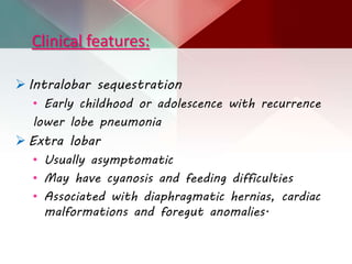
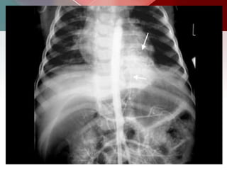
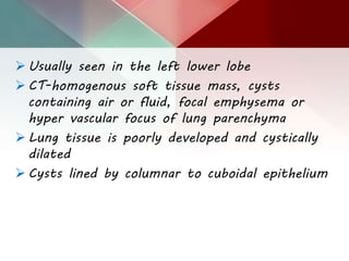
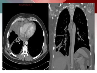
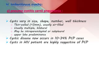
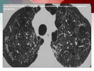
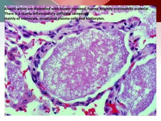
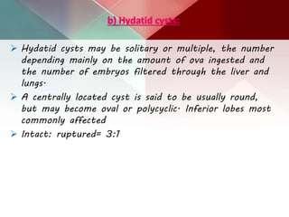
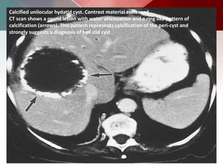
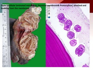
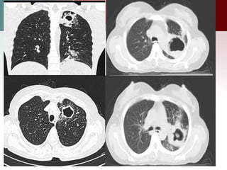
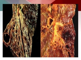
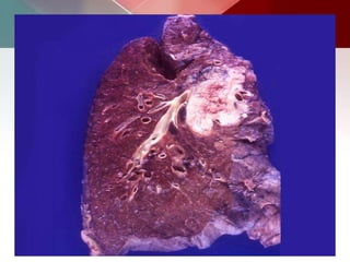
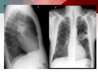
SlideShare a Scribd company logo
Presented by :
Passant Abdul Nasser Dorgham
 A cyst is a round space defined pathologically
by an epithelial or fibrous outer wall and
radiologically as a round parenchymal lucency
or low attenuation area with a well defined
interface with normal tissue.
 surrounded by a thin (2mm or less ) wall.
 A bleb or bulla has a wall that is less than 1
mm.
 Pulmonary cavities are gas-filled areas of the
lung in the center of a nodule, mass, or area of
consolidation.
 They are usually evident on plain radiography and
CT.
 They are typically thick walled and their walls
must be greater than 2-5 mm.
 They may be filled with air as well as fluid and
may also demonstrate air-fluid levels.
1) Necrosis of lung parenchyma.
2) Communication with the tracheobronchial tree.
3) Complete destruction.
4) Band of inflammation around the necrotic
material.
 They include bronchiolar check-valve mechanism, vascular
occlusion or ischemia necrosis, and dilation of the
bronchioles.
 The valve mechanism in which the entrance from a
bronchus into a lung cavity become obstructed in a
valve-like manner, presumably with a piece of necrotic
tissue.
 Allows the ingress of air during inspiration but prevents
the egress during expiration.
 The cause of pulmonary cavities is broad.
 They may develop as a chronic complication of a
pulmonary cyst or secondary to cystic degeneration of
a pulmonary mass.
 They may enlarge or involute over time.
I. Cystic focal & multi-focal lung disease:
Cystic ( wall thickness less than 4 mm ).
Bullae .
Blebs.
Pneumatocele.
Congenital cystic lesions.
 Bronchogenic cyst.
 Congenital adenomatoid malformation.
Infections.
 Hydatid disease.
 Coccidioidomycosis.
 Pneumocystis carinii.
traumatic cysts.
II. Diffuse cystic lung disease:
 Pulmonary lymphangioleiomyomatosis.
 Pulmonary Langerhans cell histiocytosis.
 Honey comb lung.
 Idiopathic pulmonary fibrosis.
 Connective tissue disease ( related pulmonary
fibrosis).
 Asbestosis.
 Chronic hypersensitivity pneumonitis.
 Advanced sarcoidosis.
 Diffuse bronchiectasis.
 Metastatic disease ( rare).
III. Pediatric & developmental causes of lung cysts:
 Bronchogenic cysts.
 Pulmonary sequestration.
 Congenital cystic adenomatoid malformation.
 Lobar emphysema .
 Bronchial atresia .
 Lymphangiectasia.
 Pleuro-pulmonary blastoma.
IV. Non developmental causes:
 Pneumatocele.
V. others:
 Lymphatic cysts.
 Eneteric cysts.
 Mesothelial cysts.
 Simple arenchymal cysts.
I. cavitating malignancy:
 primary bronchogenic carcinoma (especially
squamous cell carcinoma)
 cavitating pulmonary metastases
 Adenocarcinoma
 sarcoma
II. non-infective granuloma
 granulomatosis with polyangitis.
 rheumatoid nodules.
III. vascular
 pulmonary infarct.
IV. trauma
 pneumatocoeles (a thin walled pneumatocoele is not
really a cavity).
V. congenital (not true "cavity")
 congenital cystic adenomatoid malformation (CCAM).
 pulmonary sequestration.
 bronchogenic cyst.
 a sub-pleural collection of air within the layers of
visceral pleura caused by a ruptured alveolus.
 The air dissects through the interstitial tissue into
the thin, fibrous layer of visceral pleura where it
accumulates to form a bleb.
 Rupture of a bleb is often associated with the
development of a spontaneous pneumothorax.
 It is usually less than 1 cm in diameter.
 An air filled space within the lung parenchyma
resulting from deterioration of the alveolar tissue.
 These lesions have a fibrous wall ,trabeculated by the
remnants of alveolar septa.
 Bullae can reach substantial size and occupy an entire
lobe
 Usually seen in chronic obstructive pulmonary disease
but also seen in normal young healthy individuals
Bulla results from destruction of alveolar walls (para-septal
emphysema).
The bleb results from rupture of alveolar air into the pleura .
 Pneumatocoeles are intrapulmonary air filled cystic
spaces that can have a variety of sizes and
appearances.
 They may contain air-fluid levels and are usually the
result of ventilator-inducted lung injury in neonates
or post-pneumonic.
 Associated with infection most commonly
staphylococcus aureus.
 It characteristically increases in size over a period of
days to weeks ( probably due to ball –valve air
trapping).
Radiographic findings:
 Smooth inner margins
 Contain air fluid level if any fluid
 Wall (if visible) is thin and regular
 Persist despite absence of symptoms
A) bronchogenic cyst.
 Bronchogenic cysts are numerous foregut buds
disconnected and separated from the tracheobronchial
tree to form a cystic mass during embryogenesis
(between 4th - 6th weeks).
 Most common site is middle mediastinum (65- 90%).
 Remote locations, including the interatrial septum,
neck, abdomen, and retroperitoneal space.
Clinical features:
 Most bronchogenic cysts are found
incidentally.
 In infants- compression of the trachea or
bronchi and esophagus-wheezing, stridor,
dyspnea, and dysphagia.
 Intra-parenchymal cysts may manifest with
recurrent infection.
Thin walled spherical unilocular masses –fluid filled, air filled or with
air fluid levels
Microscopically:
 Lined by secretory respiratory epithelium
(cuboid or columnar ciliated epithelium)
 Wall-cartilage, elastic tissues, mucous glands and
smooth muscle
 They do not usually communicate with the
bronchial tree, and are therefore typically not
air filled.
 Contain fluid (water), variable amounts of
proteinaceous material, blood products, and
calcium oxalate
B) Pulmonary sequestration:
 It is characterized by a portion of lung that
does not connect to the tracheobronchial tree
and has a systemic arterial supply, usually
from the thoracic or abdominal aorta.
 Two types of sequestration have been
described: intra lobar and extra lobar.
• The extra lobar form has its own pleural
investment and systemic venous drainage.
• The intralobar form shares the pleural
investment with the normal lung and
usually (but not invariably) drains into the
pulmonary venous system.
Clinical features:
 Intralobar sequestration
• Early childhood or adolescence with recurrence
lower lobe pneumonia
 Extra lobar
• Usually asymptomatic
• May have cyanosis and feeding difficulties
• Associated with diaphragmatic hernias, cardiac
malformations and foregut anomalies.
 Usually seen in the left lower lobe
 CT-homogenous soft tissue mass, cysts
containing air or fluid, focal emphysema or
hyper vascular focus of lung parenchyma
 Lung tissue is poorly developed and cystically
dilated
 Cysts lined by columnar to cuboidal epithelium
Gross pathology:
 Pleura thickened with adhesions
 Parenchyma-cysts up to 5cm with mucinous or purulent
material and fibrosis.
microscopically:
 Loose, spongy tissue with numerous small
cystic spaces containing clear, mucoid
fluid.
 Dilated bronchi with mucous or purulent
material
 Alveoli filled with foamy macrophages
 Thick walled vessels reflecting systemic
vascular drainage with elastic stains
c) Congenital cystic adenomatoid malformation (CCAM)
 multi-cystic mass of segmental lung tissue
with abnormal bronchial proliferation.
 It is considered part of the spectrum of
bronchopulmonary foregut malformations.
 They account for ~25% of congenital lung
lesions.
 there is a male predominance.
Pathogenesis:
 Failure of normal broncho-alveolar development
with hamartomatous proliferation of terminal
respiratory units in a gland-like pattern
(adenomatoid) without proper alveolar formation.
 These lesions have intra cystic communications
and, unlike bronchogenic cysts, can also have a
connection to the tracheobronchial tree.
 Lesions are usually unilateral and involve a single
lobe. Although there is no well-documented lobar
predilection, they appear less frequently in the
middle lobe.
Subtypes:
Five subtypes are currently classified, mainly according to
cyst size:
type I
o most common: 70% of cases 3
o large cysts
o one or more dominant cysts: 2-10 cm in size
o may be surrounded by smaller cysts
type II
o 15-20% of cases 3
o cysts are <2 cm in diameter
o associated with other abnormalities
o renal agenesis or dysgenesis
o pulmonary sequestration
o congenital cardiac anomalies
type III
o ~10% of cases
o micro cysts: <5 mm in diameter
o typically involves an entire lobe
o has a poorer prognosis
type IV
o unlined cyst
o typically affects a single lobe
o indistinguishable from type I on imaging 11
type 0
o very rare, lethal postnatally
o acinar dysgenesis or dysplasia 11
o represents global arrest of lung development 12
Radiographic features:
The appearance of CPAMs will vary depending on the type.
Antenatal ultrasound
o CPAM appears as an isolated cystic or solid intrathoracic mass.
o A solid thoracic mass is usually indicative of a type III CPAM
and is typically hyperechoic.
o There can be a mass effect where the heart may appear
displaced to the opposite side.
Plain radiograph
o Chest radiographs in type I and II CPAMs may demonstrate a
multicystic (air-filled) lesion.
o Large lesions may cause a mass effect with resultant mediastinal
shift, depression, and even inversion of the diaphragm.
o In the early neonatal period, the cysts may be completely or
partially fluid-filled, in which case the lesion may appear solid or
with air-fluid levels.
o Type III lesions appear solid.
CCAM type I
CT scan shows multiple large cysts (>2cm) involving the lower lobe of left lung. The cysts
are air-filled, expand the lower lobe, cause mediastinal shift and hypoplasia of right lung
CCAM type I
CCAM type I
a) pneumo cystitis carnii pneumonia :
 Cysts vary in size, shape, number, wall thickness
o Thin-walled (<3mm), usually air-filled
o Usually multiple, bilateral
o May be intraparenchymal or subpleural
o upper lobe predominance
 Cystic disease now occurs in 10-34% PCP cases
 Cysts in HIV patient are highly suggestive of PCP
Lung cysts are usually multiple, thin walled and bilateral, but range in size, shape and
distribution
Alveoli which are distended with honey- combed, foamy, brightly eosinophilic material .
There is a scanty inflammatory infiltrate composed
mainly of monocyte, occasional plasma cells and histiocytes.
b) Hydatid cysts:
 Hydatid cysts may be solitary or multiple, the number
depending mainly on the amount of ova ingested and
the number of embryos filtered through the liver and
lungs.
 A centrally located cyst is said to be usually round,
but may become oval or polycyclic. Inferior lobes most
commonly affected
 Intact: ruptured= 3:1
Calcified unilocular hydatid cyst. Contrast material enhanced
CT scan shows a round lesion with water attenuation and a ring like pattern of
calcification (arrows). This pattern represents calcification of the peri-cyst and
strongly suggests a diagnosis of hydatid cyst
Outer acellular laminated membrane, Germinal membrane& Protoscolices, attached and
budding from the membrane
c) Cavitating Pulmonary tuberculosis:
 Post-primary infections are far more likely to cavitate
than primary infections and are seen in 20-45% of
cases.
 In the vast majority of cases, they develop in the
posterior segments of the upper lobes (85%).
 The development of an air-fluid level implies
communication with the airway, and thus the
possibility of contagion.
 Endo-bronchial spread along nearby airways is a
relatively common finding, resulting in a relatively
well-defined 2-4 mm nodules or branching lesions
(tree-in-bud sign) on CT.
a) Pulmonary Langerhans cell histiocytosis:
 PLCH is typically a disease of young adults which
predominately affects the lungs and bones
 Very strong association with smoking(90%)
o interstitial lung disease.
o Lung affected in isolation or in association with organ
systems.
Pulmonary disease in PLCH is characterized by peri-
bronchiolar 1-10 mm nodules in the early stages
In later stages of PLCH, the major pulmonary finding
is cysts (present in 80% of patients) and there may
be no nodules present
Lung bases are relatively spared at all disease stages.
Characteristic combination of diffuse cysts and centrilobular micro-nodules
Gross pathology :
 Cysts more pronounced later
in the disease usually less
than 10mm in diameter may
measure up to 2 - 3
centimeters in size
 Thin-walled, but on occasion
may be up to a few
millimeters thick
 confluence of 2 or more
cysts results in bizarre
shapes :
o bilobed
o cloverleaf
o branching
o internal septations
Histopathology:
 The earliest histologic lesion of PLCH consists of proliferation of
Langerhans’ cells along small airways. These early cellular lesions
expand to form nodules 1 to 5 mm in diameter.
 The characteristic lesion is composed of variable numbers of
Langerhans’ cells,eosinophils,plasma cells, lymphocytes, fibroblasts,
and pigmented alveolar macrophages, which form a loosely
aggregated granulomas.
 These granulomas are typically centered around distal bronchioles,
where they infiltrate and destroy airway walls.
 As these cellular granulomas evolve, peripheral fibrosis forms
resulting in traction on the central bronchiole which becomes cyst-
like
 Evolution from nodule, through cavitating nodule and thick walled
cysts, to the 'stable‘ thin-walled cysts.
B) Bronchiectasis:
 Bronchiectasis, or the dilatation and distortion of bronchi and
bronchioles, may be mistaken for cystic airspace disease .
 Bronchiectasis may be the result of either a chronic suppurative
process or accompany lung fibrosis, when it is then referred to
as traction bronchiectasis.
 Cystic bronchiectasis can be differentiated from true cystic lung
disease by the continuous relationship of the cystic structure to
bronchial tree
 Approximately uniform, medium-sized cavities are typical of
cystic bronchiectasis.
 Valsalva and Mueller maneuvers produce rapid change in the size
of cysts, which freely communicate with the airways; this change
distinguishes cystic bronchiectasis from other conditions.
a)Squamous cell
carcinoma of the lung
 (SCC) is one of the non-small cell carcinomas of the
lung, overtaken by adenocarcinoma of the lung as the
most commonly encountered lung cancer.
 Squamous cell carcinoma accounts for ~30-35% of all
lung cancers and in most instances are due to heavy
smoking.
 Macroscopically these tumors tend to be off-white in
color, arising from, and extending into a bronchus.
 They invade the surrounding lung parenchyma and can
extend into the chest wall.
 Larger tumors have a tendency to undergo central
necrosis.
Radiographic features:
 The more central lesions may appear as a bulky hilum,
representing the tumor and local nodal involvement.
 Lobar collapse may be seen due to obstruction of a
bronchus.
 When the right upper lobe is collapsed and a hilar
mass is present, this is known as the Golden S sign.
 A more peripheral location may appear as a rounded
or speculated mass.
 Cavitation may be seen as an air-fluid level
Lung cysts &amp; cavities

More Related Content

What's hot

Signs in Chest Xray
Signs in Chest Xray Signs in Chest Xray
Signs in Chest Xray
Archana Koshy
 
Congenital lung abnormalities
Congenital lung abnormalitiesCongenital lung abnormalities
Congenital lung abnormalities
airwave12
 
Solitary pulmonary nodules radiology pk ppt
Solitary pulmonary nodules  radiology pk pptSolitary pulmonary nodules  radiology pk ppt
Solitary pulmonary nodules radiology pk ppt
Dr pradeep Kumar
 
Mediastinal mass
Mediastinal massMediastinal mass
Mediastinal mass
Vikram Patil
 
aortic arch anamolies
aortic arch anamoliesaortic arch anamolies
aortic arch anamolies
Malleswara rao Dangeti
 
Cystic lung disease; Stepwise approach to diagnosis
Cystic lung disease; Stepwise approach to diagnosisCystic lung disease; Stepwise approach to diagnosis
Cystic lung disease; Stepwise approach to diagnosis
Dr Ravi Kumar Sharma
 
Pulmonary sequestration
Pulmonary sequestrationPulmonary sequestration
Pulmonary sequestration
Drhammad Rehman
 
Chest x rays
Chest x raysChest x rays
Chest x rays
Dr. Prem Mohan Jha
 
Reticular pattern
Reticular patternReticular pattern
Reticular pattern
Gamal Agmy
 
Imaging of placenta
Imaging of placentaImaging of placenta
Imaging of placenta
juhipatel1802
 
X-Ray: Calcification in CXR
X-Ray: Calcification in CXRX-Ray: Calcification in CXR
Asymptomatic palpable adnexal masses: ultrasound evaluation- warda
Asymptomatic palpable adnexal masses: ultrasound evaluation- wardaAsymptomatic palpable adnexal masses: ultrasound evaluation- warda
Asymptomatic palpable adnexal masses: ultrasound evaluation- warda
Osama Warda
 
Invertogram ANORECTAL MALFORMATION ( ARM ) PRANAYA
Invertogram ANORECTAL MALFORMATION ( ARM ) PRANAYAInvertogram ANORECTAL MALFORMATION ( ARM ) PRANAYA
Invertogram ANORECTAL MALFORMATION ( ARM ) PRANAYA
PRANAYA PANIGRAHI
 
CT CHEST ANATOMY
CT CHEST ANATOMYCT CHEST ANATOMY
CT CHEST ANATOMY
Sakher Alkhaderi
 
Presentation1, radiological imaging of scimitar syndrome
Presentation1, radiological imaging of scimitar syndromePresentation1, radiological imaging of scimitar syndrome
Presentation1, radiological imaging of scimitar syndrome
Abdellah Nazeer
 
Diagnostic Imaging of Pulmonary Tumors
Diagnostic Imaging of Pulmonary TumorsDiagnostic Imaging of Pulmonary Tumors
Diagnostic Imaging of Pulmonary Tumors
Mohamed M.A. Zaitoun
 
Cervicothoracic sign.pptx
Cervicothoracic sign.pptxCervicothoracic sign.pptx
Cervicothoracic sign.pptx
Ravindran Chetambath
 
Chest radiology in intensive care
Chest radiology in intensive careChest radiology in intensive care
Chest radiology in intensive care
Andrew Ferguson
 
20.5.pleural effusion &amp;empyema
20.5.pleural effusion &amp;empyema20.5.pleural effusion &amp;empyema
20.5.pleural effusion &amp;empyema
pediatricsmgmcri
 
Ascites and Pleural Effusion
 Ascites and Pleural Effusion Ascites and Pleural Effusion
Ascites and Pleural Effusion
Media Genie
 

What's hot (20)

Signs in Chest Xray
Signs in Chest Xray Signs in Chest Xray
Signs in Chest Xray
 
Congenital lung abnormalities
Congenital lung abnormalitiesCongenital lung abnormalities
Congenital lung abnormalities
 
Solitary pulmonary nodules radiology pk ppt
Solitary pulmonary nodules  radiology pk pptSolitary pulmonary nodules  radiology pk ppt
Solitary pulmonary nodules radiology pk ppt
 
Mediastinal mass
Mediastinal massMediastinal mass
Mediastinal mass
 
aortic arch anamolies
aortic arch anamoliesaortic arch anamolies
aortic arch anamolies
 
Cystic lung disease; Stepwise approach to diagnosis
Cystic lung disease; Stepwise approach to diagnosisCystic lung disease; Stepwise approach to diagnosis
Cystic lung disease; Stepwise approach to diagnosis
 
Pulmonary sequestration
Pulmonary sequestrationPulmonary sequestration
Pulmonary sequestration
 
Chest x rays
Chest x raysChest x rays
Chest x rays
 
Reticular pattern
Reticular patternReticular pattern
Reticular pattern
 
Imaging of placenta
Imaging of placentaImaging of placenta
Imaging of placenta
 
X-Ray: Calcification in CXR
X-Ray: Calcification in CXRX-Ray: Calcification in CXR
X-Ray: Calcification in CXR
 
Asymptomatic palpable adnexal masses: ultrasound evaluation- warda
Asymptomatic palpable adnexal masses: ultrasound evaluation- wardaAsymptomatic palpable adnexal masses: ultrasound evaluation- warda
Asymptomatic palpable adnexal masses: ultrasound evaluation- warda
 
Invertogram ANORECTAL MALFORMATION ( ARM ) PRANAYA
Invertogram ANORECTAL MALFORMATION ( ARM ) PRANAYAInvertogram ANORECTAL MALFORMATION ( ARM ) PRANAYA
Invertogram ANORECTAL MALFORMATION ( ARM ) PRANAYA
 
CT CHEST ANATOMY
CT CHEST ANATOMYCT CHEST ANATOMY
CT CHEST ANATOMY
 
Presentation1, radiological imaging of scimitar syndrome
Presentation1, radiological imaging of scimitar syndromePresentation1, radiological imaging of scimitar syndrome
Presentation1, radiological imaging of scimitar syndrome
 
Diagnostic Imaging of Pulmonary Tumors
Diagnostic Imaging of Pulmonary TumorsDiagnostic Imaging of Pulmonary Tumors
Diagnostic Imaging of Pulmonary Tumors
 
Cervicothoracic sign.pptx
Cervicothoracic sign.pptxCervicothoracic sign.pptx
Cervicothoracic sign.pptx
 
Chest radiology in intensive care
Chest radiology in intensive careChest radiology in intensive care
Chest radiology in intensive care
 
20.5.pleural effusion &amp;empyema
20.5.pleural effusion &amp;empyema20.5.pleural effusion &amp;empyema
20.5.pleural effusion &amp;empyema
 
Ascites and Pleural Effusion
 Ascites and Pleural Effusion Ascites and Pleural Effusion
Ascites and Pleural Effusion
 

Similar to Lung cysts &amp; cavities

Differential diagnosis of cavitary lung lesions
Differential diagnosis of cavitary lung lesionsDifferential diagnosis of cavitary lung lesions
Differential diagnosis of cavitary lung lesions
Dr.Bijay Yadav
 
Cystic Lung Diseases
Cystic Lung DiseasesCystic Lung Diseases
Cystic Lung Diseases
Dr Riham Hazem Raafat
 
Typical and Atypical Presentations of Sarcoidosis
Typical and Atypical Presentations of SarcoidosisTypical and Atypical Presentations of Sarcoidosis
Typical and Atypical Presentations of Sarcoidosis
Gamal Agmy
 
Cystic lung disease
Cystic lung disease   Cystic lung disease
Cystic lung disease
Dr Shami Bhagat
 
abnormal chest xray ppt
abnormal chest xray ppt abnormal chest xray ppt
abnormal chest xray ppt
shyamsobti
 
lung abscess
lung abscesslung abscess
lung abscess
dr.shameer basha
 
Ppt cyst lung
Ppt cyst lungPpt cyst lung
Ppt cyst lung
namrathrs87
 
The road to HRCT evaluation of pediatric diffuse lung diseases .part 1
The road to HRCT evaluation of pediatric diffuse lung diseases .part 1The road to HRCT evaluation of pediatric diffuse lung diseases .part 1
The road to HRCT evaluation of pediatric diffuse lung diseases .part 1
Ahmed Bahnassy
 
Congenital lung anomalies
Congenital lung anomaliesCongenital lung anomalies
Congenital lung anomalies
ebinroshan07
 
Chest X-Ray in Pediatrics.pptx
Chest X-Ray in Pediatrics.pptxChest X-Ray in Pediatrics.pptx
Chest X-Ray in Pediatrics.pptx
HurshidaShia
 
cavitatorylesionsofthelung-150316082210-conversion-gate01.pdf
cavitatorylesionsofthelung-150316082210-conversion-gate01.pdfcavitatorylesionsofthelung-150316082210-conversion-gate01.pdf
cavitatorylesionsofthelung-150316082210-conversion-gate01.pdf
PeerzadaUmair
 
Cavitatory lesions of the lung
Cavitatory lesions of the lungCavitatory lesions of the lung
Cavitatory lesions of the lung
reddyvjm
 
Casos clinico-Radiológicos
Casos clinico-RadiológicosCasos clinico-Radiológicos
Casos clinico-Radiológicos
Javier Pacheco Paternina
 
Radiological imaging of pleural diseases
Radiological imaging of pleural diseases Radiological imaging of pleural diseases
Radiological imaging of pleural diseases
Pankaj Kaira
 
unilateral hyperlucent lung in children
unilateral hyperlucent lung in childrenunilateral hyperlucent lung in children
unilateral hyperlucent lung in children
Annie Agarwal
 
tuberculosis viral infections mediastinum radiology
tuberculosis viral infections mediastinum radiologytuberculosis viral infections mediastinum radiology
tuberculosis viral infections mediastinum radiology
Dr,saket Jain
 
Interstitial lung-diseases
Interstitial lung-diseases Interstitial lung-diseases
Interstitial lung-diseases
mohamedrafi112
 
Pneumonia.pptxojasojioiosiokiiijijidjijioaj
Pneumonia.pptxojasojioiosiokiiijijidjijioajPneumonia.pptxojasojioiosiokiiijijidjijioaj
Pneumonia.pptxojasojioiosiokiiijijidjijioaj
AmarReddy75
 
Lung malformation part 1
Lung malformation part 1Lung malformation part 1
Lung malformation part 1
Faheem Andrabi
 
Thorax 00
Thorax 00Thorax 00
Thorax 00
maricar chua
 

Similar to Lung cysts &amp; cavities (20)

Differential diagnosis of cavitary lung lesions
Differential diagnosis of cavitary lung lesionsDifferential diagnosis of cavitary lung lesions
Differential diagnosis of cavitary lung lesions
 
Cystic Lung Diseases
Cystic Lung DiseasesCystic Lung Diseases
Cystic Lung Diseases
 
Typical and Atypical Presentations of Sarcoidosis
Typical and Atypical Presentations of SarcoidosisTypical and Atypical Presentations of Sarcoidosis
Typical and Atypical Presentations of Sarcoidosis
 
Cystic lung disease
Cystic lung disease   Cystic lung disease
Cystic lung disease
 
abnormal chest xray ppt
abnormal chest xray ppt abnormal chest xray ppt
abnormal chest xray ppt
 
lung abscess
lung abscesslung abscess
lung abscess
 
Ppt cyst lung
Ppt cyst lungPpt cyst lung
Ppt cyst lung
 
The road to HRCT evaluation of pediatric diffuse lung diseases .part 1
The road to HRCT evaluation of pediatric diffuse lung diseases .part 1The road to HRCT evaluation of pediatric diffuse lung diseases .part 1
The road to HRCT evaluation of pediatric diffuse lung diseases .part 1
 
Congenital lung anomalies
Congenital lung anomaliesCongenital lung anomalies
Congenital lung anomalies
 
Chest X-Ray in Pediatrics.pptx
Chest X-Ray in Pediatrics.pptxChest X-Ray in Pediatrics.pptx
Chest X-Ray in Pediatrics.pptx
 
cavitatorylesionsofthelung-150316082210-conversion-gate01.pdf
cavitatorylesionsofthelung-150316082210-conversion-gate01.pdfcavitatorylesionsofthelung-150316082210-conversion-gate01.pdf
cavitatorylesionsofthelung-150316082210-conversion-gate01.pdf
 
Cavitatory lesions of the lung
Cavitatory lesions of the lungCavitatory lesions of the lung
Cavitatory lesions of the lung
 
Casos clinico-Radiológicos
Casos clinico-RadiológicosCasos clinico-Radiológicos
Casos clinico-Radiológicos
 
Radiological imaging of pleural diseases
Radiological imaging of pleural diseases Radiological imaging of pleural diseases
Radiological imaging of pleural diseases
 
unilateral hyperlucent lung in children
unilateral hyperlucent lung in childrenunilateral hyperlucent lung in children
unilateral hyperlucent lung in children
 
tuberculosis viral infections mediastinum radiology
tuberculosis viral infections mediastinum radiologytuberculosis viral infections mediastinum radiology
tuberculosis viral infections mediastinum radiology
 
Interstitial lung-diseases
Interstitial lung-diseases Interstitial lung-diseases
Interstitial lung-diseases
 
Pneumonia.pptxojasojioiosiokiiijijidjijioaj
Pneumonia.pptxojasojioiosiokiiijijidjijioajPneumonia.pptxojasojioiosiokiiijijidjijioaj
Pneumonia.pptxojasojioiosiokiiijijidjijioaj
 
Lung malformation part 1
Lung malformation part 1Lung malformation part 1
Lung malformation part 1
 
Thorax 00
Thorax 00Thorax 00
Thorax 00
 

Recently uploaded

Histololgy of Female Reproductive System.pptx
Histololgy of Female Reproductive System.pptxHistololgy of Female Reproductive System.pptx
Histololgy of Female Reproductive System.pptx
AyeshaZaid1
 
Osteoporosis - Definition , Evaluation and Management .pdf
Osteoporosis - Definition , Evaluation and Management .pdfOsteoporosis - Definition , Evaluation and Management .pdf
Osteoporosis - Definition , Evaluation and Management .pdf
Jim Jacob Roy
 
TEST BANK For Basic and Clinical Pharmacology, 14th Edition by Bertram G. Kat...
TEST BANK For Basic and Clinical Pharmacology, 14th Edition by Bertram G. Kat...TEST BANK For Basic and Clinical Pharmacology, 14th Edition by Bertram G. Kat...
TEST BANK For Basic and Clinical Pharmacology, 14th Edition by Bertram G. Kat...
rightmanforbloodline
 
CHEMOTHERAPY_RDP_CHAPTER 2 _LEPROSY.pdf1
CHEMOTHERAPY_RDP_CHAPTER 2 _LEPROSY.pdf1CHEMOTHERAPY_RDP_CHAPTER 2 _LEPROSY.pdf1
CHEMOTHERAPY_RDP_CHAPTER 2 _LEPROSY.pdf1
rishi2789
 
Cell Therapy Expansion and Challenges in Autoimmune Disease
Cell Therapy Expansion and Challenges in Autoimmune DiseaseCell Therapy Expansion and Challenges in Autoimmune Disease
Cell Therapy Expansion and Challenges in Autoimmune Disease
Health Advances
 
Hemodialysis: Chapter 4, Dialysate Circuit - Dr.Gawad
Hemodialysis: Chapter 4, Dialysate Circuit - Dr.GawadHemodialysis: Chapter 4, Dialysate Circuit - Dr.Gawad
Hemodialysis: Chapter 4, Dialysate Circuit - Dr.Gawad
NephroTube - Dr.Gawad
 
share - Lions, tigers, AI and health misinformation, oh my!.pptx
share - Lions, tigers, AI and health misinformation, oh my!.pptxshare - Lions, tigers, AI and health misinformation, oh my!.pptx
share - Lions, tigers, AI and health misinformation, oh my!.pptx
Tina Purnat
 
Does Over-Masturbation Contribute to Chronic Prostatitis.pptx
Does Over-Masturbation Contribute to Chronic Prostatitis.pptxDoes Over-Masturbation Contribute to Chronic Prostatitis.pptx
Does Over-Masturbation Contribute to Chronic Prostatitis.pptx
walterHu5
 
Diabetic nephropathy diagnosis treatment
Diabetic nephropathy diagnosis treatmentDiabetic nephropathy diagnosis treatment
Diabetic nephropathy diagnosis treatment
arahmanzai5
 
Netter's Atlas of Human Anatomy 7.ed.pdf
Netter's Atlas of Human Anatomy 7.ed.pdfNetter's Atlas of Human Anatomy 7.ed.pdf
Netter's Atlas of Human Anatomy 7.ed.pdf
BrissaOrtiz3
 
Adhd Medication Shortage Uk - trinexpharmacy.com
Adhd Medication Shortage Uk - trinexpharmacy.comAdhd Medication Shortage Uk - trinexpharmacy.com
Adhd Medication Shortage Uk - trinexpharmacy.com
reignlana06
 
Abortion PG Seminar Power point presentation
Abortion PG Seminar Power point presentationAbortion PG Seminar Power point presentation
Abortion PG Seminar Power point presentation
AksshayaRajanbabu
 
Artificial Intelligence Symposium (THAIS)
Artificial Intelligence Symposium (THAIS)Artificial Intelligence Symposium (THAIS)
Artificial Intelligence Symposium (THAIS)
Josep Vidal-Alaball
 
The Best Ayurvedic Antacid Tablets in India
The Best Ayurvedic Antacid Tablets in IndiaThe Best Ayurvedic Antacid Tablets in India
The Best Ayurvedic Antacid Tablets in India
Swastik Ayurveda
 
The Electrocardiogram - Physiologic Principles
The Electrocardiogram - Physiologic PrinciplesThe Electrocardiogram - Physiologic Principles
The Electrocardiogram - Physiologic Principles
MedicoseAcademics
 
Muscles of Mastication by Dr. Rabia Inam Gandapore.pptx
Muscles of Mastication by Dr. Rabia Inam Gandapore.pptxMuscles of Mastication by Dr. Rabia Inam Gandapore.pptx
Muscles of Mastication by Dr. Rabia Inam Gandapore.pptx
Dr. Rabia Inam Gandapore
 
REGULATION FOR COMBINATION PRODUCTS AND MEDICAL DEVICES.pptx
REGULATION FOR COMBINATION PRODUCTS AND MEDICAL DEVICES.pptxREGULATION FOR COMBINATION PRODUCTS AND MEDICAL DEVICES.pptx
REGULATION FOR COMBINATION PRODUCTS AND MEDICAL DEVICES.pptx
LaniyaNasrink
 
Top 10 Best Ayurvedic Kidney Stone Syrups in India
Top 10 Best Ayurvedic Kidney Stone Syrups in IndiaTop 10 Best Ayurvedic Kidney Stone Syrups in India
Top 10 Best Ayurvedic Kidney Stone Syrups in India
Swastik Ayurveda
 
CHEMOTHERAPY_RDP_CHAPTER 3_ANTIFUNGAL AGENT.pdf
CHEMOTHERAPY_RDP_CHAPTER 3_ANTIFUNGAL AGENT.pdfCHEMOTHERAPY_RDP_CHAPTER 3_ANTIFUNGAL AGENT.pdf
CHEMOTHERAPY_RDP_CHAPTER 3_ANTIFUNGAL AGENT.pdf
rishi2789
 
TEST BANK For An Introduction to Brain and Behavior, 7th Edition by Bryan Kol...
TEST BANK For An Introduction to Brain and Behavior, 7th Edition by Bryan Kol...TEST BANK For An Introduction to Brain and Behavior, 7th Edition by Bryan Kol...
TEST BANK For An Introduction to Brain and Behavior, 7th Edition by Bryan Kol...
rightmanforbloodline
 

Recently uploaded (20)

Histololgy of Female Reproductive System.pptx
Histololgy of Female Reproductive System.pptxHistololgy of Female Reproductive System.pptx
Histololgy of Female Reproductive System.pptx
 
Osteoporosis - Definition , Evaluation and Management .pdf
Osteoporosis - Definition , Evaluation and Management .pdfOsteoporosis - Definition , Evaluation and Management .pdf
Osteoporosis - Definition , Evaluation and Management .pdf
 
TEST BANK For Basic and Clinical Pharmacology, 14th Edition by Bertram G. Kat...
TEST BANK For Basic and Clinical Pharmacology, 14th Edition by Bertram G. Kat...TEST BANK For Basic and Clinical Pharmacology, 14th Edition by Bertram G. Kat...
TEST BANK For Basic and Clinical Pharmacology, 14th Edition by Bertram G. Kat...
 
CHEMOTHERAPY_RDP_CHAPTER 2 _LEPROSY.pdf1
CHEMOTHERAPY_RDP_CHAPTER 2 _LEPROSY.pdf1CHEMOTHERAPY_RDP_CHAPTER 2 _LEPROSY.pdf1
CHEMOTHERAPY_RDP_CHAPTER 2 _LEPROSY.pdf1
 
Cell Therapy Expansion and Challenges in Autoimmune Disease
Cell Therapy Expansion and Challenges in Autoimmune DiseaseCell Therapy Expansion and Challenges in Autoimmune Disease
Cell Therapy Expansion and Challenges in Autoimmune Disease
 
Hemodialysis: Chapter 4, Dialysate Circuit - Dr.Gawad
Hemodialysis: Chapter 4, Dialysate Circuit - Dr.GawadHemodialysis: Chapter 4, Dialysate Circuit - Dr.Gawad
Hemodialysis: Chapter 4, Dialysate Circuit - Dr.Gawad
 
share - Lions, tigers, AI and health misinformation, oh my!.pptx
share - Lions, tigers, AI and health misinformation, oh my!.pptxshare - Lions, tigers, AI and health misinformation, oh my!.pptx
share - Lions, tigers, AI and health misinformation, oh my!.pptx
 
Does Over-Masturbation Contribute to Chronic Prostatitis.pptx
Does Over-Masturbation Contribute to Chronic Prostatitis.pptxDoes Over-Masturbation Contribute to Chronic Prostatitis.pptx
Does Over-Masturbation Contribute to Chronic Prostatitis.pptx
 
Diabetic nephropathy diagnosis treatment
Diabetic nephropathy diagnosis treatmentDiabetic nephropathy diagnosis treatment
Diabetic nephropathy diagnosis treatment
 
Netter's Atlas of Human Anatomy 7.ed.pdf
Netter's Atlas of Human Anatomy 7.ed.pdfNetter's Atlas of Human Anatomy 7.ed.pdf
Netter's Atlas of Human Anatomy 7.ed.pdf
 
Adhd Medication Shortage Uk - trinexpharmacy.com
Adhd Medication Shortage Uk - trinexpharmacy.comAdhd Medication Shortage Uk - trinexpharmacy.com
Adhd Medication Shortage Uk - trinexpharmacy.com
 
Abortion PG Seminar Power point presentation
Abortion PG Seminar Power point presentationAbortion PG Seminar Power point presentation
Abortion PG Seminar Power point presentation
 
Artificial Intelligence Symposium (THAIS)
Artificial Intelligence Symposium (THAIS)Artificial Intelligence Symposium (THAIS)
Artificial Intelligence Symposium (THAIS)
 
The Best Ayurvedic Antacid Tablets in India
The Best Ayurvedic Antacid Tablets in IndiaThe Best Ayurvedic Antacid Tablets in India
The Best Ayurvedic Antacid Tablets in India
 
The Electrocardiogram - Physiologic Principles
The Electrocardiogram - Physiologic PrinciplesThe Electrocardiogram - Physiologic Principles
The Electrocardiogram - Physiologic Principles
 
Muscles of Mastication by Dr. Rabia Inam Gandapore.pptx
Muscles of Mastication by Dr. Rabia Inam Gandapore.pptxMuscles of Mastication by Dr. Rabia Inam Gandapore.pptx
Muscles of Mastication by Dr. Rabia Inam Gandapore.pptx
 
REGULATION FOR COMBINATION PRODUCTS AND MEDICAL DEVICES.pptx
REGULATION FOR COMBINATION PRODUCTS AND MEDICAL DEVICES.pptxREGULATION FOR COMBINATION PRODUCTS AND MEDICAL DEVICES.pptx
REGULATION FOR COMBINATION PRODUCTS AND MEDICAL DEVICES.pptx
 
Top 10 Best Ayurvedic Kidney Stone Syrups in India
Top 10 Best Ayurvedic Kidney Stone Syrups in IndiaTop 10 Best Ayurvedic Kidney Stone Syrups in India
Top 10 Best Ayurvedic Kidney Stone Syrups in India
 
CHEMOTHERAPY_RDP_CHAPTER 3_ANTIFUNGAL AGENT.pdf
CHEMOTHERAPY_RDP_CHAPTER 3_ANTIFUNGAL AGENT.pdfCHEMOTHERAPY_RDP_CHAPTER 3_ANTIFUNGAL AGENT.pdf
CHEMOTHERAPY_RDP_CHAPTER 3_ANTIFUNGAL AGENT.pdf
 
TEST BANK For An Introduction to Brain and Behavior, 7th Edition by Bryan Kol...
TEST BANK For An Introduction to Brain and Behavior, 7th Edition by Bryan Kol...TEST BANK For An Introduction to Brain and Behavior, 7th Edition by Bryan Kol...
TEST BANK For An Introduction to Brain and Behavior, 7th Edition by Bryan Kol...
 

Lung cysts &amp; cavities

  • 1. Presented by : Passant Abdul Nasser Dorgham
  • 2.
  • 3.  A cyst is a round space defined pathologically by an epithelial or fibrous outer wall and radiologically as a round parenchymal lucency or low attenuation area with a well defined interface with normal tissue.  surrounded by a thin (2mm or less ) wall.  A bleb or bulla has a wall that is less than 1 mm.
  • 4.  Pulmonary cavities are gas-filled areas of the lung in the center of a nodule, mass, or area of consolidation.  They are usually evident on plain radiography and CT.  They are typically thick walled and their walls must be greater than 2-5 mm.  They may be filled with air as well as fluid and may also demonstrate air-fluid levels.
  • 5. 1) Necrosis of lung parenchyma. 2) Communication with the tracheobronchial tree. 3) Complete destruction. 4) Band of inflammation around the necrotic material.
  • 6.  They include bronchiolar check-valve mechanism, vascular occlusion or ischemia necrosis, and dilation of the bronchioles.  The valve mechanism in which the entrance from a bronchus into a lung cavity become obstructed in a valve-like manner, presumably with a piece of necrotic tissue.  Allows the ingress of air during inspiration but prevents the egress during expiration.
  • 7.
  • 8.  The cause of pulmonary cavities is broad.  They may develop as a chronic complication of a pulmonary cyst or secondary to cystic degeneration of a pulmonary mass.  They may enlarge or involute over time.
  • 9. I. Cystic focal & multi-focal lung disease: Cystic ( wall thickness less than 4 mm ). Bullae . Blebs. Pneumatocele. Congenital cystic lesions.  Bronchogenic cyst.  Congenital adenomatoid malformation. Infections.  Hydatid disease.  Coccidioidomycosis.  Pneumocystis carinii. traumatic cysts.
  • 10. II. Diffuse cystic lung disease:  Pulmonary lymphangioleiomyomatosis.  Pulmonary Langerhans cell histiocytosis.  Honey comb lung.  Idiopathic pulmonary fibrosis.  Connective tissue disease ( related pulmonary fibrosis).  Asbestosis.  Chronic hypersensitivity pneumonitis.  Advanced sarcoidosis.  Diffuse bronchiectasis.  Metastatic disease ( rare).
  • 11. III. Pediatric & developmental causes of lung cysts:  Bronchogenic cysts.  Pulmonary sequestration.  Congenital cystic adenomatoid malformation.  Lobar emphysema .  Bronchial atresia .  Lymphangiectasia.  Pleuro-pulmonary blastoma. IV. Non developmental causes:  Pneumatocele. V. others:  Lymphatic cysts.  Eneteric cysts.  Mesothelial cysts.  Simple arenchymal cysts.
  • 12. I. cavitating malignancy:  primary bronchogenic carcinoma (especially squamous cell carcinoma)  cavitating pulmonary metastases  Adenocarcinoma  sarcoma
  • 13. II. non-infective granuloma  granulomatosis with polyangitis.  rheumatoid nodules. III. vascular  pulmonary infarct. IV. trauma  pneumatocoeles (a thin walled pneumatocoele is not really a cavity). V. congenital (not true "cavity")  congenital cystic adenomatoid malformation (CCAM).  pulmonary sequestration.  bronchogenic cyst.
  • 14.  a sub-pleural collection of air within the layers of visceral pleura caused by a ruptured alveolus.  The air dissects through the interstitial tissue into the thin, fibrous layer of visceral pleura where it accumulates to form a bleb.  Rupture of a bleb is often associated with the development of a spontaneous pneumothorax.  It is usually less than 1 cm in diameter.
  • 15.  An air filled space within the lung parenchyma resulting from deterioration of the alveolar tissue.  These lesions have a fibrous wall ,trabeculated by the remnants of alveolar septa.  Bullae can reach substantial size and occupy an entire lobe  Usually seen in chronic obstructive pulmonary disease but also seen in normal young healthy individuals
  • 16.
  • 17.
  • 18. Bulla results from destruction of alveolar walls (para-septal emphysema). The bleb results from rupture of alveolar air into the pleura .
  • 19.  Pneumatocoeles are intrapulmonary air filled cystic spaces that can have a variety of sizes and appearances.  They may contain air-fluid levels and are usually the result of ventilator-inducted lung injury in neonates or post-pneumonic.  Associated with infection most commonly staphylococcus aureus.  It characteristically increases in size over a period of days to weeks ( probably due to ball –valve air trapping).
  • 20. Radiographic findings:  Smooth inner margins  Contain air fluid level if any fluid  Wall (if visible) is thin and regular  Persist despite absence of symptoms
  • 21.
  • 22. A) bronchogenic cyst.  Bronchogenic cysts are numerous foregut buds disconnected and separated from the tracheobronchial tree to form a cystic mass during embryogenesis (between 4th - 6th weeks).  Most common site is middle mediastinum (65- 90%).  Remote locations, including the interatrial septum, neck, abdomen, and retroperitoneal space.
  • 23. Clinical features:  Most bronchogenic cysts are found incidentally.  In infants- compression of the trachea or bronchi and esophagus-wheezing, stridor, dyspnea, and dysphagia.  Intra-parenchymal cysts may manifest with recurrent infection.
  • 24.
  • 25. Thin walled spherical unilocular masses –fluid filled, air filled or with air fluid levels
  • 26.
  • 27. Microscopically:  Lined by secretory respiratory epithelium (cuboid or columnar ciliated epithelium)  Wall-cartilage, elastic tissues, mucous glands and smooth muscle  They do not usually communicate with the bronchial tree, and are therefore typically not air filled.  Contain fluid (water), variable amounts of proteinaceous material, blood products, and calcium oxalate
  • 28.
  • 29. B) Pulmonary sequestration:  It is characterized by a portion of lung that does not connect to the tracheobronchial tree and has a systemic arterial supply, usually from the thoracic or abdominal aorta.  Two types of sequestration have been described: intra lobar and extra lobar. • The extra lobar form has its own pleural investment and systemic venous drainage. • The intralobar form shares the pleural investment with the normal lung and usually (but not invariably) drains into the pulmonary venous system.
  • 30.
  • 31. Clinical features:  Intralobar sequestration • Early childhood or adolescence with recurrence lower lobe pneumonia  Extra lobar • Usually asymptomatic • May have cyanosis and feeding difficulties • Associated with diaphragmatic hernias, cardiac malformations and foregut anomalies.
  • 32.
  • 33.  Usually seen in the left lower lobe  CT-homogenous soft tissue mass, cysts containing air or fluid, focal emphysema or hyper vascular focus of lung parenchyma  Lung tissue is poorly developed and cystically dilated  Cysts lined by columnar to cuboidal epithelium
  • 34.
  • 35. Gross pathology:  Pleura thickened with adhesions  Parenchyma-cysts up to 5cm with mucinous or purulent material and fibrosis.
  • 36. microscopically:  Loose, spongy tissue with numerous small cystic spaces containing clear, mucoid fluid.  Dilated bronchi with mucous or purulent material  Alveoli filled with foamy macrophages  Thick walled vessels reflecting systemic vascular drainage with elastic stains
  • 37.
  • 38. c) Congenital cystic adenomatoid malformation (CCAM)  multi-cystic mass of segmental lung tissue with abnormal bronchial proliferation.  It is considered part of the spectrum of bronchopulmonary foregut malformations.  They account for ~25% of congenital lung lesions.  there is a male predominance.
  • 39. Pathogenesis:  Failure of normal broncho-alveolar development with hamartomatous proliferation of terminal respiratory units in a gland-like pattern (adenomatoid) without proper alveolar formation.  These lesions have intra cystic communications and, unlike bronchogenic cysts, can also have a connection to the tracheobronchial tree.  Lesions are usually unilateral and involve a single lobe. Although there is no well-documented lobar predilection, they appear less frequently in the middle lobe.
  • 40. Subtypes: Five subtypes are currently classified, mainly according to cyst size: type I o most common: 70% of cases 3 o large cysts o one or more dominant cysts: 2-10 cm in size o may be surrounded by smaller cysts type II o 15-20% of cases 3 o cysts are <2 cm in diameter o associated with other abnormalities o renal agenesis or dysgenesis o pulmonary sequestration o congenital cardiac anomalies
  • 41. type III o ~10% of cases o micro cysts: <5 mm in diameter o typically involves an entire lobe o has a poorer prognosis type IV o unlined cyst o typically affects a single lobe o indistinguishable from type I on imaging 11 type 0 o very rare, lethal postnatally o acinar dysgenesis or dysplasia 11 o represents global arrest of lung development 12
  • 42. Radiographic features: The appearance of CPAMs will vary depending on the type. Antenatal ultrasound o CPAM appears as an isolated cystic or solid intrathoracic mass. o A solid thoracic mass is usually indicative of a type III CPAM and is typically hyperechoic. o There can be a mass effect where the heart may appear displaced to the opposite side. Plain radiograph o Chest radiographs in type I and II CPAMs may demonstrate a multicystic (air-filled) lesion. o Large lesions may cause a mass effect with resultant mediastinal shift, depression, and even inversion of the diaphragm. o In the early neonatal period, the cysts may be completely or partially fluid-filled, in which case the lesion may appear solid or with air-fluid levels. o Type III lesions appear solid.
  • 43. CCAM type I CT scan shows multiple large cysts (>2cm) involving the lower lobe of left lung. The cysts are air-filled, expand the lower lobe, cause mediastinal shift and hypoplasia of right lung
  • 46. a) pneumo cystitis carnii pneumonia :  Cysts vary in size, shape, number, wall thickness o Thin-walled (<3mm), usually air-filled o Usually multiple, bilateral o May be intraparenchymal or subpleural o upper lobe predominance  Cystic disease now occurs in 10-34% PCP cases  Cysts in HIV patient are highly suggestive of PCP
  • 47. Lung cysts are usually multiple, thin walled and bilateral, but range in size, shape and distribution
  • 48. Alveoli which are distended with honey- combed, foamy, brightly eosinophilic material . There is a scanty inflammatory infiltrate composed mainly of monocyte, occasional plasma cells and histiocytes.
  • 49. b) Hydatid cysts:  Hydatid cysts may be solitary or multiple, the number depending mainly on the amount of ova ingested and the number of embryos filtered through the liver and lungs.  A centrally located cyst is said to be usually round, but may become oval or polycyclic. Inferior lobes most commonly affected  Intact: ruptured= 3:1
  • 50. Calcified unilocular hydatid cyst. Contrast material enhanced CT scan shows a round lesion with water attenuation and a ring like pattern of calcification (arrows). This pattern represents calcification of the peri-cyst and strongly suggests a diagnosis of hydatid cyst
  • 51. Outer acellular laminated membrane, Germinal membrane& Protoscolices, attached and budding from the membrane
  • 52. c) Cavitating Pulmonary tuberculosis:  Post-primary infections are far more likely to cavitate than primary infections and are seen in 20-45% of cases.  In the vast majority of cases, they develop in the posterior segments of the upper lobes (85%).  The development of an air-fluid level implies communication with the airway, and thus the possibility of contagion.  Endo-bronchial spread along nearby airways is a relatively common finding, resulting in a relatively well-defined 2-4 mm nodules or branching lesions (tree-in-bud sign) on CT.
  • 53.
  • 54. a) Pulmonary Langerhans cell histiocytosis:  PLCH is typically a disease of young adults which predominately affects the lungs and bones  Very strong association with smoking(90%) o interstitial lung disease. o Lung affected in isolation or in association with organ systems. Pulmonary disease in PLCH is characterized by peri- bronchiolar 1-10 mm nodules in the early stages In later stages of PLCH, the major pulmonary finding is cysts (present in 80% of patients) and there may be no nodules present Lung bases are relatively spared at all disease stages.
  • 55. Characteristic combination of diffuse cysts and centrilobular micro-nodules
  • 56. Gross pathology :  Cysts more pronounced later in the disease usually less than 10mm in diameter may measure up to 2 - 3 centimeters in size  Thin-walled, but on occasion may be up to a few millimeters thick  confluence of 2 or more cysts results in bizarre shapes : o bilobed o cloverleaf o branching o internal septations
  • 57. Histopathology:  The earliest histologic lesion of PLCH consists of proliferation of Langerhans’ cells along small airways. These early cellular lesions expand to form nodules 1 to 5 mm in diameter.  The characteristic lesion is composed of variable numbers of Langerhans’ cells,eosinophils,plasma cells, lymphocytes, fibroblasts, and pigmented alveolar macrophages, which form a loosely aggregated granulomas.  These granulomas are typically centered around distal bronchioles, where they infiltrate and destroy airway walls.  As these cellular granulomas evolve, peripheral fibrosis forms resulting in traction on the central bronchiole which becomes cyst- like  Evolution from nodule, through cavitating nodule and thick walled cysts, to the 'stable‘ thin-walled cysts.
  • 58.
  • 59. B) Bronchiectasis:  Bronchiectasis, or the dilatation and distortion of bronchi and bronchioles, may be mistaken for cystic airspace disease .  Bronchiectasis may be the result of either a chronic suppurative process or accompany lung fibrosis, when it is then referred to as traction bronchiectasis.  Cystic bronchiectasis can be differentiated from true cystic lung disease by the continuous relationship of the cystic structure to bronchial tree  Approximately uniform, medium-sized cavities are typical of cystic bronchiectasis.  Valsalva and Mueller maneuvers produce rapid change in the size of cysts, which freely communicate with the airways; this change distinguishes cystic bronchiectasis from other conditions.
  • 60.
  • 61. a)Squamous cell carcinoma of the lung  (SCC) is one of the non-small cell carcinomas of the lung, overtaken by adenocarcinoma of the lung as the most commonly encountered lung cancer.  Squamous cell carcinoma accounts for ~30-35% of all lung cancers and in most instances are due to heavy smoking.  Macroscopically these tumors tend to be off-white in color, arising from, and extending into a bronchus.  They invade the surrounding lung parenchyma and can extend into the chest wall.  Larger tumors have a tendency to undergo central necrosis.
  • 62.
  • 63. Radiographic features:  The more central lesions may appear as a bulky hilum, representing the tumor and local nodal involvement.  Lobar collapse may be seen due to obstruction of a bronchus.  When the right upper lobe is collapsed and a hilar mass is present, this is known as the Golden S sign.  A more peripheral location may appear as a rounded or speculated mass.  Cavitation may be seen as an air-fluid level