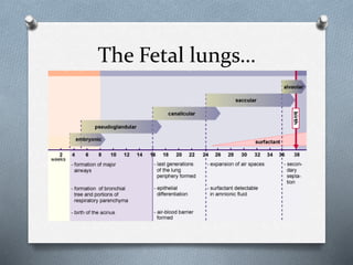
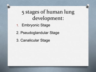
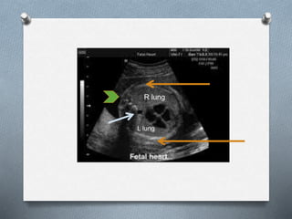
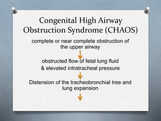
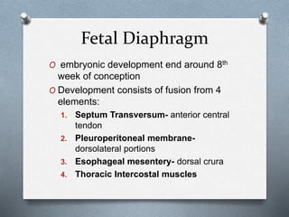
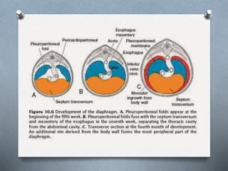
18. Lung Hypoplasia
O Incomplete or defective development of
the lung resulting in overall reduced size
due to reduced numbers or size of acini
O Factors required for adequate
development of the Lungs:
a. sufficient amniotic fluid volumes
b. adequate thoracic space size
c. normal breathing movements
d. normal fluid within the lung
19. Lung Hypoplasia
O Thoracic dimensions assesed by
measuring the TC/AC ratio and cardio-
thoracic ratio
O Decreased dimensions correlate with
Lung Hypoplasia
***TC/AC ratio NV >0.8
CT ratio NV <0.5
20. Lung Hypoplasia
O most cases of pulmonary hypoplasia
occur secondary to congenital anomalies
or secondary to pregnancy complications
while some can be a primary event
21.
22. CCAM of the Lung
O Congenital Cystic Adenomatoid
Malformation
O Benign hamartomatous or dysplastic
tumor
O Overgrowth of terminal bronchioles
resulting to a reduced number of alveoli
23. CCAM of the Lung
O 25% of all congenital lung masses
O 1 in 25,000 live births
O >95% occur in 1 lobe or segment
24. CCAM of the Lung
O 3 Categories:
Type I Type II Type III
2-10 cm cyst 0.5 - 2 cms cyst <2 cms cyst
Variable sized cyst Uniform sized cysts Appear solid
Compresses the
normal
parenchyma
Evenly distributed
and blends w/ the
normal
parenchyma
Small cystic or solid
type
25. CCAM of the Lung
O Additional Subtypes:
Type 0 Type IV
Acinar dysplasia or
agenesis
Large peripheral cyst of the
distal acinus lined
predominantly with alveolar
type cells
26. CCAM of the Lung
O Prenatal Classification based on
Ultrasound Findings:
Macrocystic Microcystic
Single or multiple cysts >/=
5 mm in diameter
<5mm diameter cysts
Seen on ultrasound as
cysts
Solid hyperechogenic mass
due to numerous acoutic
interfaces
Favourable prognosis Poorer prognosis
27. CCAM of the Lung
O depending on the size of the lesion,
esophageal compression may occur
resulting to subsequent polyhydramnios
O Its natural history is unpredictable
because some may grow or regress
29. CCAM of the Lung
Sagittal Section Transverse Image
30. CCAM of the Lung
Complications:
If with Associated findings like:
Mediastinal shift
Polyhydramnios
Presence of Hydrops
Will determine prognosis. Once Hydrops
100% mortality.
31. CCAM of the Lung
Prognosis:
- Unilateral Type I CCAM (Large cystic type) if
with no Polyhydramnios and Hydrops
GOOD Prognosis
- Poorer prognosis even if unilateral if with
associated features such as:
large sized cysts
polyhydramnios
fetal hydrops
mediastinal shift
associated anomalies
32. Bronchopulmonary
Sequestration (BPS)
O masses of non functioning lung tissue
that do not have a connection to the
normal tracheobronchial tree and is
supplied by an anomalous systemic artery
O normally situated in the lower lobes
33. Bronchopulmonary
Sequestration (BPS)
Intralobar Extralobar
Blood Supply: aortic branch Thoracic and abdominal
aorta
Venous drainage:
Pulmonary veins
Systemic veins
(hemiazygous, azygous
vein)
More common Less common
Abnormal segment of lung
tissue shares visceral
pleural covering of the
normal lung
Discrete accesory lobe with
its own pleural covering
Less associated with other
anomalies
More associated with other
anomalies e.g CDH
34. Bronchopulmonary
Sequestration (BPS)
O Other findings associated with Extralobar
category:
O typically found in the left thorax (65-90%) or
w/in or below the diaphragm (10-15%)
O associated more with male fetuses than
female (4:1 ratio)
38. Bronchopulmonary
Sequestration (BPS)
O Management:
O Close monitoring
O early delivery may be contemplated after 32-
33 weeks AOG
O thoracoamniotic shunting or thoracocentesis
can be considered in associated pleural
effusions and fetal hydrops
39. Congenital High Airway
Obstruction Syndrome (CHAOS)
complete or near complete obstruction of
the upper airway
obstructed flow of fetal lung fluid
& elevated intratracheal pressure
Distension of the tracheobronchial tree and
lung expansion
40. Congenital High Airway
Obstruction Syndrome (CHAOS)
Thinning of alveolar walls
Reduction of Type II Pneumocytes
Decreased Surfactant
Decreased venous return
Non Immune Hydrops
41. Congenital High Airway
Obstruction Syndrome (CHAOS)
Findings of:
Large hyperechoic lungs,
flattened or inverted diaphragm
dilated airways distal to the obstruction
fetal ascites or hydrops
42.
43. Other Lung Abnormalities:
O Congenital Lobar Emphysema
- Obstructed bronchus leading to progressive
hyperexpansion of the lung parenchyma
- Right, middle and upper lobes most commonly
affected
- Hard to detect on ultrasound since cellular and
fluid composition of affected lobe very similar
to normal lung upon examination
44. Other Lung Abnormalities:
O Bronchogenic cysts
- From abnormal budding of the ventricular
diverticulum
- Focal cystic duplication of the
tracheobronchial tree
- Few milliliters to >5 cm
45. Fetal Diaphragm
O embryonic development end around 8th
week of conception
O Development consists of fusion from 4
elements:
1. Septum Transversum- anterior central
tendon
2. Pleuroperitoneal membrane-
dorsolateral portions
3. Esophageal mesentery- dorsal crura
4. Thoracic Intercostal muscles
46.
47. Fetal Diaphragm
O dome shaped partition between the
thoracic and abdominal cavities
O Allows passage of organs, vessels, and
nerves from the thoracic into the
abdominal cavity
O On UTZ: smooth hypoechoic line between
the lungs and liver or spleen
48. Congenital Diaphragmatic
Hernia
O a development
defect of the
diaphragm that
allows abdominal
viscera to
herniate into the
chest
(Insert CDH gross
Picture)
52. Congenital Diaphragmatic
Hernia
O Sonographic Features:
1. Absence of gastric bubble in its normal
position
2. Displacement of the heart across the
mediastinum towards the right
3. Bowel and liver in the left chest
4. Presence of the stomach adjacent to the
heart
54. Congenital Hydrothorax
O a.k.a. Pleural effusions
O may be associated with generalized
edema and ascites or may occur in
isolation
O chylothorax is the most common cause
O rare (incidence of 1 in 15,000
pregnancies)
55. O Complications arise by the degree of fluid
accumulation
O mediastinal compression with reduced
fetal swallowing
O Polyhydramnios
In comparison with the development of the other organs the prenatal development of the lungs occupies a special position. The lungs as breathing organs are unnecessary for intrauterine existence. Nevertheless, they must be developed to such an extent that they are immediately ready to function following birth. This explains why the entire development extends from the embryonic period through the fetal period up to birth (and even afterwards). During intrauterine life, the lungs are nevertheless an important source of amniotic fluid.
By the end of the 4th week of life, during the embryonic stage of lung development, the respiratory diverticulum/lung bud (caudal end of the laryngotracheal groove grows caudally foming the primitive trachea
2 lateral outpocketings which are the lung buds. By the end of the 5th week, each of the lung bud enlarges forming the right and the left main bronchus
By the end of the 6th week, all bronchopulmonary segments have been formed
During this stage, there is repeated dichotomous branching of the main bronchus, forming (CLICK) three secondary bronchi on the right and two secondary bronchi on the left..
These in turn will become the conducting airways
By the 16th week, (CLICK) the tracheobronchial tree has been formed
(At 16-28 weeks, the gas exchanging portion of the lungs are formed and vascularized, this is when the secondary bronchioles divide continously into smaller canals.)
During the canalicular period Each terminal bronchiole divides into 2 or more respiratory bronchioles.
It is also the time when capillaries will start to establish close contact
a.k.a terminal sac period.
These ducts would later end in terminal sacs a.k.a. primitive alveoli surrounded by flat alveolar cells wherein surrounding capillaries come in close contact.
By the end of the 7th month, sufficient numbers of mature alveolar sacs and capillaries are present to guarantee adequate gas exchange, granting the premature infant to be able to survive
Starts from 36 weeks to term.
Further thinning of the blood gas barrier, increase production of surfactant, and progressive branching of the respiratory airways leading to alveolar production
To be able to determine presence of normal thoracic anatomy, assesment / measurement of the thoracic circumference at the level of the four chamber view of the heart should be obtained. Key view for the assessment of intrathoracic fetal anatomy
Fundamental plane where heart and lung are assessed
On this view, the ff structures can, and should be assessed:
Thoracic outline- consisting of 2 displayed ribs and overlying soft tissues and skin. The ribs should be uniformly echogenic, continously smooth, with convex contour and encircle 2/3 of the thoracic area.
Two lungs: which occupies 2/3 of the thoracic area. The normal lungs appear symmetric and homogenous with medium level echogenicity increasing gradually as gestation progresses. Echogenicity is slightly increased compared with the liver. Right lung slightly larger than the left. The lungs should abut the thoracic cage with no hypoechoic space in between.
Thoracic aorta; lying in the pre vertebral area just left of the midline behind the left atrium
One thoracic vertebra: on the midline posteriorly
This is the axial view of the chest. Its shape is almost circular. The ribs are shown to be uniformly echogenic smooth with convex contour and again encircling 2/3 of the chest wall.
The figure B or image B shows where the thoracic circumference should be measured, which is along the outer rib contour excluding the skin and soft tissue.
Deficiency on any of these factors may lead to pulmonary hypoplasia
Hamartoma – benign focal malformation that resembles a neoplasm in the tissue of its origin
Occurs during the pseudoglandular period resulting to expansion of conducting airways and peripheral lung tubules and acinar tubules
With this classification designation of CCAM be changed to Congenital Pulmonary Airway Malformation owing to the fact that only three of the five types were cystic and only one type (type 3) adenomatoid
Microcystic: difficult to ID normal parenchyma on affected side
Hamartoma – benign focal malformation that resembles a neoplasm in the tissue of its origin
Occurs during the pseudoglandular period resulting to expansion of conducting airways and peripheral lung tubules and acinar tubules
Large cystic lesion on the right hemithorax
In the sagittal section depicted by the arrows, the CCAM is located at the posterior chest inferior to the heart
Second image shows the transverse view of the same fetus, arrowhead points to the gastric bubble, showing the extent of the lesion situated at the left lung
Intralobar accounts for 75% of pulmonary sequestrations
Abn echogenic lung lobe at basal area of the right chest
Demonstrated on coronal view
Cross sectional view
Doppler done to establish the lobe directy supplied by the vessels from the aorta
These are images of an intralobar category
Markers: extralobar sequestration with aberrant blood supply (arrow)
CHAOS is usually a constellation of findings which arise due to obstruction of the upper airway tract.
Causes of obstruction: laryngeal atresia; stenosis; laryngeal cyst; tracheal atresia or stenosis
CHAOS is usually a constellation of findings which arise due to obstruction of the upper airway tract.
Causes of obstruction: laryngeal atresia; stenosis; laryngeal cyst; tracheal atresia or stenosis
Upper airway causing dilated trachea; large echogenic lungs, inverted diaphragm and fetal ascites
Fetal embryonic development occur between 8-12 weeks of development.
Ventrally from the septum transversum (most impt component) which forms the anterior central tendon
the pleuroperitoneal membrane fuses with the dorsal mesentery of the esophagus and the dorsal portion of the septum transversus, forming the dorsolateral portions to separate the thoracic and abdominopelvic cavities (primitive diaphragm).
Esophageal mesentery froming the dorsal crura
And the thoracic intercostal muscles(body wall)
Causes: >>>> either by primary diaphragmatic defect with secondary migration of abdominal organs into the thoracic cavity or defect in the fusion of the 4 components
Almost all of the hernias occur in the posterolaterally located Bochdalek foramen, which characteristically involve the left side.
Morgagni hernias occur in the anteromedial retrosternal part of the diaphragm as a result of the maldevelopment of the septum tranversum
… gastric bubble not seen in the abdomen but beside the heart in the thoracic cavity
Hydrothorax is the accumulation of fluid in the pleural space. It can be primary or secondary, and unilateral or bilateral.
At diagnosis, it is bilateral in about 75% of the cases and is already associated with fetal hydrops in 60-65% (1, 2). If isolated, it is commonly due to congenital primary chylothorax, with an estimated incidence of 1/10.000-15.000 pregnancies (1, 2)
After the diagnosis of fetal hydrothorax, the optimal antenatal management is controversial since some fetuses may not be significantly compromised, whereas others may develop hydrops or die at birth from pulmonary hypoplasia. Therefore, the decision to treat, deliver prematurely or follow conservatively can be difficult.
Thoracocentesis to aspirate the pleural fluid was first proposed as a treatment of primary fetal hydrothorax in 1982 (20). It is performed under continuous ultrasound guidance using a 20-gauge needle inserted percutaneously.
The main drawback of thoracocentesis is that in the majority of cases it is usually followed by rapid re-accumulation of the effusion within 24-48 hours, necessitating repeated taps.