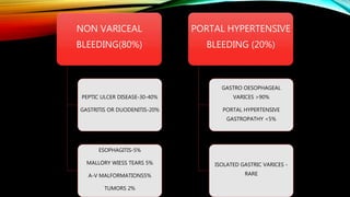
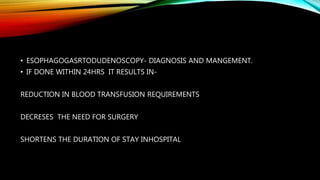
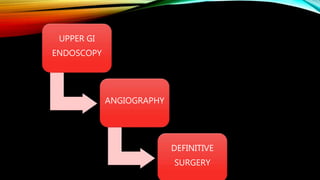
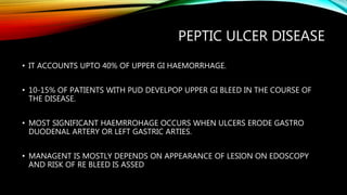
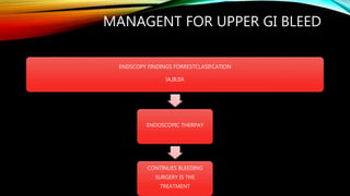
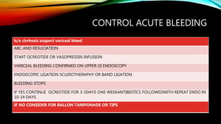
2. DEFINITION
• UPPER GI BLEEDING REFERS TO BLEEDING ARISING FROM GI TRACT PROXIMAL
TO THE LIGAMENT OF TRIETZ.
• IT ACCOUNTS UPTO 80% OF SIGNIFICANT GI HEAEMORRHAGE.
• IT MAY PRESENT AS HEMATEMISIS OR MELENA.
4. • ESOPHAGOGASRTODUDENOSCOPY- DIAGNOSIS AND MANGEMENT.
• IF DONE WITHIN 24HRS IT RESULTS IN-
REDUCTION IN BLOOD TRANSFUSION REQUIREMENTS
DECRESES THE NEED FOR SURGERY
SHORTENS THE DURATION OF STAY INHOSPITAL
6. PEPTIC ULCER DISEASE
• IT ACCOUNTS UPTO 40% OF UPPER GI HAEMORRHAGE.
• 10-15% OF PATIENTS WITH PUD DEVELPOP UPPER GI BLEED IN THE COURSE OF
THE DISEASE.
• MOST SIGNIFICANT HAEMRROHAGE OCCURS WHEN ULCERS ERODE GASTRO
DUODENAL ARTERY OR LEFT GASTRIC ARTIES.
• MANAGENT IS MOSTLY DEPENDS ON APPEARANCE OF LESION ON EDOSCOPY
AND RISK OF RE BLEED IS ASSED
7. FORREST CLASSIFICATION FOR
ENDOSCOPIC FINDINGS AND
REBLEEDING RISKS IN PUD
CLASSIFICATION REBLEEDING RISK
GRADE I A ACTIVE PULSATILE BLEEDING HIGH
GRADE IB ACTIVE NON PULSATILE
BLEEDING
HIGH
GRADE IIA NON BLEEDING VISIBLE VESSEL HIGH
GRADE IIB ADHERENT CLOT INTERMEDIATE
GRADE IIC ULCER WITH BLACK SPOT LOW
GRADE III CLEAN NON BLEEDING ULCER
BED
LOW
8. MANAGENT FOR UPPER GI BLEED
ENDSCOPY FINDINGS FORRESTCLASIFCATION
IA,IB,IIA
ENDOSCOPIC THERPAY
CONTINUES BLEEDING
SURGERY IS THE
TREATMENT
9. • MEDICAL MANAGEMENT
• PROTON PUMP INHIBITORS: REDUCE THE RISK OF RE BLEED,SURGICAL INTERVENTION
• H PYLORI TREATMENT: PPI PLUS H.PYOLRI TREATMENT DECRESES THE RISK REBLEED
• H PYLORI IS ERADICATED WE CAN STOP PPI
• NSAIDS AND SSRI ARE ULCEROGENIC THEY SHOULD BE AVOIDED.
10. ENDOSCOPIC MANAGEMENT
• EPINEPHRINE INJECTION-1IN 10,000 DILUTIONS IN 4 QUDRANTS OF ULCER.
• THERMAL ENERGY: HEAT PROBES,MONOPOLAR OR BIPOLAR COAGULATION,LASER OR
ARGON PLASMA COAGULATION.
• HAEMO CLIPS: SPURTING VESSELS.
• EPINEPHRINE INJECTION ALONG WITH COAGULATION ACHIVES HEMOSTASIS UPTO
90%.
• ARGON PLASMA COAGULATION USEFUL SUPERFICIAL LESION.
• REPEAT ENDOSCOPY DECREASES REBLEEDINGUPTO 75%
11. SURGICAL MANAGENT
• INDICATIONS:
• HEMODYNAMICALLY INSTABILITY DESPITE OF VIGOROUS RESUCIATION(>6 UNIT
TRANSFUSION)
• FAILURE TO CONTROL HEMORRHAGE AFTER ENDOSCOPIC THERAPHY.
• RECCUURENT HEMORRHAGE
• SHOCK ASSOCIATED WITH RECURRENT HEMORRHAGE
• CONTINUED SLOW BLEEDING WITH TRANSFUSION REQUIREMENT >3UNIT/DAY
12. SURGICAL OPTIONS
FOR DUODENAL ULCER
DUODENO PYLOROMYOTOMY.
TRUNCAL VAGOTOMY
HIGHLY SELECTUVE VAGOTOMY
ANTECTOMY WITH TRUNCAL VAGOTOMY
14. MALLORY WEISS TEARS
• Mallory-wiess tears are mucosal and submucosal tears that occur at gastroesophageal
junction.
• Forcefull contraction of abdominal wall against an unrelaxed cardia resulting in mucosal
laceration.
• Alcoholics and poeople with repeated vomittings.
• Most of the tears occur along the lesser curvation.
• 90% bleeding are self limited and mucosa heals within 72 hours.
• If endotheraphy fails variceal bleeding should be ruled out
• Recuurence is uncommon.
15. STRESS GASTRITIS
• Characterised by multiple superficial erosionsof entire stomach most commonly in
the body.
• Acid and pepsin injury in acontext of ischemia duringhypoperfusion.
• Curling ulcers a type of stress ulcer.
• Even in astate of significant bleeding acid suppressive theraphy is often sucessful in
controlling the hemorrhage.
16. OESOPHAGITIS
• Rarely it present as a haemorrhage.
• It usually occurs in immunocompromised patients .
• Commonly presents as anemia due to chronic blood loss.
• Due to repeat inflammtory response of mucosa to acid secretions aggrevated with infection
leads to hemorrhage.
• Acid suppression in main stay of treatment.
• Rarely crohns disease ,radiation certain mefdications may cause oesophageal hemorrhage.
17. DIEUFLOY LESION
• Vascular malformationsfound primarly along lesser curvature with in 6cm from GE
junction.
• They present rupture of large vesels of 1-3mm size in submucosa with mucosal
erosions overlying them.
• Usually mucosal defects are of size2-5mm in size and difficult to identify.
• Application of thermal or sclerosant therphay effective in 80-100%
18. GASTRIC ANTRAL
VASCULARECTASIA
• Charecterised by a coolection of dilated venules appearing as linear red streaks
converging on to the antrum in longitudinal fashion giving it the appearance of
watermelon.
• Bleeding is rare,presentation, usually pesents as anemia.
• Endscoic theraphy with argon plasma coagulation is effective in 90% patients.
• Rarely patients require antrectomy.
19. MALIGNANCY
• Gastrointestinal stromal tumour commonly presses upper GI bleed followed by
leiomyoma and lymphoma.
• If patient is stable standard resection for tumour should be done as curative
procudure.
• As palliative procedure wedge resection of the tumour mass should be done.
20. AORTOENTERIC FISTULA
• Primary aortoduodenal fistula is arare entity.
• Common entity seen is graft –enteric erosion after aortic graft cases.
• Patient with h/o of aortic aneurysm or prosthetic aneurysm repair
May develop psudoaneurysm at proximal anastomotic site due to infection.
This ruptures into duodenum causing massive hemorrhage.
Typicall they present asself limiting episode of sentinel bleed followed by massive
hemorrhage typically from third or fouth part of duodenum on endoscopic
visulisation.
A ct scan with intravenous contrsat may show air around aorta sugesting psudo
aneurysm rarely contrast material in duodenum.
Treatment include ligation of proximal aorta and distal aorta and bypass graft should
be done follwed by fistula repair
21. HEMOBILIA
• Unusual cause of GI bleed suspected in patient with right upper quadrant pain jaundice and
hemorrhage.
• It is typicaly associated with trauma,recent instrumentation of billiary tree or hepatic
neoplasm.
• Endoscopy may be helpful by demonstating blood at ampulla of vater.
• Angiography is diagnostic procedure of choice.
• Angiographic embolisation is the treatment of choice.
22. HEMOSUCUS PANCREATICUS
rare cause of upper GI bleed due to bleeding from pancreatic duct.
Commonly because of rupture of psudocyst into spleenic artery.
Presents as abdominal pain with blood loss with h/o pancreatitis.
Angiography is diagnostic andpermits embolisation.
Rarely distal paancreatectomy should be considered.
23. IATROGENIC BLEEDING
Percutaneous transhepatic procedures , endoscopic sphincterotomy, percutaneous
gastrotomy are some of the procedure associated with iatrogenic bleeding.
Most commonly they are self limitng late bledding occurs after 48hrs.
Some times pateints upper GI surgery with anastomosis may present with suture or
stapler site hemorrhage.
Commonly this is manged endoscopically.
24. BLEEDING RELATED TO PORTAL
HYPERTENSION
• Result from bleeding from varices.
• They are dilated submucosal veins developed to decompress portal system into
systemic circulation.
• They dilate upto 2cm causing tension on overlying mucosa which ruptures on
minimal trauma.
• More common in distal oesophagus.
25. • Variceal hemorrhage is associated with increased risk of rebleeding.
• Increased need for blood transfusion.
• Increased duration of hospital stay.
• Increases mortality.
• Treatment focuses to control acute attack and to preventrebleeding.
26. CONTROL ACUTE BLEEDING
h/o cirrhosis suspect variceal bleed
ABC AND RESUCIATION
START OCREOTIDE OR VASOPRESSIN INFUSION
VARICEAL BLEEDING CONFIRMED ON UPPER GI ENDOSCOPY
ENDOSCOPIC LIGATION SCLEROTHERAPHY OR BAND LIGATION
BLEEDING STOPS
IF YES CONTINUE OCREOTIDE FOR 3-5DAYS ONE WEEKANTIBIOTICS FOLLOWEDWITH REPEAT ENDO IN
10-14 DAYS.
IF NO CONSIDER FOR BALLON TAMPONADE OR TIPS
27. MEDICAL MANAGEMENT
• Pharmacotheraphy is directed to decrease portal hyper tension.
• Vasopressin decreases splanchnic blood flow,it may cause cardiac ischemia.
• Ocreotide ,a synthetic somatostatin is also used.
• Telprisin is a newer vasopressin with decreased side effects.
28. ENDOSCOPY
• Endoscopic band ligation is the treatment of choice for variceal bleed.
• Endoscopic sclerotheraphy can be used but it as side effects like
mediastinits,sticture and perforation.
Endosopy controls hemorrhage upto 90% of patients.
29. BALLOON TAMPONADE
• It controls acute hemorrage.
• The sengstaken –blakemore tube consists of gastric tube with two bulbs.
• Gastric bulb is inflated first, if beeding not controlled oesophageal tube is controlled.
• These tube are associated with complications like aspiration and inappropiate placement
resulting in perforation.
• Minnesota tube includes proximal oesophagus to aspirate swalloed srection.
30. • This tubes compresses the varices and secures hemostasis.
• Reccurence rate is veryhigh upto 50%.
Bleeding is refractory then trans jugular intrahepatic shunt is used.
Then surgical shunt are tried.
Lastly transplantation is considered.