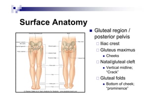
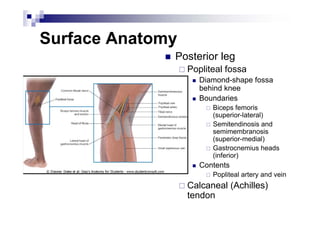
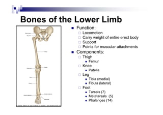
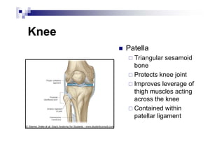
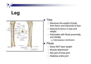
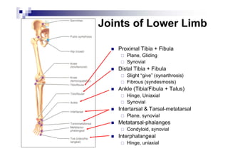
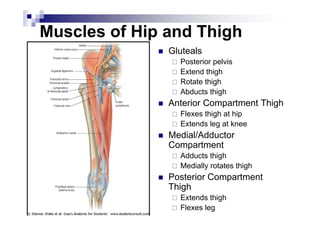
The Lower Limb
Pelvis, Thigh, Leg
and Foot
Surface Anatomy
Surface Anatomy
„ Gluteal region /
posterior pelvis
Iliac crest
Gluteus maximus
„ Cheeks
Natal/gluteal cleft
„ Vertical midline;
“Crack”
Gluteal folds
„ Bottom of cheek;
“prominence”
Surface Anatomy
„ Anterior thigh and leg
Palpate
„ Patella
„ Condyles of femur
Femoral Triangle
„ Boundaries:
Sartorius (lateral)
Adductor longus (medial)
Inguinal ligament (superior)
„ Contents:
Femoral artery, vein and
nerve, lymph nodes
Surface Anatomy
„ Posterior leg
Popliteal fossa
„ Diamond-shape fossa
behind knee
„ Boundaries
Biceps femoris
(superior-lateral)
Semitendinosis and
semimembranosis
(superior-medial)
Gastrocnemius heads
(inferior)
„ Contents
Popliteal artery and vein
Calcaneal (Achilles)
tendon
Surface Anatomy
„ Anterior leg bones
Tibia
„ Tibial tuberosity
„ Anterior crest
„ Medial surface
„ Medial malleolus
Fibula
„ Lateral malleolus
Skeletal
Composition
Bones of the Lower Limb
„ Function:
Locomotion
Carry weight of entire erect body
Support
Points for muscular attachments
„ Components:
Thigh
„ Femur
Knee
„ Patella
Leg
„ Tibia (medial)
„ Fibula (lateral)
Foot
„ Tarsals (7)
„ Metatarsals (5)
„ Phalanges (14)
Thigh
„ Femur
Largest, longest,
strongest bone in the
body!!
Receives a lot of
stress
Courses medially
„ More in women!
Articulates with
acetabulum proximally
Articulates with tibia
and patella distally
Knee
„ Patella
Triangular sesamoid
bone
Protects knee joint
Improves leverage of
thigh muscles acting
across the knee
Contained within
patellar ligament
Leg
„ Tibia
Receives the weight of body
from femur and transmits to foot
Second to femur in size and
weight
Articulates with fibula proximally
and distally
„ Interosseous membrane
„ Fibula
Does NOT bear weight
Muscle attachment
Not part of knee joint
Stabilize ankle joint
Foot „ Function:
Supports the weight of the
body
Act as a lever to propel the
body forward
„ Parts:
Tarsals
„ Talus = ankle
Between tibia and fibula
Articulates with both
„ Calcaneus = heel
Attachment for Calcaneal
tendon
Carries talus
„ Navicular
„ Cuboid
„ Medial, lateral and
intermediate cuneiforms
Metatarsals
Phalanges
Foot
„ 3 arches
Medial
Lateral
Transverse
„ Has tendons that run
inferior to foot bones
Help support arches
of foot
„ Function
Recoil after stepping
Longitudinal
Joints of Lower Limb
„ Hip (femur + acetabulum)
Ball + socket
Multiaxial
Synovial
„ Knee (femur + tibia)
Hinge (modified)
Biaxial
Synovial
Contains menisci, bursa, many
ligaments
„ Knee (femur + patella)
Plane
Gliding of patella
Synovial
Joints of Lower Limb
„ Proximal Tibia + Fibula
Plane, Gliding
Synovial
„ Distal Tibia + Fibula
Slight “give” (synarthrosis)
Fibrous (syndesmosis)
„ Ankle (Tibia/Fibula + Talus)
Hinge, Uniaxial
Synovial
„ Intertarsal & Tarsal-metatarsal
Plane, synovial
„ Metatarsal-phalanges
Condyloid, synovial
„ Interphalangeal
Hinge, uniaxial
Muscles
Muscles of Hip and Thigh
„ Gluteals
Posterior pelvis
Extend thigh
Rotate thigh
Abducts thigh
„ Anterior Compartment Thigh
Flexes thigh at hip
Extends leg at knee
„ Medial/Adductor
Compartment
Adducts thigh
Medially rotates thigh
„ Posterior Compartment
Thigh
Extends thigh
Flexes leg
Gluteals
„ Gluteus maximus
Origin - Ilium, sacrum and coccyx
Insertion - Gluteal tuberosity of femur,
iliotibial tract
Action - Extends thigh, some lateral
rotation and abduction
Innervation - Inferior gluteal nerve
„ Gluteus medius
„ Gluteus minimus
Origin - Ilium
Insertion - Greater trochanter of femur
Action - Abduction, medial rotation
Innervation - Superior gluteal nerve
„ Lesser Gluteals help stabilize hip
to allow fluent bipedal walking
Posterior Pelvis
„ Tensor fasciae latae
Origin – iliac crest and
anterior inferior iliac spine
Insertion – iliotibial tract
Action - Flex thigh, abduct
thigh, medial rotation of
thigh
Innervation – Superior
gluteal nerve
Anterior Compartment Thigh
„ Quadriceps femoris
Rectus femoris
„ Origin – anterior inferior iliac
spine, margin of acetabulum
„ Insertion – patella and tibial
tuberosity via the patellar
ligament
„ Action – extends knee, flexes
thigh
Vastus lateralis
Vastus medialis
Vastus intermedius
„ Origin - femur
„ Insertion – patella and tibial
tuberosity via the patellar
ligament
„ Action – extends knee
„ Sartorius
„ Origin - anterior superior iliac
spine
„ Insertion – medial tibia
„ Action - flex, abduct, lat rotate
thigh; weak knee flexor
All above innervated by the femoral nerve!!!
Anterior Compartment Thigh
„ Iliopsoas
Origin - Ilia, sacrum,
lumbar vertebrae
Insertion – lesser
trochanter
Action – flexor of thigh
Innervation – femoral
nerve
Adductors
„ Adductor longus
„ Adductor brevis
„ Adductor magnus
Origin – inferior pelvis
Insertion - femur
Action – adducts and medial rotates
Innervation – Obturator nerve
„ Pectineus
Origin - pubis
Insertion – lesser trochanter
Action – adducts, medial rotates
Innervation – femoral, sometimes
obturator
„ Gracilis
Origin - pubis
Insertion – medial tibia
Action – adducts thigh, flex, medial,
rotates leg
Innervation – Obturator nerve
Posterior Compartment - Hamstring
„ Biceps femoris (2 heads)
Origin – ischial tuberosity, distal
femur
Insertion - lateral tibia, head
fibula
Action - thigh extension, knee
flexion, lateral rotation
„ Semitendinosus
„ Semimembranosus
Origin - ischial tuberosity
Insertion - medial tibia
Action - thigh extension, knee
flexion, medial rotation
Sciatic nerve innervates all of the above muscles!!!
Muscles of the Leg
„ Anterior Compartment
Dorsiflex ankle, invert foot, extend toes
Innervation: Deep fibular nerve
„ Lateral Compartment
Plantarflex, evert foot
Innervation: Superficial Fibular nerve
„ Posterior Compartment
Superficial and deep layers
Plantarflex foot, flex toes
Innervation: Tibial nerve
Anterior Compartment
„ Tibialis anterior
Origin - tibia
Insertion - tarsals
Action - dorsiflexion, foot inversion
„ Extensor digitorum longus
Origin – tibia and fibula
Insertion - phalanges
Action – toe extension
„ Extensor hallucis longus
Origin – fibula, interosseous
membrane
Insertion – big toe
Action - extend big toe, dorsiflex
foot
All innervated by deep fibular nerve
Lateral Compartment
„ Fibularis (peroneus) longus
Origin – lateral fibula
Insertion – 5th metatarsal,
tarsal
Action - plantarflex, evert
foot
„ Fibularis (peroneus) brevis
Origin – distal fibula
Insertion - proximal fifth
metatarsal
Action – same as above!!
All innervated by the superficial fibular nerve
Superficial Posterior Compartment
„ Triceps surae
Gastrocnemius (2 heads)
„ Origin - medial and lateral condyles of
femur
„ Insertion - posterior calcaneus via
Achilles tendon
Soleus
„ Origin – tibia and fibula
„ Insertion – same as above
Action of both – plantarflex foot
„ Plantaris (variable)
Origin – posterior femur
Insertion – same as above!
Action – plantarflex foot, week knee
flexion
All innervated by the tibial nerve
Deep Posterior Compartment
„ Popliteus
Origin - lateral condyle femur
and lateral meniscus
Insertion – proximal tibia
Action – flex and medially rotate leg
„ Flexor digitorum longus
Origin - tibia
Insertion - distal phalanges of toe 2-5
Action – plantarflex and invert foot, flex toe
„ Flexor hallucis longus
Origin - fibula
Insertion - distal phalanx of hallux
Action - plantarflex and invert foot, flex toe
„ Tibialis posterior
Origin – tibia, fibula, and interosseous
membrane
Insertion - tarsals and metatarsals
Action - plantarflex and invert foot
All innervated by the tibial nerve
Innervation
Plexuses of the Lower Limb
„ “Lumbosacral plexus”
„ Lumbar Plexus
Arises from L1-L4
Lies within the psoas major
muscle
Mostly anterior structures
„ Sacral Plexus
Arises from spinal nerve
L4-S4
Lies caudal to the lumbar
plexus
Mostly posterior structures
Lumbar Plexus
„ Femoral nerve
Cutaneous branches
„ Thigh, leg, foot (e.g. saphenous nerve)
Motor branches
„ Anterior thigh muscles (e.g. quadriceps,
sartorius, iliopsoas)
„ Obturator nerve
Sensory
„ Skin medial thigh; hip, knee joints
Motor
„ Adductor muscles
„ Lateral femoral cutaneous
Sensory
„ Skin lateral thigh
„ Genitofemoral
Sensory
„ Skin scrotum, labia major, anterior thigh
Motor
„ Cremaster muscle
Sacral Plexus
„ Sciatic
Motor:
„ Hamstring
Branches into:
„ Tibial nerve
Cutaneous
ƒ Posterior leg and sole of foot
Motor
ƒ Posterior leg, foot
„ Common fibular (peroneal) nerve
Cutaneous
ƒ Anterior and lateral leg, dorsum foot
Motor
ƒ Lateral compartment, tibialis anterior,
toe extensors
„ Superior gluteal nerve
Motor
„ Gluteus medius and minimus, tensor
fasciae latae
Sacral Plexus (continued)
„ Inferior gluteal nerve
Motor
„ Gluteus maximus
„ Posterior femoral
cutaneous nerve
Sensory
„ Inferior buttocks, posterior
thigh, popliteal fossa
„ Pudendal nerve
Sensory
„ External genitalia, anus
Motor
„ Muscles of perineum
Vasculature
Arteries
„ Common iliac (from
aorta) branches into:
Internal iliac
„ Supplies pelvic organs
External iliac
„ Supplies lower limb
Arteries
„ Internal iliac branches into:
Cranial and Caudal Gluteals
(Superior and Inferior)
„ Gluteals
Internal Pudendal
„ Perineum, external genitalia
Obturator
„ Adductor muscles
Other branches supply rectum,
bladder, uterus, vagina, male
reproductive glands
Arteries
„ External iliac becomes…….
Femoral
„ Once passes the inguinal ligament
„ Lower limb
„ Branches into Deep femoral
Adductors, hamstrings, quadriceps
Branches into Medial/lateral femoral
circumflex
ƒ Head and neck of femur
„ Femoral becomes……
Popliteal (continuation of femoral)
„ Branches into:
Geniculars
ƒ Knee
„ Splits into:
Anterior Tibial
ƒ Anterior leg muscles, further branches to
feet
Posterior Tibial
ƒ Flexor muscles, plantar arch, branches to
toes
Veins „ Deep Veins: Mostly share names of
arteries
Ultimately empty into Inferior Vena
Cava
„ Plantar
„ Tibial
„ Fibular
„ Popliteal
„ Femoral
„ External/internal iliac
„ Common iliac
„ Superficial Veins
Dorsal venous arch (foot)
Great saphenous (empties into femoral)
Small saphenous (empties into
popliteal)

Lower extremity.pdf

  • 1.
    The Lower Limb Pelvis,Thigh, Leg and Foot
  • 2.
  • 3.
    Surface Anatomy „ Glutealregion / posterior pelvis Iliac crest Gluteus maximus „ Cheeks Natal/gluteal cleft „ Vertical midline; “Crack” Gluteal folds „ Bottom of cheek; “prominence”
  • 4.
    Surface Anatomy „ Anteriorthigh and leg Palpate „ Patella „ Condyles of femur Femoral Triangle „ Boundaries: Sartorius (lateral) Adductor longus (medial) Inguinal ligament (superior) „ Contents: Femoral artery, vein and nerve, lymph nodes
  • 5.
    Surface Anatomy „ Posteriorleg Popliteal fossa „ Diamond-shape fossa behind knee „ Boundaries Biceps femoris (superior-lateral) Semitendinosis and semimembranosis (superior-medial) Gastrocnemius heads (inferior) „ Contents Popliteal artery and vein Calcaneal (Achilles) tendon
  • 6.
    Surface Anatomy „ Anteriorleg bones Tibia „ Tibial tuberosity „ Anterior crest „ Medial surface „ Medial malleolus Fibula „ Lateral malleolus
  • 7.
  • 8.
    Bones of theLower Limb „ Function: Locomotion Carry weight of entire erect body Support Points for muscular attachments „ Components: Thigh „ Femur Knee „ Patella Leg „ Tibia (medial) „ Fibula (lateral) Foot „ Tarsals (7) „ Metatarsals (5) „ Phalanges (14)
  • 9.
    Thigh „ Femur Largest, longest, strongestbone in the body!! Receives a lot of stress Courses medially „ More in women! Articulates with acetabulum proximally Articulates with tibia and patella distally
  • 10.
    Knee „ Patella Triangular sesamoid bone Protectsknee joint Improves leverage of thigh muscles acting across the knee Contained within patellar ligament
  • 11.
    Leg „ Tibia Receives theweight of body from femur and transmits to foot Second to femur in size and weight Articulates with fibula proximally and distally „ Interosseous membrane „ Fibula Does NOT bear weight Muscle attachment Not part of knee joint Stabilize ankle joint
  • 12.
    Foot „ Function: Supportsthe weight of the body Act as a lever to propel the body forward „ Parts: Tarsals „ Talus = ankle Between tibia and fibula Articulates with both „ Calcaneus = heel Attachment for Calcaneal tendon Carries talus „ Navicular „ Cuboid „ Medial, lateral and intermediate cuneiforms Metatarsals Phalanges
  • 13.
    Foot „ 3 arches Medial Lateral Transverse „Has tendons that run inferior to foot bones Help support arches of foot „ Function Recoil after stepping Longitudinal
  • 14.
    Joints of LowerLimb „ Hip (femur + acetabulum) Ball + socket Multiaxial Synovial „ Knee (femur + tibia) Hinge (modified) Biaxial Synovial Contains menisci, bursa, many ligaments „ Knee (femur + patella) Plane Gliding of patella Synovial
  • 15.
    Joints of LowerLimb „ Proximal Tibia + Fibula Plane, Gliding Synovial „ Distal Tibia + Fibula Slight “give” (synarthrosis) Fibrous (syndesmosis) „ Ankle (Tibia/Fibula + Talus) Hinge, Uniaxial Synovial „ Intertarsal & Tarsal-metatarsal Plane, synovial „ Metatarsal-phalanges Condyloid, synovial „ Interphalangeal Hinge, uniaxial
  • 16.
  • 17.
    Muscles of Hipand Thigh „ Gluteals Posterior pelvis Extend thigh Rotate thigh Abducts thigh „ Anterior Compartment Thigh Flexes thigh at hip Extends leg at knee „ Medial/Adductor Compartment Adducts thigh Medially rotates thigh „ Posterior Compartment Thigh Extends thigh Flexes leg
  • 18.
    Gluteals „ Gluteus maximus Origin- Ilium, sacrum and coccyx Insertion - Gluteal tuberosity of femur, iliotibial tract Action - Extends thigh, some lateral rotation and abduction Innervation - Inferior gluteal nerve „ Gluteus medius „ Gluteus minimus Origin - Ilium Insertion - Greater trochanter of femur Action - Abduction, medial rotation Innervation - Superior gluteal nerve „ Lesser Gluteals help stabilize hip to allow fluent bipedal walking
  • 19.
    Posterior Pelvis „ Tensorfasciae latae Origin – iliac crest and anterior inferior iliac spine Insertion – iliotibial tract Action - Flex thigh, abduct thigh, medial rotation of thigh Innervation – Superior gluteal nerve
  • 20.
    Anterior Compartment Thigh „Quadriceps femoris Rectus femoris „ Origin – anterior inferior iliac spine, margin of acetabulum „ Insertion – patella and tibial tuberosity via the patellar ligament „ Action – extends knee, flexes thigh Vastus lateralis Vastus medialis Vastus intermedius „ Origin - femur „ Insertion – patella and tibial tuberosity via the patellar ligament „ Action – extends knee „ Sartorius „ Origin - anterior superior iliac spine „ Insertion – medial tibia „ Action - flex, abduct, lat rotate thigh; weak knee flexor All above innervated by the femoral nerve!!!
  • 21.
    Anterior Compartment Thigh „Iliopsoas Origin - Ilia, sacrum, lumbar vertebrae Insertion – lesser trochanter Action – flexor of thigh Innervation – femoral nerve
  • 22.
    Adductors „ Adductor longus „Adductor brevis „ Adductor magnus Origin – inferior pelvis Insertion - femur Action – adducts and medial rotates Innervation – Obturator nerve „ Pectineus Origin - pubis Insertion – lesser trochanter Action – adducts, medial rotates Innervation – femoral, sometimes obturator „ Gracilis Origin - pubis Insertion – medial tibia Action – adducts thigh, flex, medial, rotates leg Innervation – Obturator nerve
  • 23.
    Posterior Compartment -Hamstring „ Biceps femoris (2 heads) Origin – ischial tuberosity, distal femur Insertion - lateral tibia, head fibula Action - thigh extension, knee flexion, lateral rotation „ Semitendinosus „ Semimembranosus Origin - ischial tuberosity Insertion - medial tibia Action - thigh extension, knee flexion, medial rotation Sciatic nerve innervates all of the above muscles!!!
  • 24.
    Muscles of theLeg „ Anterior Compartment Dorsiflex ankle, invert foot, extend toes Innervation: Deep fibular nerve „ Lateral Compartment Plantarflex, evert foot Innervation: Superficial Fibular nerve „ Posterior Compartment Superficial and deep layers Plantarflex foot, flex toes Innervation: Tibial nerve
  • 25.
    Anterior Compartment „ Tibialisanterior Origin - tibia Insertion - tarsals Action - dorsiflexion, foot inversion „ Extensor digitorum longus Origin – tibia and fibula Insertion - phalanges Action – toe extension „ Extensor hallucis longus Origin – fibula, interosseous membrane Insertion – big toe Action - extend big toe, dorsiflex foot All innervated by deep fibular nerve
  • 26.
    Lateral Compartment „ Fibularis(peroneus) longus Origin – lateral fibula Insertion – 5th metatarsal, tarsal Action - plantarflex, evert foot „ Fibularis (peroneus) brevis Origin – distal fibula Insertion - proximal fifth metatarsal Action – same as above!! All innervated by the superficial fibular nerve
  • 27.
    Superficial Posterior Compartment „Triceps surae Gastrocnemius (2 heads) „ Origin - medial and lateral condyles of femur „ Insertion - posterior calcaneus via Achilles tendon Soleus „ Origin – tibia and fibula „ Insertion – same as above Action of both – plantarflex foot „ Plantaris (variable) Origin – posterior femur Insertion – same as above! Action – plantarflex foot, week knee flexion All innervated by the tibial nerve
  • 28.
    Deep Posterior Compartment „Popliteus Origin - lateral condyle femur and lateral meniscus Insertion – proximal tibia Action – flex and medially rotate leg „ Flexor digitorum longus Origin - tibia Insertion - distal phalanges of toe 2-5 Action – plantarflex and invert foot, flex toe „ Flexor hallucis longus Origin - fibula Insertion - distal phalanx of hallux Action - plantarflex and invert foot, flex toe „ Tibialis posterior Origin – tibia, fibula, and interosseous membrane Insertion - tarsals and metatarsals Action - plantarflex and invert foot All innervated by the tibial nerve
  • 30.
  • 31.
    Plexuses of theLower Limb „ “Lumbosacral plexus” „ Lumbar Plexus Arises from L1-L4 Lies within the psoas major muscle Mostly anterior structures „ Sacral Plexus Arises from spinal nerve L4-S4 Lies caudal to the lumbar plexus Mostly posterior structures
  • 32.
    Lumbar Plexus „ Femoralnerve Cutaneous branches „ Thigh, leg, foot (e.g. saphenous nerve) Motor branches „ Anterior thigh muscles (e.g. quadriceps, sartorius, iliopsoas) „ Obturator nerve Sensory „ Skin medial thigh; hip, knee joints Motor „ Adductor muscles „ Lateral femoral cutaneous Sensory „ Skin lateral thigh „ Genitofemoral Sensory „ Skin scrotum, labia major, anterior thigh Motor „ Cremaster muscle
  • 33.
    Sacral Plexus „ Sciatic Motor: „Hamstring Branches into: „ Tibial nerve Cutaneous ƒ Posterior leg and sole of foot Motor ƒ Posterior leg, foot „ Common fibular (peroneal) nerve Cutaneous ƒ Anterior and lateral leg, dorsum foot Motor ƒ Lateral compartment, tibialis anterior, toe extensors „ Superior gluteal nerve Motor „ Gluteus medius and minimus, tensor fasciae latae
  • 34.
    Sacral Plexus (continued) „Inferior gluteal nerve Motor „ Gluteus maximus „ Posterior femoral cutaneous nerve Sensory „ Inferior buttocks, posterior thigh, popliteal fossa „ Pudendal nerve Sensory „ External genitalia, anus Motor „ Muscles of perineum
  • 35.
  • 36.
    Arteries „ Common iliac(from aorta) branches into: Internal iliac „ Supplies pelvic organs External iliac „ Supplies lower limb
  • 37.
    Arteries „ Internal iliacbranches into: Cranial and Caudal Gluteals (Superior and Inferior) „ Gluteals Internal Pudendal „ Perineum, external genitalia Obturator „ Adductor muscles Other branches supply rectum, bladder, uterus, vagina, male reproductive glands
  • 38.
    Arteries „ External iliacbecomes……. Femoral „ Once passes the inguinal ligament „ Lower limb „ Branches into Deep femoral Adductors, hamstrings, quadriceps Branches into Medial/lateral femoral circumflex ƒ Head and neck of femur „ Femoral becomes…… Popliteal (continuation of femoral) „ Branches into: Geniculars ƒ Knee „ Splits into: Anterior Tibial ƒ Anterior leg muscles, further branches to feet Posterior Tibial ƒ Flexor muscles, plantar arch, branches to toes
  • 39.
    Veins „ DeepVeins: Mostly share names of arteries Ultimately empty into Inferior Vena Cava „ Plantar „ Tibial „ Fibular „ Popliteal „ Femoral „ External/internal iliac „ Common iliac „ Superficial Veins Dorsal venous arch (foot) Great saphenous (empties into femoral) Small saphenous (empties into popliteal)