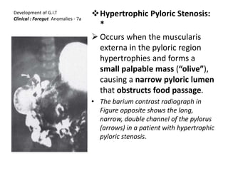
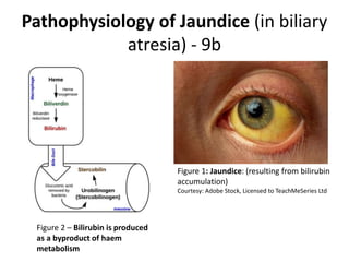
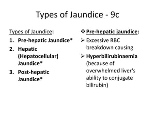
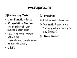
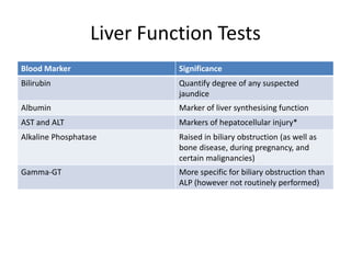
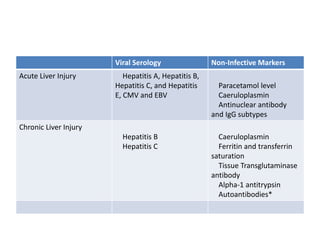
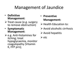
LECTURE SEVEN OVERVIEW
(Lecture 7b)
Gastrointestinal Tract Development
and Malformations
© 2020. Dr. Joseph K. Choge, PhD
Development of G.I.T
Clinical : Foregut Anomalies - 7a
Hypertrophic Pyloric Stenosis:
*
 Occurs when the muscularis
externa in the pyloric region
hypertrophies and forms a
small palpable mass (“olive”),
causing a narrow pyloric lumen
that obstructs food passage.
• The barium contrast radiograph in
Figure opposite shows the long,
narrow, double channel of the pylorus
(arrows) in a patient with hypertrophic
pyloric stenosis.
Hypertrophic Pyloric Stenosis
Clinical Presentation - 7b
• Vomiting, which is:
 Projectile,
 Non-bilious
 Post-prandial (occurs
after feeding)
 Infants treated with
erythromycin have
increased incidence
• O/E:
 Palpable small mass at
the right costal margin
Development of G.I.T
Clinical : Foregut Anomalies – 8a
Developmental anomalies of the gall bladder
anatomy:
Are fairly common in which are found:
Bi-lobed gall bladder
Diverticula and
Septated gall bladder
(the latter likely due to incomplete
recanalization of the gall bladder lumen)
Development of G.I.T
Clinical: Foregut Anomalies – 8b
a. Intrahepatic gall bladder :
 Occurs when the gallbladder rudiment advances
beyond the hepatic diverticulum and becomes buried
within the substance of the liver.
b. Floating gall bladder:
 Occurs when the gallbladder rudiment lags behind the
hepatic diverticulum and thereby becomes suspended
from the liver by a mesentery.
 A floating gall bladder is at risk for torsion (i.e., a
twisting or rotation around the axis of the mesentery).
Development of G.I.T
Clinical : Foregut Anomalies - 9a
c. Developmental anomalies
of the cystic duct anatomy:
 Are fairly common.
• Biliary atresia:
 Defined as the obliteration
of extra-hepatic and/or
intrahepatic ducts.
 Ducts are replaced by
fibrotic tissue due to acute
and chronic inflammation
Clinical Features (Biliary atresia):
 Progressive neonatal jaundice
(onset soon after birth)
Blockage of bile pigments (refer to
haem metabolism by-products)
results in:
 white clay–colored stool (from
missing stercobilin)
 dark-colored urine (from missing
urobilin)
 Average survival time is 12–19
months with a 100% mortality
rate*
Pathophysiology of Jaundice (in biliary
atresia) - 9b
Figure 1: Jaundice: (resulting from bilirubin
accumulation)
Courtesy: Adobe Stock, Licensed to TeachMeSeries Ltd
Figure 2 – Bilirubin is produced
as a byproduct of haem
metabolism
Types of Jaundice - 9c
Types of Jaundice:
1. Pre-hepatic Jaundice*
2. Hepatic
(Hepatocellular)
Jaundice*
3. Post-hepatic
Jaundice*
Pre-hepatic jaundice:
 Excessive RBC
breakdown causing
 Hyperbilirubinaemia
(because of
overwhelmed liver's
ability to conjugate
bilirubin)
Types of Jaundice - 9d
 Hepatocellular Jaundice:
• Due to hepatic cell
dysfunction (from disease)
• Liver loses ability to
conjugate bilirubin
• In liver cirrhosis, liver cells
also compress, causing:
biliary obstruction; mixing
of conjugated &
conjugated bilirubin in
blood
Post-hepatic Jaundice:
• Due to obstruction of
biliary drainage
• Non-excreted bilirubin
will have been
conjugated by the liver,
resulting in conjugated
hyperbilirubinaemia
Common Causes of Jaundice
Pre-hepatic Causes:
• Haemolytic anaemia
• Gilbert’s syndrome
• Criggler-Najjar
syndrome
 Hepatocellular Causes:
• Alcoholic liver disease
• Viral hepatitis
• Iatrogenic, e.g. medication
• Hereditary
haemochromatosis
• Autoimmune hepatitis
• Primary biliary cirrhosis or
primary sclerosing
cholangitis
• Hepatocellular carcinoma
Causes (Continued)
 Post-hepatic Causes:
• Intra-luminal causes:
 Gallstones
• Mural causes:
 Cholangiocarcinoma,
 Biliary strictures,
 Biliary atresia
 Drug-induced cholestasis
• Extra-mural causes,
 Pancreatic cancer or
 Abdominal masses (e.g.
lymphomas)
• Bilirubinuria: bilirubin in urine
• Bilirubinuria can give clues of
type of jaundice
• Conjugated bilirubin can be
excreted via urine (hence
darkening of urine) but
• Unconjugated bilirubin is
insoluble in water (hence
normal urine observed)
• Pale stools: due to reduced
stercobilin entering GIT
Investigations
(1)Laboratory Tests:
• Liver Function Tests
• Coagulation Studies
(PT marker of liver
synthesis function)
• FBC (Anaemia, raised
MCV and
thrombocytopenia seen
in liver disease),
• U&E's
(2) Imaging:
 Abdominal Ultrasound
 Magnetic Resonance
CholangioPancreatogra
phy (MRCP)
(3) Liver Biopsy
Liver Function Tests
Blood Marker Significance
Bilirubin Quantify degree of any suspected
jaundice
Albumin Marker of liver synthesising function
AST and ALT Markers of hepatocellular injury*
Alkaline Phosphatase Raised in biliary obstruction (as well as
bone disease, during pregnancy, and
certain malignancies)
Gamma-GT More specific for biliary obstruction than
ALP (however not routinely performed)
Viral Serology Non-Infective Markers
Acute Liver Injury Hepatitis A, Hepatitis B,
Hepatitis C, and Hepatitis
E, CMV and EBV
Paracetamol level
Caeruloplasmin
Antinuclear antibody
and IgG subtypes
Chronic Liver Injury
Hepatitis B
Hepatitis C
Caeruloplasmin
Ferritin and transferrin
saturation
Tissue Transglutaminase
antibody
Alpha-1 antitrypsin
Autoantibodies*
Management of Jaundice
• Definitive
Management:
 Treat cause (e.g. surgery
to remove obstruction)
• Symptomatic
Management:
 e.g. Anti-histamines for
itching; treat
hypoglycaemia, monitor
coagulopathy (Vitamin
K, FFP prn);
• Preventive
Management:
 Health Education to:
 Avoid alcoholic cirrhosis
 Avoid hepatitis
 etc

Z-L7-B123-GIDb(0).pptx

  • 1.
    LECTURE SEVEN OVERVIEW (Lecture7b) Gastrointestinal Tract Development and Malformations © 2020. Dr. Joseph K. Choge, PhD
  • 2.
    Development of G.I.T Clinical: Foregut Anomalies - 7a Hypertrophic Pyloric Stenosis: *  Occurs when the muscularis externa in the pyloric region hypertrophies and forms a small palpable mass (“olive”), causing a narrow pyloric lumen that obstructs food passage. • The barium contrast radiograph in Figure opposite shows the long, narrow, double channel of the pylorus (arrows) in a patient with hypertrophic pyloric stenosis.
  • 3.
    Hypertrophic Pyloric Stenosis ClinicalPresentation - 7b • Vomiting, which is:  Projectile,  Non-bilious  Post-prandial (occurs after feeding)  Infants treated with erythromycin have increased incidence • O/E:  Palpable small mass at the right costal margin
  • 4.
    Development of G.I.T Clinical: Foregut Anomalies – 8a Developmental anomalies of the gall bladder anatomy: Are fairly common in which are found: Bi-lobed gall bladder Diverticula and Septated gall bladder (the latter likely due to incomplete recanalization of the gall bladder lumen)
  • 5.
    Development of G.I.T Clinical:Foregut Anomalies – 8b a. Intrahepatic gall bladder :  Occurs when the gallbladder rudiment advances beyond the hepatic diverticulum and becomes buried within the substance of the liver. b. Floating gall bladder:  Occurs when the gallbladder rudiment lags behind the hepatic diverticulum and thereby becomes suspended from the liver by a mesentery.  A floating gall bladder is at risk for torsion (i.e., a twisting or rotation around the axis of the mesentery).
  • 6.
    Development of G.I.T Clinical: Foregut Anomalies - 9a c. Developmental anomalies of the cystic duct anatomy:  Are fairly common. • Biliary atresia:  Defined as the obliteration of extra-hepatic and/or intrahepatic ducts.  Ducts are replaced by fibrotic tissue due to acute and chronic inflammation Clinical Features (Biliary atresia):  Progressive neonatal jaundice (onset soon after birth) Blockage of bile pigments (refer to haem metabolism by-products) results in:  white clay–colored stool (from missing stercobilin)  dark-colored urine (from missing urobilin)  Average survival time is 12–19 months with a 100% mortality rate*
  • 7.
    Pathophysiology of Jaundice(in biliary atresia) - 9b Figure 1: Jaundice: (resulting from bilirubin accumulation) Courtesy: Adobe Stock, Licensed to TeachMeSeries Ltd Figure 2 – Bilirubin is produced as a byproduct of haem metabolism
  • 8.
    Types of Jaundice- 9c Types of Jaundice: 1. Pre-hepatic Jaundice* 2. Hepatic (Hepatocellular) Jaundice* 3. Post-hepatic Jaundice* Pre-hepatic jaundice:  Excessive RBC breakdown causing  Hyperbilirubinaemia (because of overwhelmed liver's ability to conjugate bilirubin)
  • 9.
    Types of Jaundice- 9d  Hepatocellular Jaundice: • Due to hepatic cell dysfunction (from disease) • Liver loses ability to conjugate bilirubin • In liver cirrhosis, liver cells also compress, causing: biliary obstruction; mixing of conjugated & conjugated bilirubin in blood Post-hepatic Jaundice: • Due to obstruction of biliary drainage • Non-excreted bilirubin will have been conjugated by the liver, resulting in conjugated hyperbilirubinaemia
  • 10.
    Common Causes ofJaundice Pre-hepatic Causes: • Haemolytic anaemia • Gilbert’s syndrome • Criggler-Najjar syndrome  Hepatocellular Causes: • Alcoholic liver disease • Viral hepatitis • Iatrogenic, e.g. medication • Hereditary haemochromatosis • Autoimmune hepatitis • Primary biliary cirrhosis or primary sclerosing cholangitis • Hepatocellular carcinoma
  • 11.
    Causes (Continued)  Post-hepaticCauses: • Intra-luminal causes:  Gallstones • Mural causes:  Cholangiocarcinoma,  Biliary strictures,  Biliary atresia  Drug-induced cholestasis • Extra-mural causes,  Pancreatic cancer or  Abdominal masses (e.g. lymphomas) • Bilirubinuria: bilirubin in urine • Bilirubinuria can give clues of type of jaundice • Conjugated bilirubin can be excreted via urine (hence darkening of urine) but • Unconjugated bilirubin is insoluble in water (hence normal urine observed) • Pale stools: due to reduced stercobilin entering GIT
  • 12.
    Investigations (1)Laboratory Tests: • LiverFunction Tests • Coagulation Studies (PT marker of liver synthesis function) • FBC (Anaemia, raised MCV and thrombocytopenia seen in liver disease), • U&E's (2) Imaging:  Abdominal Ultrasound  Magnetic Resonance CholangioPancreatogra phy (MRCP) (3) Liver Biopsy
  • 13.
    Liver Function Tests BloodMarker Significance Bilirubin Quantify degree of any suspected jaundice Albumin Marker of liver synthesising function AST and ALT Markers of hepatocellular injury* Alkaline Phosphatase Raised in biliary obstruction (as well as bone disease, during pregnancy, and certain malignancies) Gamma-GT More specific for biliary obstruction than ALP (however not routinely performed)
  • 14.
    Viral Serology Non-InfectiveMarkers Acute Liver Injury Hepatitis A, Hepatitis B, Hepatitis C, and Hepatitis E, CMV and EBV Paracetamol level Caeruloplasmin Antinuclear antibody and IgG subtypes Chronic Liver Injury Hepatitis B Hepatitis C Caeruloplasmin Ferritin and transferrin saturation Tissue Transglutaminase antibody Alpha-1 antitrypsin Autoantibodies*
  • 15.
    Management of Jaundice •Definitive Management:  Treat cause (e.g. surgery to remove obstruction) • Symptomatic Management:  e.g. Anti-histamines for itching; treat hypoglycaemia, monitor coagulopathy (Vitamin K, FFP prn); • Preventive Management:  Health Education to:  Avoid alcoholic cirrhosis  Avoid hepatitis  etc

Editor's Notes

  • #3 Stomach (Pylorus) Anomalies: Hypertrophic pyloric stenosis (Figure 10.8): Hypertrophic pyloric stenosis occurs when the muscularis externa in the pyloric region hypertrophies and forms a small palpable mass (“olive”), causing a narrow pyloric lumen that obstructs food passage. It is associated clinically with projectile, non-bilious vomiting after feeding and a small, palpable mass at the right costal margin. Increased incidence has been found in infants treated with the antibiotic erythromycin. The barium contrast radiograph in Figure 10.8 shows the long, narrow, double channel of the pylorus (arrows) in a patient with hypertrophic pyloric stenosis. ======================================================================== Clinical box: Congenital Hypertrophic Pyloric Stenosis In the condition known as congenital hypertrophic pyloric stenosis the smooth muscle of the pylorus is overdeveloped, thereby causing an obstruction. The stenosis can be relieved surgically. Failure of recanalization of the duodenum results in duodenal stenosis, and in projectile vomiting after birth. Complete occlusion of the intestinal lumen or atresia can occur anywhere along the length of the intestine.
  • #5 3. Clinical considerations a. Developmental anomalies of the gall bladder (Figure 10.10) anatomy are fairly common in which two, bilobed, diverticula, and septated gall bladders are found. Figure 10.10 shows the developmental anomalies of the gall bladder. Septated gall bladder is most likely due to incomplete recanalization of the lumen. b. Developmental anomalies of the cystic duct (Figure 10.11) anatomy are fairly common. Figure 10.11 shows various developmental anomalies of the cystic duct. c. Biliary atresia (Figure 10.12) is defined as the obliteration of extrahepatic and/or intrahepatic ducts. The ducts are replaced by fibrotic tissue due to acute and chronic inflammation. It is associated clinically with progressive neonatal jaundice with onset soon after birth, white clay–colored stool, and dark-colored urine. The average survival time is 12–19 months with a 100% mortality rate. Figure 10.12 shows different forms of extrahepatic biliary atresia. d. Intrahepatic gall bladder occurs when the gallbladder rudiment advances beyond the hepatic diverticulum and becomes buried within the substance of the liver. e. Floating gall bladder occurs when the gallbladder rudiment lags behind the hepatic diverticulum and thereby becomes suspended from the liver by a mesentery. A floating gall bladder is at risk for torsion (i.e., a twisting or rotation around the axis of the mesentery).
  • #6 3. Clinical considerations a. Developmental anomalies of the gall bladder (Figure 10.10) anatomy are fairly common in which two, bilobed, diverticula, and septated gall bladders are found. Figure 10.10 shows the developmental anomalies of the gall bladder. Septated gall bladder is most likely due to incomplete recanalization of the lumen. b. Developmental anomalies of the cystic duct (Figure 10.11) anatomy are fairly common. Figure 10.11 shows various developmental anomalies of the cystic duct. c. Biliary atresia (Figure 10.12) is defined as the obliteration of extrahepatic and/or intrahepatic ducts. The ducts are replaced by fibrotic tissue due to acute and chronic inflammation. It is associated clinically with progressive neonatal jaundice with onset soon after birth, white clay–colored stool, and dark-colored urine. The average survival time is 12–19 months with a 100% mortality rate. Figure 10.12 shows different forms of extrahepatic biliary atresia. d. Intrahepatic gall bladder occurs when the gallbladder rudiment advances beyond the hepatic diverticulum and becomes buried within the substance of the liver. e. Floating gall bladder occurs when the gallbladder rudiment lags behind the hepatic diverticulum and thereby becomes suspended from the liver by a mesentery. A floating gall bladder is at risk for torsion (i.e., a twisting or rotation around the axis of the mesentery).
  • #7 3. Clinical considerations a. Developmental anomalies of the gall bladder (Figure 10.10) anatomy are fairly common in which two, bilobed, diverticula, and septated gall bladders are found. Figure 10.10 shows the developmental anomalies of the gall bladder. Septated gall bladder is most likely due to incomplete recanalization of the lumen. b. Developmental anomalies of the cystic duct (Figure 10.11) anatomy are fairly common. Figure 10.11 shows various developmental anomalies of the cystic duct. c. Biliary atresia (Figure 10.12) is defined as the obliteration of extrahepatic and/or intrahepatic ducts. The ducts are replaced by fibrotic tissue due to acute and chronic inflammation. It is associated clinically with progressive neonatal jaundice with onset soon after birth, white clay–colored stool, and dark-colored urine. The average survival time is 12–19 months with a 100% mortality rate. Figure 10.12 shows different forms of extrahepatic biliary atresia. d. Intrahepatic gall bladder occurs when the gallbladder rudiment advances beyond the hepatic diverticulum and becomes buried within the substance of the liver. e. Floating gall bladder occurs when the gallbladder rudiment lags behind the hepatic diverticulum and thereby becomes suspended from the liver by a mesentery. A floating gall bladder is at risk for torsion (i.e., a twisting or rotation around the axis of the mesentery).
  • #8 Pathophysiology of Jaundice Jaundice refers to the yellow discolouration of the sclera and skin (Fig. 1) that is due to hyperbilirubinaemia, occurring at bilirubin levels roughly greater than 50 µmol/L. Jaundice results from high levels of bilirubin in the blood. Bilirubin is the normal breakdown product from the catabolism of haem, and thus is formed from the destruction of red blood cells. Under normal circumstances, bilirubin undergoes conjugation within the liver, making it water-soluble. It is then excreted via the bile into the GI tract, the majority of which is egested in the faeces as urobilinogen and stercobilin (the metabolic breakdown product of urobilingoen). Around 10% of urobilinogen is reabsorbed into the bloodstream and excreted through the kidneys. Jaundice occurs when this pathway is disrupted. By Johndheathcote [CC BY-SA 3.0], via Wikimedia Commons Fig 1 - Bilirubin is produced as a byproduct of haem metabolism. Figure 2 – Bilirubin is produced as a byproduct of haem metabolism
  • #9 Types of Jaundice There are three main types of jaundice: pre-hepatic, hepatocellular, and post-hepatic. Pre-Hepatic Jaundice: In pre-hepatic jaundice, there is excessive red cell breakdown which overwhelms the liver’s ability to conjugate bilirubin. This causes an unconjugated hyperbilirubinaemia. Any bilirubin that manages to become conjugated will be excreted normally, yet it is the unconjugated bilirubin that remains in the blood stream to cause the jaundice. Hepatocellular Jaundice: In hepatocellular (or intrahepatic) jaundice, there is dysfunction of the hepatic cells. The liver loses the ability to conjugate bilirubin, but in cases where it also may become cirrhotic, it compresses the intra-hepatic portions of the biliary tree to cause a degree of obstruction. This leads to both unconjugated and conjugated bilirubin in the blood, termed a ‘mixed picture’. Post-Hepatic Post-hepatic jaundice refers to obstruction of biliary drainage. The bilirubin that is not excreted will have been conjugated by the liver, hence the result is a conjugated hyperbilirubinaemia.
  • #10 Types of Jaundice There are three main types of jaundice: pre-hepatic, hepatocellular, and post-hepatic. Pre-Hepatic Jaundice: In pre-hepatic jaundice, there is excessive red cell breakdown which overwhelms the liver’s ability to conjugate bilirubin. This causes an unconjugated hyperbilirubinaemia. Any bilirubin that manages to become conjugated will be excreted normally, yet it is the unconjugated bilirubin that remains in the blood stream to cause the jaundice. Hepatocellular Jaundice: In hepatocellular (or intrahepatic) jaundice, there is dysfunction of the hepatic cells. The liver loses the ability to conjugate bilirubin, but in cases where it also may become cirrhotic, it compresses the intra-hepatic portions of the biliary tree to cause a degree of obstruction. This leads to both unconjugated and conjugated bilirubin in the blood, termed a ‘mixed picture’. Post-Hepatic Jaundice: Post-hepatic jaundice refers to obstruction of biliary drainage. The bilirubin that is not excreted will have been conjugated by the liver, hence the result is a conjugated hyperbilirubinaemia.
  • #12 Bilirubinuria A good estimation of which type of jaundice is present (prior to any further investigation) can be made from observing the colour of the urine. Conjugated bilirubin can be excreted via the urine (as it is water soluble), whereas unconjugated cannot. Consequently, dark (‘coca-cola’) urine manifests in conjugated or mixed hyperbilirubinaemias, whereas normal urine is seen in unconjugated disease. Moreover, those with an obstructive picture will likely note pale stools, due to the reduced levels of stercobilin entering the GI tract, which normally colours the stool. Investigations In many cases, the likely underlying cause can be elicited from the history, with the investigations simply confirming suspicions. Hence, whilst a complete list of investigations is given below, these should be tailored to the clinical features of the patient. Laboratory Tests Any patient presenting with jaundice should have the following bloods taken: Liver function tests (LFTs), as summarised in Table 2 Coagulation studies (PT can be used as a marker of liver synthesis function) FBC (anaemia, raised MCV, and thrombocytopenia all seen in liver disease) and U&Es Specialist blood tests, as summarised below as part of a liver screen Imaging The imaging used will depend on the presumed aetiology. An ultrasound abdomen is usually first line, identifying any obstructive pathology present or gross liver pathology (albeit often user dependent). Magnetic Resonance Cholangiopancreatography (MRCP) is used to visual the biliary tree, typically performed if the jaundice is obstructive, but US abdomen was inconclusive or limited, or as further work-up for surgical intervention. A liver biopsy can be performed when the diagnosis has not been made despite the above investigations.
  • #13 Investigations In many cases, the likely underlying cause can be elicited from the history, with the investigations simply confirming suspicions. Hence, whilst a complete list of investigations is given below, these should be tailored to the clinical features of the patient. Laboratory Tests Any patient presenting with jaundice should have the following bloods taken: Liver function tests (LFTs), as summarised in Table 2 Coagulation studies (PT can be used as a marker of liver synthesis function) FBC (anaemia, raised MCV, and thrombocytopenia all seen in liver disease) and U&Es Specialist blood tests, as summarised below as part of a liver screen Imaging The imaging used will depend on the presumed aetiology. An ultrasound abdomen is usually first line, identifying any obstructive pathology present or gross liver pathology (albeit often user dependent). Magnetic Resonance Cholangiopancreatography (MRCP) is used to visual the biliary tree, typically performed if the jaundice is obstructive, but US abdomen was inconclusive or limited, or as further work-up for surgical intervention. A liver biopsy can be performed when the diagnosis has not been made despite the above investigations.
  • #14 Table 2 – LFT serum markers. *as an estimate, if the AST:ALT ratio >2, this is likely alcoholic liver disease, whilst if AST:ALT is around 1, then likely viral hepatitis as the cause Liver Screen A liver screen can be performed for patients whereby there is no initial cause for liver dysfunction, tailored to whether acute or chronic liver failure
  • #15 Table 3 – Acute and Chronic Liver Screens *Autoantibodies include anti-mitochondrial antibody (AMA), anti-smooth-muscle antibody (Anti-SMA), and anti-nuclear antibody (ANA), used to identify a variety of autoimmune liver conditions, such as primary sclerosing cholangitis (PSC)
  • #16 Management The definitive treatment of jaundice will be dependent on the underlying cause. Obstructive causes may require removal of a gallstone through Endoscopic Retrograde CholangioPancreatography (ERCP) or stenting of the common bile duct. Symptomatic treatment is often needed for the itching caused by hyperbilirubinaemia. An obstructive cause may warrant cholestyramine (acting to increase biliary drainage), whilst other causes may respond to simple anti-histamines. Identify and manage any complications where possible. Monitor for coagulopathy, treating promptly (either vitamin K or fresh frozen plasma (FFP) is needed) if any evidence of bleeding or rapid coagulopathy, and treat hypoglycaemia orally if possible (otherwise 5% dextrose is needed). Where patients become confused from decompensating chronic liver disease (‘hepatic encephalopathy’), laxatives (lactulose or senna) +/- neomycin or rifaximin may be used, in attempt to reduce the number of ammonia-producing bacteria in the bowel.