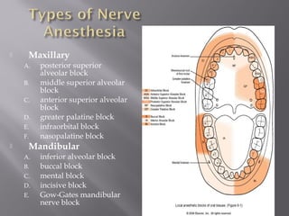
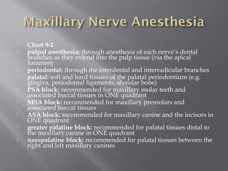
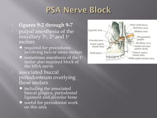
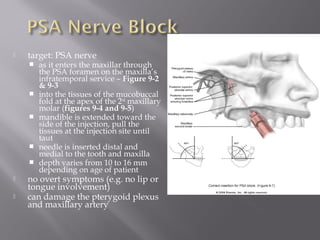
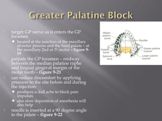
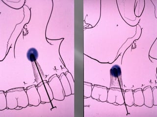
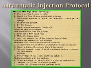
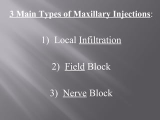
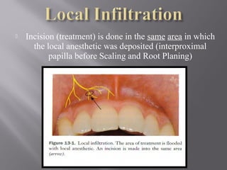
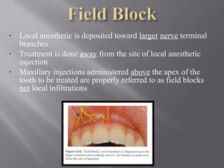
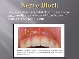
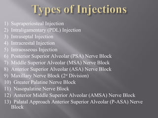
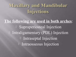
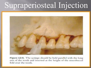
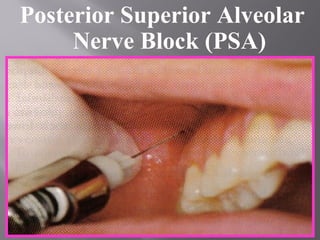
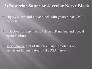
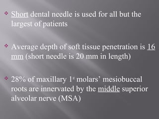
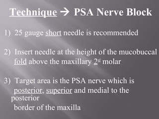
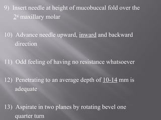
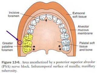
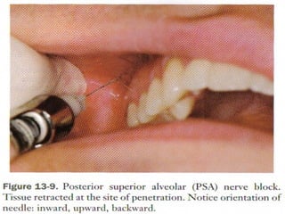
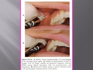
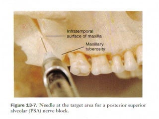
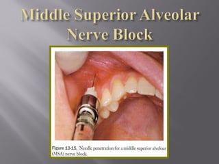
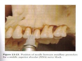
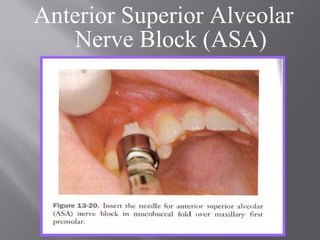
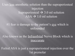
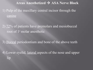
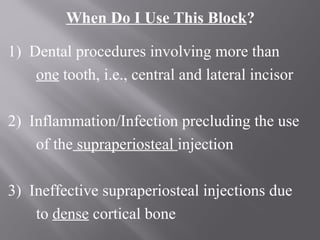
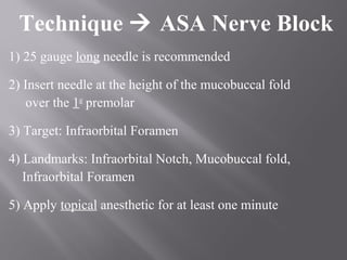
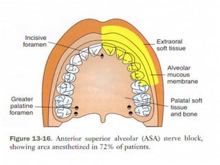
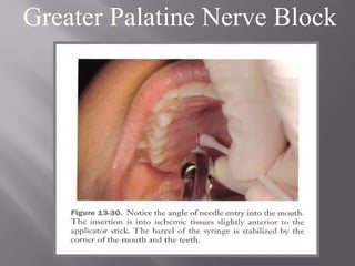
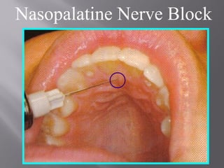
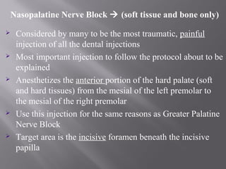
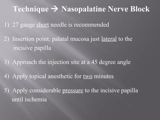
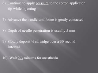
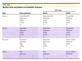
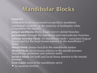
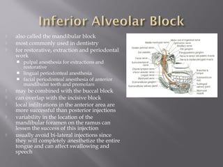
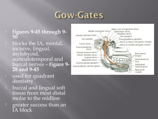
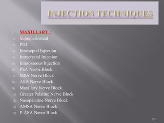
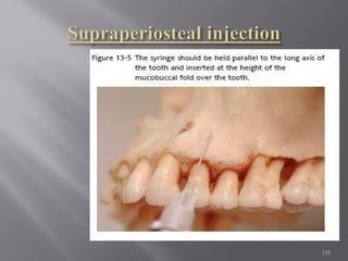
www.drhadihoseini.com
:
http://www.slideshare.net/hadidezyan
1. Local infiltration
- type of injection that anesthetizes a small area (one
or two teeth and asscociated areas)
- anesthesia deposited at nerve terminals
1. Nerve block
- type of injection that anesthetizes a larger area
- anesthesia deposited near larger nerve trunks
 Methods:
 Reducing temperature.
 Is used only to produce surface anaesthesia e.g. ethyl chloride
spray.
 Physical damage to nerve trunk e.g. nerve sectioning.
 Unsafe for therapeutic uses, only in Trigeminal Neuralgia.
 Chemical damage to nerve trunk e.g. neurolytic agents.
 Silver nitrate, Phenol - Unsafe for therapeutic use.
 Methods: Cont
 Anoxia or hypoxia resulting in lack of oxygen to
nerve.
 Unsafe as well.
 Stimulation of large nerve fibres, blocking the
perception of smaller diameter fibres.
 includes Acupuncture and TENS (Transcutaneous
Electronic Nerve Stimulation)
 Drugs that block transmission at sensory nerve
endings or along nerve fibres.
 There action is fully reversible and without permanent
damage to the tissues.
 Classified according to their chemical structures and
the determining factor is the intermediate chain, into
two groups:
Ester Amide
 They differ in two important respect:
 Their ability to induce hypersensitivity reaction.
 Their pharmacokinetics - fate and metabolism.
 Maxillary
A. posterior superior
alveolar block
B. middle superior alveolar
block
C. anterior superior alveolar
block
D. greater palatine block
E. infraorbital block
F. nasopalatine block
 Mandibular
A. inferior alveolar block
B. buccal block
C. mental block
D. incisive block
E. Gow-Gates mandibular
nerve block
 dental procedures can usually commence after
3 – 5 minutes
 failure requires re-administration using
another method
 never re-administer using the same method
 keep in mind the total # of injections and the
dosages
 never inject into an area with an abcess, or
other type of abnormality
 Chart 9-1
 pulpal anesthesia: through anesthesia of each nerve’s dental
branches as they extend into the pulp tissue (via the apical
foramen)
 periodontal: through the interdental and interradicular branches
 palatal: soft and hard tissues of the palatal periodontium (e.g.
gingiva, periodontal ligaments, alveolar bone)
 PSA block: recommended for maxillary molar teeth and
associated buccal tissues in ONE quadrant
 MSA block: recommended for maxillary premolars and
associated buccal tissues
 ASA block: recommended for maxillary canine and the incisors in
ONE quadrant
 greater palatine block: recommended for palatal tissues distal to
the maxillary canine in ONE quadrant
 nasopalatine block: recommended for palatal tissues between the
right and left maxillary canines
 figures 9-2 through 9-7
 pulpal anesthesia of the
maxillary 3rd
, 2nd
and 1st
molars
 required for procedures
involving two or more molars
 sometimes anesthesia of the 1st
molar also required block of
the MSA nerve
 associated buccal
periodonteum overlying
these molars
 including the associated
buccal gingiva, periodontal
ligament and alveolar bone
 useful for periodontal work
on this area
 target: PSA nerve
 as it enters the maxillar through
the PSA foramen on the maxilla’s
infratemporal service – Figure 9-2
& 9-3
 into the tissues of the mucobuccal
fold at the apex of the 2nd
maxillary
molar (figures 9-4 and 9-5)
 mandible is extended toward the
side of the injection, pull the
tissues at the injection site until
taut
 needle is inserted distal and
medial to the tooth and maxilla
 depth varies from 10 to 16 mm
depending on age of patient
 no overt symptoms (e.g. no lip or
tongue involvement)
 can damage the pterygoid plexus
and maxillary artery
 limited clinical usefulness
 can be used to extend the infraorbital
block distal to the maxillary canine
 can be indicated for work on maxillary
pre-molars and mesiobuccal root of 1st
molar (Figure 9-8)
 if the MSA is absent – area is innervated
by the ASA
 blocks the pulp tissue of the 1st
and 2nd
maxillary premolars and possibly the 1st
molar + associated buccal tissues and
alveolar bone
 useful for periodontal work in this area
 to block the palatine tissues in this area
– may require a greater palatine block
 target area: MSA nerve at the apex of the
maxillary 2nd
premolar (figures 9-8 and 9-9)
 mandible extended towards injection site
 stretch the upper lip to tighten the injection site
 needle is inserted into the mucobuccal fold
 tip is located well above the apex of the 2nd
premolar
 figure 9-11
 harmless tingling or numbness of the upper lip
 overinsertion is rare
 figures 9-12 through 9-14
 can be considered a local
infiltration
 used in conjunction with an MSA
block
 the ASA nerve can cross the
midline of the maxilla onto the
opposite side!
 used in procedures involving the
maxillary canines and incisors and
their associated facial tissues
 pulpal and facial tissues involved –
restorative and periodontal work
 blocks the pulp tissue + the
gingiva, periodontal ligaments
and alveolar bone in that area
 target: ASA nerve at the apex of the maxillary
canine – figures 9-12 & 9-13
 at the mucobuccal fold at the apex of the
maxillary canine – figure 9-13
 harmless tingling or numbness of the upper lip
 overinsertion is rare
 figures 9-15 through 9-17
 anesthetizes both the MSA and
ASA
 used for anesthesia of the
maxillary premolars, canine and
incisors
 indicated when more than one
premolar or anterior teeth
 pulpal tissues – for restorative work
 facial tissues – for periodontal work
 also numbs the gingiva,
periodontal ligaments and
alveolar bone in that area
 the maxillary central incisor may
also be innervated by the
nasopalatine nerve branches
 target: union of the ASA and MSA with the IO nerve
after the IO enters the IO foramen – figure 9-15
 also anesthesizes the lower eyelid, side of nose and
upper lip
 IO foramen is gently palpated along the IO rim
 move slightly down about 10mm until you feel the depression
of the IO foramen – figure 9-16
 locate the tissues at the mucobuccal fold at the apex of the 1st
premolar
 place one finger at the IO foramen and the other on the injection site
– figure 9-17
 locate the IO foramen, retract the upper lip and pull the tissues taut
 the needle is inserted parallel to the long axis of the tooth to avoid
hitting the bone
 harmless tingling or numbness of the upper lip, side of
nose and eyelid
 figures 9-19 through 9-21
 used in restorative procedures that involve more than
two maxillary posterior teeth or palatal tissues distal to
the canine
 also used in periodontal work – since it blocks the
associated lingual tissues
 anesthetizes the posterior portion of the hard palate –
from the 1st
premolar to the molars and medially to the
palate midline
 does NOT provide pulpal anesthesia – may also need
to use ASA, PSA, MSA or IO blocks
 may also need to be combined with nasopalatine block
 target: GP nerve as it enters the GP
foramen
 located at the junction of the maxillary
alveolar process and the hard palate – at
the maxillary 2nd or 3rd
molar – figure 9-
19
 palpate the GP foramen – midway
between the median palatine raphe
and lingual gingival margin of the
molar tooth – figure 9-21
 can reduce discomfort by applying
pressure to the site before and during
the injection
 produces a dull ache to block pain
impulses
 also slow deposition of anesthesia will
also help
 needle is inserted at a 90 degree angle
to the palate – figure 9-22
 figure 9-23 through 9-26
 useful for anesthesia of the bilateral portion of the hard
palate
 from the mesial of the right maxillary 1st
premolar to the mesial
of the left 1st
premolar
 for palatal soft tissue anesthesia
 periodontal treatment
 required for two or more anterior maxillary teeth
 for restorative procedures or extraction of the anterior
maxillary teeth – may need an ASA or MSA block also
 blocks both right and left nerves
 target: both right and left nerves as they enter the incisive foramen
from the mucosa of the anterior hard palate – figure 9-23 & 9-25
 posterior to the incisive papilla
 injection site is lateral to the incisive papilla – figure 9-26
 head turned to the left or right
 inserted at a 45 degree angle about 6-10 mm – gently contact the
maxillary bone and withdraw about 1mm before administering
 can reduce discomfort by applying pressure to the site before and
during the injection
 produces a dull ache to block pain impulses
 also slow deposition of anesthesia will also help
 can anesthetize the labial tissues between the central incisors prior
to palatal block
 can block some branches of the nasopalatine prior to injection
3 Main Types of Maxillary Injections:
1) Local Infiltration
2) Field Block
3) Nerve Block
 Incision (treatment) is done in the same area in which
the local anesthetic was deposited (interproximal
papilla before Scaling and Root Planing)
• Local anesthetic is deposited toward larger nerve terminal
branches
• Treatment is done away from the site of local anesthetic
injection
• Maxillary injections administered above the apex of the
tooth to be treated are properly referred to as field blocks
not local infiltrations
• Local anesthetic is deposited close to a main nerve
trunk, usually at a site removed from the area of
treatment (PSA, IANB, NPB)
1) Supraperiosteal Injection
2) Intraligamentary (PDL) Injection
3) Intraseptal Injection
4) Intracrestal Injection
5) Intraosseous Injection
6) Posterior Superior Alveolar (PSA) Nerve Block
7) Middle Superior Alveolar (MSA) Nerve Block
8) Anterior Superior Alveolar (ASA) Nerve Block
9) Maxillary Nerve Block (2nd
Division)
10) Greater Palatine Nerve Block
11) Nasopalatine Nerve Block
12) Anterior Middle Superior Alveolar (AMSA) Nerve Block
13) Palatal Approach Anterior Superior Alveolar (P-ASA) Nerve
Block
The following are used in both arches:
 Supraperiosteal Injection
 Intraligamentary (PDL) Injection
 Intraseptal Injection
 Intraosseous Injection
Supraperiosteal Injection
1) Supraperiosteal Injection
 Used for pulpal anthesia in maxillary teeth
 Anesthetizes large terminal branches of the
dental plexus
 Greater than 95% success rate
 1 or 2 teeth
 Dense bone covering the apices of the teeth can lead to
failure
-maxillary molar of children (zygomatic bone
obscures)
-central incisor of adults (nasal spine obscures)
 Negligible positive aspiration rate (less than 1%)
 Should not be used for large areas (multiple sticks/large
amount of local anesthetic solution must be used)
Technique  Supraperiosteal
Injection
1) 25 or 27 gauge short needle is recommended
2) Insert needle at height of mucobuccal fold
over apex of desired tooth
3) Apply topical anesthetic for at least one
minute
4) Orient bevel toward bone; lift lip pulling
tissues taut
5) Hold syringe parallel to long axis of the tooth
being anesthetized
6) No resistance to penetration should be felt and no
patient discomfort
7) Aspirate twice
8) Deposit .6 ml (one-third of a cartridge) into tissue
over 20 seconds
9) Do not allow tissues to balloon
10) Wait 3 to 5 minutes to begin dental treatment
Problems/Failures
 If tooth does not anesthetize the needle tip could be below
the apex of the tooth resulting in inadequate anesthesia
 If the needle lies too far from the bone then anesthesia will
be inadequate because the solution was deposited in the
soft tissue (lip)
 The needle must be oriented toward the periosteum but
should be managed properly to avoid tearing the highly
innervated periosteum
These two words are used incorrectly;
what most practitioners refer to as an
infiltration injection is actually a field
block
Posterior Superior Alveolar
Nerve Block (PSA)
2) Posterior Superior Alveolar Nerve Block
 Highly successful nerve block with greater than 95%
success
 Effective for maxillary 1st
, 2nd
and 3rd
molars and buccal
periodontium
 Mesiobuccal root of the maxillary 1st
molar is not
consistently innervated by the PSA nerve
 Short dental needle is used for all but the
largest of patients
 Average depth of soft tissue penetration is 16
mm (short needle is 20 mm in length)
 28% of maxillary 1st
molars’ mesiobuccal
roots are innervated by the middle superior
alveolar nerve (MSA)
 When the risk of hemorrhage is too great as
with a hemophiliac, you should use the
supraperiosteal or PDL injections
 Patient should feel no pain with this injection
because bone is not contacted and there is a
large area of soft tissue into which the
solution is deposited
 Positive aspiration risk is 3.1%
 Patient will often say that they do not feel
numb; reason why is because they are
accustomed to the intense feeling of
anesthesia experienced by the IANB;
reassure patient that you are going to make
sure they are comfortable during the
procedure
Technique  PSA Nerve Block
1) 25 gauge short needle is recommended
2) Insert needle at the height of the mucobuccal
fold above the maxillary 2nd
molar
3) Target area is the PSA nerve which is
posterior, superior and medial to the
posterior
border of the maxilla
4) Apply topical anesthetic for at least one minute
5) Have patient open their mouth half way which
makes more room
6) Retract the patient’s cheek with mirror
7) Pull the tissues taut
8) Orient bevel toward bone
9) Insert needle at height of mucobuccal fold over the
2nd
maxillary molar
10) Advance needle upward, inward and backward
direction
11) Odd feeling of having no resistance whatsoever
12) Penetrating to an average depth of 10-14 mm is
adequate
13) Aspirate in two planes by rotating bevel one
quarter turn
14) Deposit 0.9 ml of a cartridge (1/2
cartridge)
15) Wait 3 to 5 minutes to start treatment
Advance the needle in one movement, not three
separate movements; usually atraumatic to most patients
Problems/Failures (PSA)
 Hematoma formation if needle is overinserted too far
posteriorly
 Pterygoid plexus of veins leads to this hematoma
 Visible intraoral hematoma develops within minutes;
bleeds until the pressure of the extravascular blood equals
that of the intravascular blood which can result in a large,
unsightly hematoma
 Patients will usually claim that they do not
feel any anesthesia which is not uncommon
because patients can not reach this area to
gauge their own level of anesthesia
 If using a long dental needle the maximum
insertion should be one-half on its length or
16 mm
Problems/Failures (PSA)
Middle Superior Alveolar
Nerve Block (MSA)
3) Middle Superior Alveolar Nerve Block
 Middle Superior Alveolar Nerve is not present in 28% of
the population
 When the infraorbital nerve block fails to provide
anesthesia to teeth distal to the maxillary canines, the
MSA is indicated
 MSA provides anesthesia to 1st
and 2nd
premolars and
mesiobuccal root of maxillary 1st
molar; anesthetizes
buccal periodontium and bone
 If MSA is absent the premolars and mesiobuccal root of
maxillary 1st
molar is innervated by the ASA
 Positive aspiration risk is less than 3% (negligible)
 Infraorbital nerve block can block 1st
premolar, 2nd
premolar
and mesiobuccal root of the maxillary 1st
molar if you need
an alternative block when the MSA is not adequate
Technique  MSA Nerve Block
1) 25 or 27 gauge long or short needle
2) Insert needle at the height of the mucobuccal
fold above 2nd
maxillary premolar
3) Target is the maxillary bone above the
apex of the 2nd
maxillary premolar
4) Orient bevel toward bone to avoid
tearing periosteum
5) Apply topical anesthetic for one minute
6) Pull tissues taut
7) Penetrate tissues placing bevel of needle
well above the apex of the 2nd
maxillary
premolar
Technique- Middle Superior Alveolar Nerve Block
8) Aspirate
9) Slowly deposit 0.9-1.2 ml of solution
10) Wait 3 to 5 minutes before starting
treatment
Problems/Failures MSA
 Anesthetic not deposited above the apex of
the 2nd
premolar
 Solution deposited into the soft tissue too far
from the periosteum (lip)
 Hematoma may develop; Dentist should
apply pressure to the area with gauze for at
least sixty (60) seconds; up to 2 to 3 minutes
Anterior Superior Alveolar
Nerve Block (ASA)
 Highly successful extremely safe block that
causes hesitation in most clinicians
 Provides profound pulpal and soft tissue
anesthesia from the maxillary central incisor
distal to the premolars in 72% of patients
 Used in place of the supraperiosteal injection
Uses less anesthetic solution than the supraperiosteal
injection
Supraperiosteal  3.0 ml solution
ASA  1.0 ml solution
#1 fear is damage to the patient’s eye which is
unfounded
Also known as the Infraorbital Nerve Block which is
inaccurate
Failed ASA is just a supraperiosteal injection over the
1st premolar
Areas Anesthetized  ASA Nerve Block
1) Pulp of the maxillary central incisor through the
canine
2) 72% of patients have premolars and mesiobuccal
root of 1st
molar anesthetic
3) Buccal periodontium and bone of the above teeth
4) Lower eyelid, lateral aspects of the nose and upper
lip
When Do I Use This Block?
1) Dental procedures involving more than
one tooth, i.e., central and lateral incisor
2) Inflammation/Infection precluding the use
of the supraperiosteal injection
3) Ineffective supraperiosteal injections due
to dense cortical bone
Technique  ASA Nerve Block
1) 25 gauge long needle is recommended
2) Insert needle at the height of the mucobuccal fold
over the 1st
premolar
3) Target: Infraorbital Foramen
4) Landmarks: Infraorbital Notch, Mucobuccal fold,
Infraorbital Foramen
5) Apply topical anesthetic for at least one minute
6) Feel the infraorbital notch moving your finger
down the notch palpating the tissues gently; the
outward bulge is the lower border of the orbit
which is the roof of the infraorbital foramen;
continue the finger inferiorly until a depression
is felt which is the infraorbital foramen
7) Maintain pressure over the foramen while
inserting the needle down the long axis of the
1st
premolar
8) Advance the needle slowly until bone is contacted
gently which is the upper rim of the infraorbital
foramen
9) 16 mm total advancement of needle;1/2 of long
needle length
10) Estimate the distance between the infraorbital
foramen and mucobuccal fold
11) Aspirate
12) Deposit 1.0 ml of anesthetic solution
13) Administrator can feel the anesthetic expanding
the tissue with finger tip
14) Maintain finger pressure over the foramen for
at least one minute to disperse the anesthetic
solution
15) Needle should not be palpable in most patients
16) Wait 3 to 5 minutes for anesthesia to result
Problems/Failures (ASA)
 Failure is from the needle deviating to the medial or lateral
away from the infraorbital foramen
 Failure to reach the infraorbital foramen will result in
anesthesia of the lateral side of the nose, upper lip and
lower eyelid but not the teeth
 Hematoma formation can result although rarely; apply
pressure to area for 2 to 3 minutes; at least 60 seconds
Palatal Anesthesia
Palatal Anesthesia
 Easily one of the most traumatic experiences
for dentists due to the pain that is sometimes
elicited from the patients
 Palatal injections can be administered
atraumatically
STEPS- Results in painless palatal injections
1) Apply topical for two minutes
2) Apply pressure to site both before and
during deposition of the solution
3) Deposit solution slowly
5 PALATAL INJECTIONS
1) Anterior (Greater) Palatine Nerve Block:
no pulpal anesthesia
2) Nasopalatine Nerve Block: no pulpal anesthesia
3) Local Infiltration: no pulpal anesthesia
4) P-AMSA: pulpal and soft tissue
5) P-ASA: pulpal and soft tissue
Greater Palatine Nerve Block
GP Nerve Block (soft tissue and bone only)
Anesthetizes palatal soft tissue distal and
medially to the canine
(posterior portion of the palate)
Tissues around the Greater Palatine Foramen
are able to accommodate a larger volume of
solution than the tissue in the vicinity of the
Nasopalatine Foramen  less patient
discomfort
Indications for palatal injections:
1) Scaling and root planing
2) Subgingival restorations
3) Deep placed matrix bands
4) Extractions (oral surgery)
Technique  Greater Palatine Nerve Block
1) 27 gauge short needle
2) Insert needle in soft tissue slightly anterior
to the greater palatine foramen
3) Target is the greater palatine nerve as it
passes from the foramen between the soft
tissue and bone of the hard palate
Locate the Greater Palatine Foramen:
-use cotton swab/mirror handle
-place a cotton swab at the junction of the
maxillary alveolar process and the hard palate
-press firmly into tissues moving posteriorly
from the maxillary 1st
molar
-swab “falls” into the depression of the
greater palatine foramen
4) Foramen is most often located distal to the
2nd
maxillary molar
5) Apply considerable pressure to cotton swab
in area of foramen until a noticeable ischemia
occurs; hold pressure for 30 seconds before
injection
6) Continue to apply pressure throughout the
injection with the cotton swab
7) Slowly advance the needle until bone is
gently contacted
8) Depth of penetration is usually less than 10 mm
9) Aspirate
10) Deposit solution very slowly
Do not enter the greater palatine canal
There is no reason to have the needle
penetrate the canal
There is no negative repercussion except
post-operative pain
Nasopalatine Nerve Block
Nasopalatine Nerve Block  (soft tissue and bone only)
 Considered by many to be the most traumatic, painful
injection of all the dental injections
 Most important injection to follow the protocol about to be
explained
 Anesthetizes the anterior portion of the hard palate (soft
and hard tissues) from the mesial of the left premolar to
the mesial of the right premolar
 Use this injection for the same reasons as Greater Palatine
Nerve Block
 Target area is the incisive foramen beneath the incisive
papilla
Technique  Nasopalatine Nerve Block
1) 27 gauge short needle is recommended
2) Insertion point: palatal mucosa just lateral to the
incisive papilla
3) Approach the injection site at a 45 degree angle
4) Apply topical anesthetic for two minutes
5) Apply considerable pressure to the incisive papilla
until ischemia
6) Continue to apply pressure to the cotton applicator
tip while injecting
7) Advance the needle until bone is gently contacted
8) Depth of needle penetration is usually 5 mm
9) Slowly deposit ¼ cartridge over a 30 second
interval
10) Wait 2-3 minutes for anesthesia
There is no reason to enter the Greater
Palatine Foramen or the Nasopalatine
Foramen when providing these injections
do not advance needle more than 5 mm
into the incisive canal because it could
enter the floor of the nose causing infection
During palatal injections, the pressure
generated within the syringe will cause
the solution to spray into your mask/face;
always wear the appropriate safety
glasses and mask when giving any injection
regardless of how trivial it may seem
at the time
Technique 2nd
Example of Nasopalatine Injection
Insertion Points:
1) Labial frenum; midline of maxilla (0.3 ml over
15 seconds)
2) Interdental papilla of #8 and #9 (0.3 ml over
15 seconds)
3) Palatal soft tissues lateral to the incisal
papilla (contact bone)
Important Points:
• Topical and pressure anesthesia on the palate are not
necessary because the first injection anesthetized the
palatal
tissues
• Contact bone on the 3rd
injection (incisive papilla) only
• Interdental papilla between maxillary central incisors is
sore for a few days
• Greater palatine nerve may overlap and lead to inadequate
anesthesia of the canine and 1st
premolar
Local Infiltration of the Palate
 Anesthetizes the terminal branches of the Greater Palatine
Nerve and Nasopalatine Nerve
 Anesthetizes the soft tissue in the immediate vicinity of
the injection
Indications for Palatal Anesthesia:
1) Hemostasis during procedures of a minimal
area of tissue
2) Palatogingival pain control for rubber dam
clamps, retraction cord placement and small
surgical procedures
Important Points:
-Gate control method (inhibitory neuron prevents the
projection neuron from sending signals to the brain
(gate is closed)) of pain removal is used with
-these injections using a cotton swab for pressure
resulting in blanching tissue
-Target area is the palatal tissue 5 to 10 mm from the
free gingival margin
-Masticatory mucosa of the hard palate is only
3 to 5 mm thick
-Palatal Infiltrations are safe areas anatomically to
deposit anesthetic
P-ASA
P-ASA  Palatal Approach Anterior Superior Alveolar
Nerve Block
• Described in the 1990s by the inventors of the CCLAD
systems
• Comparative to the Nasopalatine Nerve Block
• Insertion: lateral point of the incisive papilla but the big
difference:
NEEDLE TIP IS POSITIONED IN THE
INCISIVE CANAL
• Deposit 1.4 – 1.8 ml of solution at
0.5 ml per minute
• Primary method of achieving bilateral
pulpal anesthesia of the maxillary
anterior six teeth; anterior palatal 1/3rd
• Provides profound soft tissue anesthesia
of the gingiva and mucoperiosteum
• Soft tissue of the facial attached gingiva
is achieved anterior to the maxillary
anterior six teeth
• P-ASA is the 1st
injection to produce
bilateral pulpal anesthesia of the
maxillary anterior six teeth from a single
injection
MAIN POINT OF THIS INJECTION:
P-ASA is designed to provide pulpal anesthesia of the
maxillary anterior six teeth in addition to the facial
gingival soft tissue and mucoperiosteum
it does not anesthetize the lip as with the regular mucobuccal
fold approach; esthetic Dentistry can then be assessed
without dealing with lip anesthesia when smiling
• Palatal approach allows anesthesia to be limited to the
subneural plexus for the maxillary anterior teeth and
nasopalatine nerve
• Minimum volume for injection is 1.8 ml (full cartridge)
over 0.5 ml/minute
• Insert needle very slowly
• 4% anesthetics should have volume reduced by ½
(Prilocaine/Articaine)
• Do not use 1:50,000 epinephrine
• May need supplemental mucobuccal fold
injections for canines because of their
very long roots
• Palatal ulcers develop from ischemia
1-2 days after treatment and are self-
limiting; healing occurs in 5-10 days
Technique  P-ASA
1) 27 gauge short needle is recommended
2) Insert needle just lateral to the incisive
papilla in the papillary groove
3) Target is the nasopalatine foramen
4) Needle held at 45 degree angle to the palate
(same as central incisors)
5) Insert needle 6 to 10 mm; if resistance is found do
not force needle
6) Insert needle 1-2 mm every 4-6 seconds while
administering solution
7) Resistance means you have to reinsert the needle;
careful of nose floor
8) Aspirate
9) Deposit 1.8 ml of anesthetic solution very slowly
0.5 ml/minute
10) Patient may feel “needle shock” very disturbing to
patient
Maxillary Nerve Block
1) Greater Palatine Approach
2) High Tuberosity Approach
 Also known as a 2nd
Division block
 Anesthetizes the maxillary division
of the trigeminal nerve
Areas Anesthetized:
1) Pulpal anesthesia of all teeth on the side of
injection (ipsalateral)
2) Buccal periodontium and bone on the side of
injection
3) Soft tissues and bone of the hard palate/soft palate
medial to midline
4) Skin of lower eyelid, side of the nose, cheek and
upper lip
 It would require 4 other injections to get the
effect of the Maxillary Nerve Block i.e.,
PSA, Infraorbital, Greater Palatine and
Nasopalatine
2 Approaches:
1) Greater Palatine Approach
2) High Tuberosity Approach
1) Greater Palatine Approach
Technique
 25 gauge long needle recommended
 Insert into palatal soft tissue over greater palatine foramen
 Target is the maxillary nerve as it passes through the
Pterygo-palatine Fossa; the needle passes through the
Greater Palatine Canal to reach the Pterygopalatine Fossa
 Find the foramen by using a cotton swab until
it “falls into” the foramen
 Most often found at distal of the maxillary 2nd
molar
 Topical anesthetic for at least two minutes
 Inject into the area adjacent to the Greater
Palatine Foramen in order to block the nerve
before probing into the actual foramen itself
1) Greater Palatine Approach
Technique
 Remember to apply constant pressure into this area until
the
tissue blanches which will lessen the discomfort of the
needle penetration
 Probe gently for the foramen with the needle tip at a
45 degree angle
 After finding the canal advance the needle 30 mm
 5 to 15% of foramens have boney obstructions, so if you
encounter an obstruction do not force the needle, try again
then abort
1) Greater Palatine Approach Complications
 Penetration of the orbit leading to a myriad of
complications
periorbital swelling or proptosis (bulging eye)
block of 6th
cranial nerve producing diplopia
(double vision)
Retrobulbar (behind the eye) hemorrhage,
corneal anesthesia
optic nerve anesthesia  loss of vision
Penetration of the nasal cavity (medial wall
of the pterygopalatine fossa is paper thin):
-patient complains of something draining
down their throat
-large amounts of air will be aspirated into
the cartridge
2) High Tuberosity Approach
25 gauge long needle recommended
Insert to the height of the mucobuccal fold distal
to the 2nd
molar
Target is maxillary nerve as it passes through the
pterygopalatine fossa
Superior and medial to the target site of the PSA
Again, advance the needle to a depth of 30 mm
Upward, inward and backward direction same as
PSA
Resistance should not be felt, if it is, the
angulation is too medial
At 30 mm the needle tip should lie within the
pterygopalatine fossa
Aspirate several times and inject 1.8 ml (one
cartridge) slowly
2) High Tuberosity Approach Complications
 Hematoma develops rapidly if the maxillary
artery is punctured with the needle tip
Thin, porous substance of the maxillary bone allows for
rapid diffusion of solutions into the cancellous bone
Most Dentists rely solely on the supraperiosteal injection
to provide anesthesia in the maxilla
PSA and ASA combined can deliver safe anesthesia to
virtually all patients requiring maxillary anesthesia
Universal:
-applying topical anesthetic for
one minute
-proper patient positioning
-aspiration
-making the needle safe after each
injection with the scoop technique
 Chart 9-2
 infiltration is not as successful as maxillary anesthesia
 substantial variability in the anatomy of landmarks when
compared to the maxilla
 pulpal anesthesia: block of each nerve’s dental branches
 periodontal: through the interdental and interradicular branches
 Inferior Alveolar block: for mandibular teeth + associated lingual
tissues and for the facial tissues anterior to the mandibular 1st
molar
 Buccal block: tissues buccal to the mandibular molars
 Mental block: facial tissues anterior to the mental foramen
(mandibular premolars and anterior teeth)
 Incisive block: for teeth and facial tissue anterior to the mental
foramen
 Gow-Gates: most of the mandibular nerve
 for quadrant dentistry
 also called the mandibular block
 most commonly used in dentistry
 for restorative, extraction and periodontal
work
 pulpal anesthesia for extractions and
restorative
 lingual periodonteal anesthesia
 facial periodonteal anesthesia of anterior
mandibular teeth and premolars
 may be combined with the buccal block
 can overlap with the incisive block
 local infiltrations in the anterior area are
more successful than posterior injections
 variability in the location of the
mandibular foramen on the ramus can
lessen the success of this injection
 usually avoid bi-lateral injections since
they will completely anesthetize the entire
tongue and can affect swallowing and
speech
 target: slightly superior to the mandibular
foramen – figure 9-27
 the medial border of the ramus
 will also anesthetize the adjacent anterior
lingual nerve – figure 9-30
 injection site is found using hard
landmarks
 palpate the coronoid notch – above the 3rd
molar
 imagine a horizontal line from the coronoid
notch to the pterygomandibular fold which
covers the pterygomandibular raphe – figure
9-32
 this fold becomes more prominent as the
patient opens their mouth wider
 refer to video notes
 figure 9-33
 needle is inserted into the
pterygomandibular space until the
mandible is felt – retract about 1 mm
 average depth: 20-25mm
 diffusion of anesthesia will affect the
lingual nerve
 symptoms: harmless tingling and numbness of the
lower lip due to block of the mental nerve
 tingling and numbness of the body of the tongue and
floor of mouth – lingual nerve involvement
 complications:
 failure to penetrate enough can numb the tongue but not block
sufficiently
 lingual shock – involuntary movement as the needle passes the
lingual nerve
 transient facial paralysis – facial nerve involvement if inserted
into the deeper parotid gland – figure 9-34
 inability to close the eye and drooping of the lips on the affected side
 hematoma can occur
 some muscle soreness
 patient-inflicted trauma – lip biting etc...
 figures 9-36 and 9-37
 for buccal periodonteum of mandibular molars,
gingiva, periodontal ligament and alveolar
bone
 for restorative and periodontal work
 buccal nerve is readily located on the surface of
the tissue and not within bone
 target: buccal nerve as it passes
over the anterior border of the
ramus through the buccinator
– figure 9-36
 injection site is the buccal
tissues distal and buccal to the
most distal molar – on the
anterior border of the ramus as
it meets the body – figure 9-37
 pull the buccal tissue tight and
advance the needle until you
feel bone – only about 1 to
2mm
figure 9-38
 patient-inflicted trauma – lip
biting etc...
 figures 9-39 through 9-
41
 for facial periodonteum
of mandibular
premolars and anterior
teeth on one side
 for restorative work –
incisive block should be
considered instead
 target site: mental nerve before it enters
the mental foramen where it joins with
the incisive nerve to form the IA nerve
– figure 9-39
 palpate the foramen between the apices
of the 1st
and 2nd
premolars
 palpate it intraorally – find the
mucobuccal fold between the apices of
the 1st
and 2nd
premolars – figure 9-42
 in adults, the foramen faces
posterosuperiorly
 may be anterior or posterior
 can be found using radiographs
 insertion site is the mucobuccal fold
tissue directly over or slight anterior to
the foramen site
 avoid contact with the mandible with
the needle
 depth is 5 to 6mm
 no need to enter the foramen
 for pulp and facial tissues of the teeth anterior
to the mental foramen
 same as the mental block except pulpal anesthesia is
provided also
 restorative and periodontal work
 IA block indicated for extractions – no lingual
anesthesia with an incisive block
 target: mental foramen – figure 9-43
 injection site: figure 9-44
 same as for the mental block
 directly over or anterior to the
mental foramen
 in the mucobuccal fold at the
apices of the 1st
and 2nd
premolars
 pull the buccal tissues laterally
 more anesthesia is used for this
block when compared to the
mental block
 pressure is applied during the
injection – forces for anesthetic
solution into the foramen and
block the deeper incisive nerve
 the increased injection solution
may balloon the facial tissues
 figures 9-45 through 9-
50
 blocks the IA, mental,
incisive, lingual,
mylohyoid,
auriculotemporal and
buccal nerves – figure 9-
28 and 9-45
 used for quadrant
dentistry
 buccal and lingual soft
tissue from most distal
molar to the midline
 greater success than an
IA block
 target site: anteromedial border of the
mandibular condylar neck – figure 9-46
 just inferior to the insertion of the
lateral pterygoid muscle
 injection site is intraoral
 locate the intertragic notch and labial
commisure extraorally
 draw a line from the tragus/intertragic
notch to the labial commisure – figure 9-
47
 place your thumb on the condyle (just in
front of the tragus when the mouth is
open)
 pull buccal tissue away
 place the needle inferior to the
mesiolingual cusp of the MAXILLARY
2nd
molar
 the needle penetrates distal to the
maxillary 2nd
molar
 see the video
 MAXILLARY :
1) Supraperiosteal
2) PDL
3) Intraseptal Injection
4) Intracrestal Injection
5) Intraosseous Injection
6) PSA Nerve Block
7) MSA Nerve Block
8) ASA Nerve Block
9) Maxillary Nerve Block
10) Greater Palatine Nerve Block
11) Nasopalatine Nerve Block
12) AMSA Nerve Block
13) P-ASA Nerve Block
157
158
159
160
 Greater palatine
nerve block
 Nasopalatine
nerve block
161
 MANDIBULAR INJECTION
TECHNIQUES:
1) IANB Nerve block
2) Buccal Nerve Block
3) Mandibular nerve block techniques:
- Gow Gates technique
- Vazirani Akinosi closed mouth
mandibular block
4) Mental Nerve block
5) Incisive nerve block
162
163
164
165
166
167
INCISIVE NERVE BLOCK
168
169
surface anesthesia for intact skin.
 DentiPatch (lidocaine transoral delivery system)
Preinjection – 10-15 minutes exposure prior to
injection - Root scaling/planing – apply 5-10
minutes prior to beginning procedure.
170
 PRESSURE SYRINGE :
 Used in IL injection techniques,
especially in mandibular teeth (types:
pistol-grip, pen-grip).
171
172
173
174
175
local anesthesia in dentistry 6 techinques

local anesthesia in dentistry 6 techinques

  • 2.
  • 3.
    1. Local infiltration -type of injection that anesthetizes a small area (one or two teeth and asscociated areas) - anesthesia deposited at nerve terminals 1. Nerve block - type of injection that anesthetizes a larger area - anesthesia deposited near larger nerve trunks
  • 4.
     Methods:  Reducingtemperature.  Is used only to produce surface anaesthesia e.g. ethyl chloride spray.  Physical damage to nerve trunk e.g. nerve sectioning.  Unsafe for therapeutic uses, only in Trigeminal Neuralgia.  Chemical damage to nerve trunk e.g. neurolytic agents.  Silver nitrate, Phenol - Unsafe for therapeutic use.
  • 5.
     Methods: Cont Anoxia or hypoxia resulting in lack of oxygen to nerve.  Unsafe as well.  Stimulation of large nerve fibres, blocking the perception of smaller diameter fibres.  includes Acupuncture and TENS (Transcutaneous Electronic Nerve Stimulation)  Drugs that block transmission at sensory nerve endings or along nerve fibres.  There action is fully reversible and without permanent damage to the tissues.
  • 6.
     Classified accordingto their chemical structures and the determining factor is the intermediate chain, into two groups: Ester Amide  They differ in two important respect:  Their ability to induce hypersensitivity reaction.  Their pharmacokinetics - fate and metabolism.
  • 7.
     Maxillary A. posteriorsuperior alveolar block B. middle superior alveolar block C. anterior superior alveolar block D. greater palatine block E. infraorbital block F. nasopalatine block  Mandibular A. inferior alveolar block B. buccal block C. mental block D. incisive block E. Gow-Gates mandibular nerve block
  • 8.
     dental procedurescan usually commence after 3 – 5 minutes  failure requires re-administration using another method  never re-administer using the same method  keep in mind the total # of injections and the dosages  never inject into an area with an abcess, or other type of abnormality
  • 9.
     Chart 9-1 pulpal anesthesia: through anesthesia of each nerve’s dental branches as they extend into the pulp tissue (via the apical foramen)  periodontal: through the interdental and interradicular branches  palatal: soft and hard tissues of the palatal periodontium (e.g. gingiva, periodontal ligaments, alveolar bone)  PSA block: recommended for maxillary molar teeth and associated buccal tissues in ONE quadrant  MSA block: recommended for maxillary premolars and associated buccal tissues  ASA block: recommended for maxillary canine and the incisors in ONE quadrant  greater palatine block: recommended for palatal tissues distal to the maxillary canine in ONE quadrant  nasopalatine block: recommended for palatal tissues between the right and left maxillary canines
  • 10.
     figures 9-2through 9-7  pulpal anesthesia of the maxillary 3rd , 2nd and 1st molars  required for procedures involving two or more molars  sometimes anesthesia of the 1st molar also required block of the MSA nerve  associated buccal periodonteum overlying these molars  including the associated buccal gingiva, periodontal ligament and alveolar bone  useful for periodontal work on this area
  • 11.
     target: PSAnerve  as it enters the maxillar through the PSA foramen on the maxilla’s infratemporal service – Figure 9-2 & 9-3  into the tissues of the mucobuccal fold at the apex of the 2nd maxillary molar (figures 9-4 and 9-5)  mandible is extended toward the side of the injection, pull the tissues at the injection site until taut  needle is inserted distal and medial to the tooth and maxilla  depth varies from 10 to 16 mm depending on age of patient  no overt symptoms (e.g. no lip or tongue involvement)  can damage the pterygoid plexus and maxillary artery
  • 12.
     limited clinicalusefulness  can be used to extend the infraorbital block distal to the maxillary canine  can be indicated for work on maxillary pre-molars and mesiobuccal root of 1st molar (Figure 9-8)  if the MSA is absent – area is innervated by the ASA  blocks the pulp tissue of the 1st and 2nd maxillary premolars and possibly the 1st molar + associated buccal tissues and alveolar bone  useful for periodontal work in this area  to block the palatine tissues in this area – may require a greater palatine block
  • 13.
     target area:MSA nerve at the apex of the maxillary 2nd premolar (figures 9-8 and 9-9)  mandible extended towards injection site  stretch the upper lip to tighten the injection site  needle is inserted into the mucobuccal fold  tip is located well above the apex of the 2nd premolar  figure 9-11  harmless tingling or numbness of the upper lip  overinsertion is rare
  • 14.
     figures 9-12through 9-14  can be considered a local infiltration  used in conjunction with an MSA block  the ASA nerve can cross the midline of the maxilla onto the opposite side!  used in procedures involving the maxillary canines and incisors and their associated facial tissues  pulpal and facial tissues involved – restorative and periodontal work  blocks the pulp tissue + the gingiva, periodontal ligaments and alveolar bone in that area
  • 15.
     target: ASAnerve at the apex of the maxillary canine – figures 9-12 & 9-13  at the mucobuccal fold at the apex of the maxillary canine – figure 9-13  harmless tingling or numbness of the upper lip  overinsertion is rare
  • 16.
     figures 9-15through 9-17  anesthetizes both the MSA and ASA  used for anesthesia of the maxillary premolars, canine and incisors  indicated when more than one premolar or anterior teeth  pulpal tissues – for restorative work  facial tissues – for periodontal work  also numbs the gingiva, periodontal ligaments and alveolar bone in that area  the maxillary central incisor may also be innervated by the nasopalatine nerve branches
  • 17.
     target: unionof the ASA and MSA with the IO nerve after the IO enters the IO foramen – figure 9-15  also anesthesizes the lower eyelid, side of nose and upper lip  IO foramen is gently palpated along the IO rim  move slightly down about 10mm until you feel the depression of the IO foramen – figure 9-16  locate the tissues at the mucobuccal fold at the apex of the 1st premolar  place one finger at the IO foramen and the other on the injection site – figure 9-17  locate the IO foramen, retract the upper lip and pull the tissues taut  the needle is inserted parallel to the long axis of the tooth to avoid hitting the bone  harmless tingling or numbness of the upper lip, side of nose and eyelid
  • 18.
     figures 9-19through 9-21  used in restorative procedures that involve more than two maxillary posterior teeth or palatal tissues distal to the canine  also used in periodontal work – since it blocks the associated lingual tissues  anesthetizes the posterior portion of the hard palate – from the 1st premolar to the molars and medially to the palate midline  does NOT provide pulpal anesthesia – may also need to use ASA, PSA, MSA or IO blocks  may also need to be combined with nasopalatine block
  • 19.
     target: GPnerve as it enters the GP foramen  located at the junction of the maxillary alveolar process and the hard palate – at the maxillary 2nd or 3rd molar – figure 9- 19  palpate the GP foramen – midway between the median palatine raphe and lingual gingival margin of the molar tooth – figure 9-21  can reduce discomfort by applying pressure to the site before and during the injection  produces a dull ache to block pain impulses  also slow deposition of anesthesia will also help  needle is inserted at a 90 degree angle to the palate – figure 9-22
  • 20.
     figure 9-23through 9-26  useful for anesthesia of the bilateral portion of the hard palate  from the mesial of the right maxillary 1st premolar to the mesial of the left 1st premolar  for palatal soft tissue anesthesia  periodontal treatment  required for two or more anterior maxillary teeth  for restorative procedures or extraction of the anterior maxillary teeth – may need an ASA or MSA block also  blocks both right and left nerves
  • 21.
     target: bothright and left nerves as they enter the incisive foramen from the mucosa of the anterior hard palate – figure 9-23 & 9-25  posterior to the incisive papilla  injection site is lateral to the incisive papilla – figure 9-26  head turned to the left or right  inserted at a 45 degree angle about 6-10 mm – gently contact the maxillary bone and withdraw about 1mm before administering  can reduce discomfort by applying pressure to the site before and during the injection  produces a dull ache to block pain impulses  also slow deposition of anesthesia will also help  can anesthetize the labial tissues between the central incisors prior to palatal block  can block some branches of the nasopalatine prior to injection
  • 29.
    3 Main Typesof Maxillary Injections: 1) Local Infiltration 2) Field Block 3) Nerve Block
  • 30.
     Incision (treatment)is done in the same area in which the local anesthetic was deposited (interproximal papilla before Scaling and Root Planing)
  • 31.
    • Local anestheticis deposited toward larger nerve terminal branches • Treatment is done away from the site of local anesthetic injection • Maxillary injections administered above the apex of the tooth to be treated are properly referred to as field blocks not local infiltrations
  • 32.
    • Local anestheticis deposited close to a main nerve trunk, usually at a site removed from the area of treatment (PSA, IANB, NPB)
  • 33.
    1) Supraperiosteal Injection 2)Intraligamentary (PDL) Injection 3) Intraseptal Injection 4) Intracrestal Injection 5) Intraosseous Injection 6) Posterior Superior Alveolar (PSA) Nerve Block 7) Middle Superior Alveolar (MSA) Nerve Block 8) Anterior Superior Alveolar (ASA) Nerve Block 9) Maxillary Nerve Block (2nd Division) 10) Greater Palatine Nerve Block 11) Nasopalatine Nerve Block 12) Anterior Middle Superior Alveolar (AMSA) Nerve Block 13) Palatal Approach Anterior Superior Alveolar (P-ASA) Nerve Block
  • 34.
    The following areused in both arches:  Supraperiosteal Injection  Intraligamentary (PDL) Injection  Intraseptal Injection  Intraosseous Injection
  • 35.
  • 36.
    1) Supraperiosteal Injection Used for pulpal anthesia in maxillary teeth  Anesthetizes large terminal branches of the dental plexus  Greater than 95% success rate  1 or 2 teeth
  • 37.
     Dense bonecovering the apices of the teeth can lead to failure -maxillary molar of children (zygomatic bone obscures) -central incisor of adults (nasal spine obscures)  Negligible positive aspiration rate (less than 1%)  Should not be used for large areas (multiple sticks/large amount of local anesthetic solution must be used)
  • 38.
    Technique  Supraperiosteal Injection 1)25 or 27 gauge short needle is recommended 2) Insert needle at height of mucobuccal fold over apex of desired tooth 3) Apply topical anesthetic for at least one minute 4) Orient bevel toward bone; lift lip pulling tissues taut
  • 39.
    5) Hold syringeparallel to long axis of the tooth being anesthetized 6) No resistance to penetration should be felt and no patient discomfort 7) Aspirate twice 8) Deposit .6 ml (one-third of a cartridge) into tissue over 20 seconds 9) Do not allow tissues to balloon 10) Wait 3 to 5 minutes to begin dental treatment
  • 40.
    Problems/Failures  If toothdoes not anesthetize the needle tip could be below the apex of the tooth resulting in inadequate anesthesia  If the needle lies too far from the bone then anesthesia will be inadequate because the solution was deposited in the soft tissue (lip)  The needle must be oriented toward the periosteum but should be managed properly to avoid tearing the highly innervated periosteum
  • 43.
    These two wordsare used incorrectly; what most practitioners refer to as an infiltration injection is actually a field block
  • 45.
  • 46.
    2) Posterior SuperiorAlveolar Nerve Block  Highly successful nerve block with greater than 95% success  Effective for maxillary 1st , 2nd and 3rd molars and buccal periodontium  Mesiobuccal root of the maxillary 1st molar is not consistently innervated by the PSA nerve
  • 47.
     Short dentalneedle is used for all but the largest of patients  Average depth of soft tissue penetration is 16 mm (short needle is 20 mm in length)  28% of maxillary 1st molars’ mesiobuccal roots are innervated by the middle superior alveolar nerve (MSA)
  • 48.
     When therisk of hemorrhage is too great as with a hemophiliac, you should use the supraperiosteal or PDL injections  Patient should feel no pain with this injection because bone is not contacted and there is a large area of soft tissue into which the solution is deposited
  • 49.
     Positive aspirationrisk is 3.1%  Patient will often say that they do not feel numb; reason why is because they are accustomed to the intense feeling of anesthesia experienced by the IANB; reassure patient that you are going to make sure they are comfortable during the procedure
  • 50.
    Technique  PSANerve Block 1) 25 gauge short needle is recommended 2) Insert needle at the height of the mucobuccal fold above the maxillary 2nd molar 3) Target area is the PSA nerve which is posterior, superior and medial to the posterior border of the maxilla
  • 51.
    4) Apply topicalanesthetic for at least one minute 5) Have patient open their mouth half way which makes more room 6) Retract the patient’s cheek with mirror 7) Pull the tissues taut 8) Orient bevel toward bone
  • 52.
    9) Insert needleat height of mucobuccal fold over the 2nd maxillary molar 10) Advance needle upward, inward and backward direction 11) Odd feeling of having no resistance whatsoever 12) Penetrating to an average depth of 10-14 mm is adequate 13) Aspirate in two planes by rotating bevel one quarter turn
  • 53.
    14) Deposit 0.9ml of a cartridge (1/2 cartridge) 15) Wait 3 to 5 minutes to start treatment Advance the needle in one movement, not three separate movements; usually atraumatic to most patients
  • 54.
    Problems/Failures (PSA)  Hematomaformation if needle is overinserted too far posteriorly  Pterygoid plexus of veins leads to this hematoma  Visible intraoral hematoma develops within minutes; bleeds until the pressure of the extravascular blood equals that of the intravascular blood which can result in a large, unsightly hematoma
  • 55.
     Patients willusually claim that they do not feel any anesthesia which is not uncommon because patients can not reach this area to gauge their own level of anesthesia  If using a long dental needle the maximum insertion should be one-half on its length or 16 mm Problems/Failures (PSA)
  • 60.
  • 61.
    3) Middle SuperiorAlveolar Nerve Block  Middle Superior Alveolar Nerve is not present in 28% of the population  When the infraorbital nerve block fails to provide anesthesia to teeth distal to the maxillary canines, the MSA is indicated  MSA provides anesthesia to 1st and 2nd premolars and mesiobuccal root of maxillary 1st molar; anesthetizes buccal periodontium and bone
  • 62.
     If MSAis absent the premolars and mesiobuccal root of maxillary 1st molar is innervated by the ASA  Positive aspiration risk is less than 3% (negligible)  Infraorbital nerve block can block 1st premolar, 2nd premolar and mesiobuccal root of the maxillary 1st molar if you need an alternative block when the MSA is not adequate
  • 63.
    Technique  MSANerve Block 1) 25 or 27 gauge long or short needle 2) Insert needle at the height of the mucobuccal fold above 2nd maxillary premolar 3) Target is the maxillary bone above the apex of the 2nd maxillary premolar
  • 64.
    4) Orient beveltoward bone to avoid tearing periosteum 5) Apply topical anesthetic for one minute 6) Pull tissues taut 7) Penetrate tissues placing bevel of needle well above the apex of the 2nd maxillary premolar
  • 65.
    Technique- Middle SuperiorAlveolar Nerve Block 8) Aspirate 9) Slowly deposit 0.9-1.2 ml of solution 10) Wait 3 to 5 minutes before starting treatment
  • 66.
    Problems/Failures MSA  Anestheticnot deposited above the apex of the 2nd premolar  Solution deposited into the soft tissue too far from the periosteum (lip)  Hematoma may develop; Dentist should apply pressure to the area with gauze for at least sixty (60) seconds; up to 2 to 3 minutes
  • 70.
  • 71.
     Highly successfulextremely safe block that causes hesitation in most clinicians  Provides profound pulpal and soft tissue anesthesia from the maxillary central incisor distal to the premolars in 72% of patients  Used in place of the supraperiosteal injection
  • 72.
    Uses less anestheticsolution than the supraperiosteal injection Supraperiosteal  3.0 ml solution ASA  1.0 ml solution #1 fear is damage to the patient’s eye which is unfounded Also known as the Infraorbital Nerve Block which is inaccurate Failed ASA is just a supraperiosteal injection over the 1st premolar
  • 73.
    Areas Anesthetized ASA Nerve Block 1) Pulp of the maxillary central incisor through the canine 2) 72% of patients have premolars and mesiobuccal root of 1st molar anesthetic 3) Buccal periodontium and bone of the above teeth 4) Lower eyelid, lateral aspects of the nose and upper lip
  • 74.
    When Do IUse This Block? 1) Dental procedures involving more than one tooth, i.e., central and lateral incisor 2) Inflammation/Infection precluding the use of the supraperiosteal injection 3) Ineffective supraperiosteal injections due to dense cortical bone
  • 75.
    Technique  ASANerve Block 1) 25 gauge long needle is recommended 2) Insert needle at the height of the mucobuccal fold over the 1st premolar 3) Target: Infraorbital Foramen 4) Landmarks: Infraorbital Notch, Mucobuccal fold, Infraorbital Foramen 5) Apply topical anesthetic for at least one minute
  • 76.
    6) Feel theinfraorbital notch moving your finger down the notch palpating the tissues gently; the outward bulge is the lower border of the orbit which is the roof of the infraorbital foramen; continue the finger inferiorly until a depression is felt which is the infraorbital foramen 7) Maintain pressure over the foramen while inserting the needle down the long axis of the 1st premolar
  • 77.
    8) Advance theneedle slowly until bone is contacted gently which is the upper rim of the infraorbital foramen 9) 16 mm total advancement of needle;1/2 of long needle length 10) Estimate the distance between the infraorbital foramen and mucobuccal fold 11) Aspirate
  • 78.
    12) Deposit 1.0ml of anesthetic solution 13) Administrator can feel the anesthetic expanding the tissue with finger tip 14) Maintain finger pressure over the foramen for at least one minute to disperse the anesthetic solution 15) Needle should not be palpable in most patients 16) Wait 3 to 5 minutes for anesthesia to result
  • 79.
    Problems/Failures (ASA)  Failureis from the needle deviating to the medial or lateral away from the infraorbital foramen  Failure to reach the infraorbital foramen will result in anesthesia of the lateral side of the nose, upper lip and lower eyelid but not the teeth  Hematoma formation can result although rarely; apply pressure to area for 2 to 3 minutes; at least 60 seconds
  • 84.
  • 85.
    Palatal Anesthesia  Easilyone of the most traumatic experiences for dentists due to the pain that is sometimes elicited from the patients  Palatal injections can be administered atraumatically
  • 86.
    STEPS- Results inpainless palatal injections 1) Apply topical for two minutes 2) Apply pressure to site both before and during deposition of the solution 3) Deposit solution slowly
  • 87.
    5 PALATAL INJECTIONS 1)Anterior (Greater) Palatine Nerve Block: no pulpal anesthesia 2) Nasopalatine Nerve Block: no pulpal anesthesia 3) Local Infiltration: no pulpal anesthesia 4) P-AMSA: pulpal and soft tissue 5) P-ASA: pulpal and soft tissue
  • 88.
  • 89.
    GP Nerve Block(soft tissue and bone only) Anesthetizes palatal soft tissue distal and medially to the canine (posterior portion of the palate) Tissues around the Greater Palatine Foramen are able to accommodate a larger volume of solution than the tissue in the vicinity of the Nasopalatine Foramen  less patient discomfort
  • 90.
    Indications for palatalinjections: 1) Scaling and root planing 2) Subgingival restorations 3) Deep placed matrix bands 4) Extractions (oral surgery)
  • 91.
    Technique  GreaterPalatine Nerve Block 1) 27 gauge short needle 2) Insert needle in soft tissue slightly anterior to the greater palatine foramen 3) Target is the greater palatine nerve as it passes from the foramen between the soft tissue and bone of the hard palate
  • 92.
    Locate the GreaterPalatine Foramen: -use cotton swab/mirror handle -place a cotton swab at the junction of the maxillary alveolar process and the hard palate -press firmly into tissues moving posteriorly from the maxillary 1st molar -swab “falls” into the depression of the greater palatine foramen
  • 94.
    4) Foramen ismost often located distal to the 2nd maxillary molar 5) Apply considerable pressure to cotton swab in area of foramen until a noticeable ischemia occurs; hold pressure for 30 seconds before injection
  • 95.
    6) Continue toapply pressure throughout the injection with the cotton swab 7) Slowly advance the needle until bone is gently contacted 8) Depth of penetration is usually less than 10 mm 9) Aspirate 10) Deposit solution very slowly
  • 96.
    Do not enterthe greater palatine canal There is no reason to have the needle penetrate the canal There is no negative repercussion except post-operative pain
  • 100.
  • 101.
    Nasopalatine Nerve Block (soft tissue and bone only)  Considered by many to be the most traumatic, painful injection of all the dental injections  Most important injection to follow the protocol about to be explained  Anesthetizes the anterior portion of the hard palate (soft and hard tissues) from the mesial of the left premolar to the mesial of the right premolar  Use this injection for the same reasons as Greater Palatine Nerve Block  Target area is the incisive foramen beneath the incisive papilla
  • 102.
    Technique  NasopalatineNerve Block 1) 27 gauge short needle is recommended 2) Insertion point: palatal mucosa just lateral to the incisive papilla 3) Approach the injection site at a 45 degree angle 4) Apply topical anesthetic for two minutes 5) Apply considerable pressure to the incisive papilla until ischemia
  • 103.
    6) Continue toapply pressure to the cotton applicator tip while injecting 7) Advance the needle until bone is gently contacted 8) Depth of needle penetration is usually 5 mm 9) Slowly deposit ¼ cartridge over a 30 second interval 10) Wait 2-3 minutes for anesthesia
  • 104.
    There is noreason to enter the Greater Palatine Foramen or the Nasopalatine Foramen when providing these injections do not advance needle more than 5 mm into the incisive canal because it could enter the floor of the nose causing infection
  • 105.
    During palatal injections,the pressure generated within the syringe will cause the solution to spray into your mask/face; always wear the appropriate safety glasses and mask when giving any injection regardless of how trivial it may seem at the time
  • 106.
    Technique 2nd Example ofNasopalatine Injection Insertion Points: 1) Labial frenum; midline of maxilla (0.3 ml over 15 seconds) 2) Interdental papilla of #8 and #9 (0.3 ml over 15 seconds) 3) Palatal soft tissues lateral to the incisal papilla (contact bone)
  • 107.
    Important Points: • Topicaland pressure anesthesia on the palate are not necessary because the first injection anesthetized the palatal tissues • Contact bone on the 3rd injection (incisive papilla) only • Interdental papilla between maxillary central incisors is sore for a few days • Greater palatine nerve may overlap and lead to inadequate anesthesia of the canine and 1st premolar
  • 109.
  • 110.
     Anesthetizes theterminal branches of the Greater Palatine Nerve and Nasopalatine Nerve  Anesthetizes the soft tissue in the immediate vicinity of the injection
  • 111.
    Indications for PalatalAnesthesia: 1) Hemostasis during procedures of a minimal area of tissue 2) Palatogingival pain control for rubber dam clamps, retraction cord placement and small surgical procedures
  • 112.
    Important Points: -Gate controlmethod (inhibitory neuron prevents the projection neuron from sending signals to the brain (gate is closed)) of pain removal is used with -these injections using a cotton swab for pressure resulting in blanching tissue -Target area is the palatal tissue 5 to 10 mm from the free gingival margin -Masticatory mucosa of the hard palate is only 3 to 5 mm thick -Palatal Infiltrations are safe areas anatomically to deposit anesthetic
  • 115.
  • 116.
    P-ASA  PalatalApproach Anterior Superior Alveolar Nerve Block • Described in the 1990s by the inventors of the CCLAD systems • Comparative to the Nasopalatine Nerve Block • Insertion: lateral point of the incisive papilla but the big difference: NEEDLE TIP IS POSITIONED IN THE INCISIVE CANAL
  • 117.
    • Deposit 1.4– 1.8 ml of solution at 0.5 ml per minute • Primary method of achieving bilateral pulpal anesthesia of the maxillary anterior six teeth; anterior palatal 1/3rd • Provides profound soft tissue anesthesia of the gingiva and mucoperiosteum
  • 118.
    • Soft tissueof the facial attached gingiva is achieved anterior to the maxillary anterior six teeth • P-ASA is the 1st injection to produce bilateral pulpal anesthesia of the maxillary anterior six teeth from a single injection
  • 119.
    MAIN POINT OFTHIS INJECTION: P-ASA is designed to provide pulpal anesthesia of the maxillary anterior six teeth in addition to the facial gingival soft tissue and mucoperiosteum it does not anesthetize the lip as with the regular mucobuccal fold approach; esthetic Dentistry can then be assessed without dealing with lip anesthesia when smiling
  • 120.
    • Palatal approachallows anesthesia to be limited to the subneural plexus for the maxillary anterior teeth and nasopalatine nerve • Minimum volume for injection is 1.8 ml (full cartridge) over 0.5 ml/minute • Insert needle very slowly • 4% anesthetics should have volume reduced by ½ (Prilocaine/Articaine)
  • 121.
    • Do notuse 1:50,000 epinephrine • May need supplemental mucobuccal fold injections for canines because of their very long roots • Palatal ulcers develop from ischemia 1-2 days after treatment and are self- limiting; healing occurs in 5-10 days
  • 122.
    Technique  P-ASA 1)27 gauge short needle is recommended 2) Insert needle just lateral to the incisive papilla in the papillary groove 3) Target is the nasopalatine foramen 4) Needle held at 45 degree angle to the palate (same as central incisors)
  • 123.
    5) Insert needle6 to 10 mm; if resistance is found do not force needle 6) Insert needle 1-2 mm every 4-6 seconds while administering solution 7) Resistance means you have to reinsert the needle; careful of nose floor 8) Aspirate 9) Deposit 1.8 ml of anesthetic solution very slowly 0.5 ml/minute 10) Patient may feel “needle shock” very disturbing to patient
  • 126.
    Maxillary Nerve Block 1)Greater Palatine Approach 2) High Tuberosity Approach
  • 127.
     Also knownas a 2nd Division block  Anesthetizes the maxillary division of the trigeminal nerve
  • 128.
    Areas Anesthetized: 1) Pulpalanesthesia of all teeth on the side of injection (ipsalateral) 2) Buccal periodontium and bone on the side of injection 3) Soft tissues and bone of the hard palate/soft palate medial to midline 4) Skin of lower eyelid, side of the nose, cheek and upper lip
  • 129.
     It wouldrequire 4 other injections to get the effect of the Maxillary Nerve Block i.e., PSA, Infraorbital, Greater Palatine and Nasopalatine 2 Approaches: 1) Greater Palatine Approach 2) High Tuberosity Approach
  • 130.
    1) Greater PalatineApproach Technique  25 gauge long needle recommended  Insert into palatal soft tissue over greater palatine foramen  Target is the maxillary nerve as it passes through the Pterygo-palatine Fossa; the needle passes through the Greater Palatine Canal to reach the Pterygopalatine Fossa
  • 131.
     Find theforamen by using a cotton swab until it “falls into” the foramen  Most often found at distal of the maxillary 2nd molar  Topical anesthetic for at least two minutes  Inject into the area adjacent to the Greater Palatine Foramen in order to block the nerve before probing into the actual foramen itself
  • 132.
    1) Greater PalatineApproach Technique  Remember to apply constant pressure into this area until the tissue blanches which will lessen the discomfort of the needle penetration  Probe gently for the foramen with the needle tip at a 45 degree angle  After finding the canal advance the needle 30 mm  5 to 15% of foramens have boney obstructions, so if you encounter an obstruction do not force the needle, try again then abort
  • 133.
    1) Greater PalatineApproach Complications  Penetration of the orbit leading to a myriad of complications periorbital swelling or proptosis (bulging eye) block of 6th cranial nerve producing diplopia (double vision) Retrobulbar (behind the eye) hemorrhage, corneal anesthesia optic nerve anesthesia  loss of vision
  • 134.
    Penetration of thenasal cavity (medial wall of the pterygopalatine fossa is paper thin): -patient complains of something draining down their throat -large amounts of air will be aspirated into the cartridge
  • 138.
    2) High TuberosityApproach 25 gauge long needle recommended Insert to the height of the mucobuccal fold distal to the 2nd molar Target is maxillary nerve as it passes through the pterygopalatine fossa Superior and medial to the target site of the PSA
  • 139.
    Again, advance theneedle to a depth of 30 mm Upward, inward and backward direction same as PSA Resistance should not be felt, if it is, the angulation is too medial At 30 mm the needle tip should lie within the pterygopalatine fossa Aspirate several times and inject 1.8 ml (one cartridge) slowly
  • 140.
    2) High TuberosityApproach Complications  Hematoma develops rapidly if the maxillary artery is punctured with the needle tip
  • 142.
    Thin, porous substanceof the maxillary bone allows for rapid diffusion of solutions into the cancellous bone Most Dentists rely solely on the supraperiosteal injection to provide anesthesia in the maxilla PSA and ASA combined can deliver safe anesthesia to virtually all patients requiring maxillary anesthesia
  • 143.
    Universal: -applying topical anestheticfor one minute -proper patient positioning -aspiration -making the needle safe after each injection with the scoop technique
  • 145.
     Chart 9-2 infiltration is not as successful as maxillary anesthesia  substantial variability in the anatomy of landmarks when compared to the maxilla  pulpal anesthesia: block of each nerve’s dental branches  periodontal: through the interdental and interradicular branches  Inferior Alveolar block: for mandibular teeth + associated lingual tissues and for the facial tissues anterior to the mandibular 1st molar  Buccal block: tissues buccal to the mandibular molars  Mental block: facial tissues anterior to the mental foramen (mandibular premolars and anterior teeth)  Incisive block: for teeth and facial tissue anterior to the mental foramen  Gow-Gates: most of the mandibular nerve  for quadrant dentistry
  • 146.
     also calledthe mandibular block  most commonly used in dentistry  for restorative, extraction and periodontal work  pulpal anesthesia for extractions and restorative  lingual periodonteal anesthesia  facial periodonteal anesthesia of anterior mandibular teeth and premolars  may be combined with the buccal block  can overlap with the incisive block  local infiltrations in the anterior area are more successful than posterior injections  variability in the location of the mandibular foramen on the ramus can lessen the success of this injection  usually avoid bi-lateral injections since they will completely anesthetize the entire tongue and can affect swallowing and speech
  • 147.
     target: slightlysuperior to the mandibular foramen – figure 9-27  the medial border of the ramus  will also anesthetize the adjacent anterior lingual nerve – figure 9-30  injection site is found using hard landmarks  palpate the coronoid notch – above the 3rd molar  imagine a horizontal line from the coronoid notch to the pterygomandibular fold which covers the pterygomandibular raphe – figure 9-32  this fold becomes more prominent as the patient opens their mouth wider  refer to video notes  figure 9-33  needle is inserted into the pterygomandibular space until the mandible is felt – retract about 1 mm  average depth: 20-25mm  diffusion of anesthesia will affect the lingual nerve
  • 148.
     symptoms: harmlesstingling and numbness of the lower lip due to block of the mental nerve  tingling and numbness of the body of the tongue and floor of mouth – lingual nerve involvement  complications:  failure to penetrate enough can numb the tongue but not block sufficiently  lingual shock – involuntary movement as the needle passes the lingual nerve  transient facial paralysis – facial nerve involvement if inserted into the deeper parotid gland – figure 9-34  inability to close the eye and drooping of the lips on the affected side  hematoma can occur  some muscle soreness  patient-inflicted trauma – lip biting etc...
  • 149.
     figures 9-36and 9-37  for buccal periodonteum of mandibular molars, gingiva, periodontal ligament and alveolar bone  for restorative and periodontal work  buccal nerve is readily located on the surface of the tissue and not within bone
  • 150.
     target: buccalnerve as it passes over the anterior border of the ramus through the buccinator – figure 9-36  injection site is the buccal tissues distal and buccal to the most distal molar – on the anterior border of the ramus as it meets the body – figure 9-37  pull the buccal tissue tight and advance the needle until you feel bone – only about 1 to 2mm figure 9-38  patient-inflicted trauma – lip biting etc...
  • 151.
     figures 9-39through 9- 41  for facial periodonteum of mandibular premolars and anterior teeth on one side  for restorative work – incisive block should be considered instead
  • 152.
     target site:mental nerve before it enters the mental foramen where it joins with the incisive nerve to form the IA nerve – figure 9-39  palpate the foramen between the apices of the 1st and 2nd premolars  palpate it intraorally – find the mucobuccal fold between the apices of the 1st and 2nd premolars – figure 9-42  in adults, the foramen faces posterosuperiorly  may be anterior or posterior  can be found using radiographs  insertion site is the mucobuccal fold tissue directly over or slight anterior to the foramen site  avoid contact with the mandible with the needle  depth is 5 to 6mm  no need to enter the foramen
  • 153.
     for pulpand facial tissues of the teeth anterior to the mental foramen  same as the mental block except pulpal anesthesia is provided also  restorative and periodontal work  IA block indicated for extractions – no lingual anesthesia with an incisive block  target: mental foramen – figure 9-43
  • 154.
     injection site:figure 9-44  same as for the mental block  directly over or anterior to the mental foramen  in the mucobuccal fold at the apices of the 1st and 2nd premolars  pull the buccal tissues laterally  more anesthesia is used for this block when compared to the mental block  pressure is applied during the injection – forces for anesthetic solution into the foramen and block the deeper incisive nerve  the increased injection solution may balloon the facial tissues
  • 155.
     figures 9-45through 9- 50  blocks the IA, mental, incisive, lingual, mylohyoid, auriculotemporal and buccal nerves – figure 9- 28 and 9-45  used for quadrant dentistry  buccal and lingual soft tissue from most distal molar to the midline  greater success than an IA block
  • 156.
     target site:anteromedial border of the mandibular condylar neck – figure 9-46  just inferior to the insertion of the lateral pterygoid muscle  injection site is intraoral  locate the intertragic notch and labial commisure extraorally  draw a line from the tragus/intertragic notch to the labial commisure – figure 9- 47  place your thumb on the condyle (just in front of the tragus when the mouth is open)  pull buccal tissue away  place the needle inferior to the mesiolingual cusp of the MAXILLARY 2nd molar  the needle penetrates distal to the maxillary 2nd molar  see the video
  • 157.
     MAXILLARY : 1)Supraperiosteal 2) PDL 3) Intraseptal Injection 4) Intracrestal Injection 5) Intraosseous Injection 6) PSA Nerve Block 7) MSA Nerve Block 8) ASA Nerve Block 9) Maxillary Nerve Block 10) Greater Palatine Nerve Block 11) Nasopalatine Nerve Block 12) AMSA Nerve Block 13) P-ASA Nerve Block 157
  • 158.
  • 159.
  • 160.
  • 161.
     Greater palatine nerveblock  Nasopalatine nerve block 161
  • 162.
     MANDIBULAR INJECTION TECHNIQUES: 1)IANB Nerve block 2) Buccal Nerve Block 3) Mandibular nerve block techniques: - Gow Gates technique - Vazirani Akinosi closed mouth mandibular block 4) Mental Nerve block 5) Incisive nerve block 162
  • 163.
  • 164.
  • 165.
  • 166.
  • 167.
  • 168.
  • 169.
  • 170.
     DentiPatch (lidocainetransoral delivery system) Preinjection – 10-15 minutes exposure prior to injection - Root scaling/planing – apply 5-10 minutes prior to beginning procedure. 170
  • 171.
     PRESSURE SYRINGE:  Used in IL injection techniques, especially in mandibular teeth (types: pistol-grip, pen-grip). 171
  • 172.
  • 173.
  • 174.
  • 175.