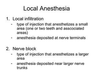
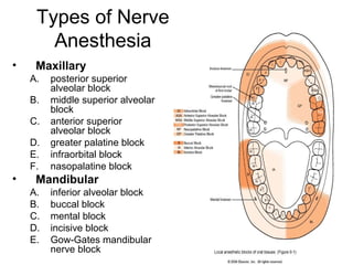
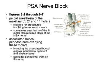
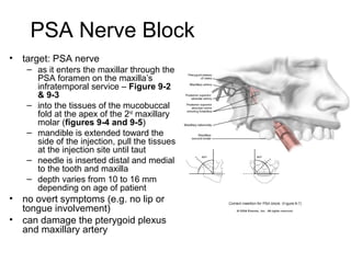
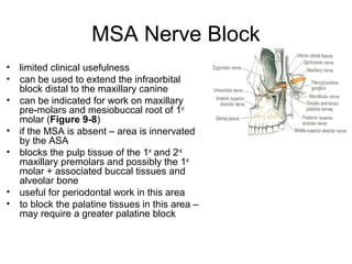
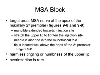
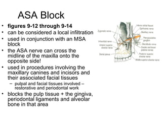
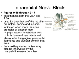
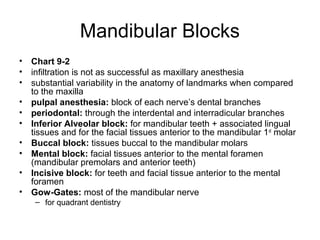
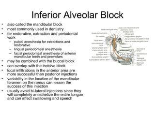
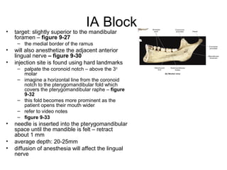
Local Anesthesia
1. Local infiltration
  -   type of injection that anesthetizes a small
      area (one or two teeth and asscociated
      areas)
  -   anesthesia deposited at nerve terminals

2. Nerve block
  -   type of injection that anesthetizes a larger
      area
  -   anesthesia deposited near larger nerve
      trunks
Types of Nerve
       Anesthesia
•    Maxillary
    A.   posterior superior
         alveolar block
    B.   middle superior alveolar
         block
    C.   anterior superior
         alveolar block
    D.   greater palatine block
    E.   infraorbital block
    F.   nasopalatine block
•    Mandibular
    A.   inferior alveolar block
    B.   buccal block
    C.   mental block
    D.   incisive block
    E.   Gow-Gates mandibular
         nerve block
Considerations
• dental procedures can usually commence after 3
  – 5 minutes
• failure requires re-administration using another
  method
• never re-administer using the same method
• keep in mind the total # of injections and the
  dosages
• never inject into an area with an abcess, or other
  type of abnormality
Maxillary Nerve Anesthesia
• Chart 9-1
• pulpal anesthesia: through anesthesia of each nerve’s dental
  branches as they extend into the pulp tissue (via the apical foramen)
• periodontal: through the interdental and interradicular branches
• palatal: soft and hard tissues of the palatal periodontium (e.g.
  gingiva, periodontal ligaments, alveolar bone)
• PSA block: recommended for maxillary molar teeth and associated
  buccal tissues in ONE quadrant
• MSA block: recommended for maxillary premolars and associated
  buccal tissues
• ASA block: recommended for maxillary canine and the incisors in
  ONE quadrant
• greater palatine block: recommended for palatal tissues distal to
  the maxillary canine in ONE quadrant
• nasopalatine block: recommended for palatal tissues between the
  right and left maxillary canines
PSA Nerve Block
• figures 9-2 through 9-7
• pulpal anesthesia of the
  maxillary 3rd, 2nd and 1st molars
   – required for procedures
     involving two or more molars
   – sometimes anesthesia of the 1st
     molar also required block of the
     MSA nerve
• associated buccal
  periodonteum overlying
  these molars
   – including the associated buccal
     gingiva, periodontal ligament
     and alveolar bone
   – useful for periodontal work on
     this area
PSA Nerve Block
•   target: PSA nerve
     – as it enters the maxillar through the
       PSA foramen on the maxilla’s
       infratemporal service – Figure 9-2
       & 9-3
     – into the tissues of the mucobuccal
       fold at the apex of the 2nd maxillary
       molar (figures 9-4 and 9-5)
     – mandible is extended toward the
       side of the injection, pull the tissues
       at the injection site until taut
     – needle is inserted distal and medial
       to the tooth and maxilla
     – depth varies from 10 to 16 mm
       depending on age of patient
•   no overt symptoms (e.g. no lip or
    tongue involvement)
•   can damage the pterygoid plexus
    and maxillary artery
MSA Nerve Block
•   limited clinical usefulness
•   can be used to extend the infraorbital
    block distal to the maxillary canine
•   can be indicated for work on maxillary
    pre-molars and mesiobuccal root of 1st
    molar (Figure 9-8)
•   if the MSA is absent – area is innervated
    by the ASA
•   blocks the pulp tissue of the 1st and 2nd
    maxillary premolars and possibly the 1st
    molar + associated buccal tissues and
    alveolar bone
•   useful for periodontal work in this area
•   to block the palatine tissues in this area –
    may require a greater palatine block
MSA Block
• target area: MSA nerve at the apex of the
  maxillary 2nd premolar (figures 9-8 and 9-9)
  –   mandible extended towards injection site
  –   stretch the upper lip to tighten the injection site
  –   needle is inserted into the mucobuccal fold
  –   tip is located well above the apex of the 2nd premolar
       • figure 9-11
• harmless tingling or numbness of the upper lip
• overinsertion is rare
ASA Block
• figures 9-12 through 9-14
• can be considered a local infiltration
• used in conjunction with an MSA
  block
• the ASA nerve can cross the
  midline of the maxilla onto the
  opposite side!
• used in procedures involving the
  maxillary canines and incisors and
  their associated facial tissues
   – pulpal and facial tissues involved –
     restorative and periodontal work
• blocks the pulp tissue + the gingiva,
  periodontal ligaments and alveolar
  bone in that area
ASA Block
• target: ASA nerve at the apex of the
  maxillary canine – figures 9-12 & 9-13
• at the mucobuccal fold at the apex of the
  maxillary canine – figure 9-13
• harmless tingling or numbness of the
  upper lip
• overinsertion is rare
Infraorbital Nerve Block
• figures 9-15 through 9-17
• anesthetizes both the MSA and
  ASA
• used for anesthesia of the maxillary
  premolars, canine and incisors
• indicated when more than one
  premolar or anterior teeth
   – pulpal tissues – for restorative work
   – facial tissues – for periodontal work
• also numbs the gingiva, periodontal
  ligaments and alveolar bone in that
  area
• the maxillary central incisor may
  also be innervated by the
  nasopalatine nerve branches
IO Block
• target: union of the ASA and MSA with the IO nerve
  after the IO enters the IO foramen – figure 9-15
• also anesthesizes the lower eyelid, side of nose and
  upper lip
• IO foramen is gently palpated along the IO rim
   – move slightly down about 10mm until you feel the depression of
     the IO foramen – figure 9-16
   – locate the tissues at the mucobuccal fold at the apex of the 1 st
     premolar
       • place one finger at the IO foramen and the other on the injection site
         – figure 9-17
       • locate the IO foramen, retract the upper lip and pull the tissues taut
       • the needle is inserted parallel to the long axis of the tooth to avoid
         hitting the bone
• harmless tingling or numbness of the upper lip, side of
  nose and eyelid
Greater Palatine Block
• figures 9-19 through 9-21
• used in restorative procedures that involve more than
  two maxillary posterior teeth or palatal tissues distal to
  the canine
• also used in periodontal work – since it blocks the
  associated lingual tissues
• anesthetizes the posterior portion of the hard palate –
  from the 1st premolar to the molars and medially to the
  palate midline
• does NOT provide pulpal anesthesia – may also need to
  use ASA, PSA, MSA or IO blocks
• may also need to be combined with nasopalatine block
Greater Palatine Block
• target: GP nerve as it enters the GP
  foramen
    – located at the junction of the maxillary
      alveolar process and the hard palate – at
      the maxillary 2nd or 3rd molar – figure 9-
      19
• palpate the GP foramen – midway
  between the median palatine raphe and
  lingual gingival margin of the molar tooth
  – figure 9-21
• can reduce discomfort by applying
  pressure to the site before and during the
  injection
    – produces a dull ache to block pain
      impulses
    – also slow deposition of anesthesia will
      also help
• needle is inserted at a 90 degree angle
  to the palate – figure 9-22
Nasopalatine Block
• figure 9-23 through 9-26
• useful for anesthesia of the bilateral portion of the hard
  palate
   – from the mesial of the right maxillary 1st premolar to the mesial of
     the left 1st premolar
• for palatal soft tissue anesthesia
   – periodontal treatment
• required for two or more anterior maxillary teeth
• for restorative procedures or extraction of the anterior
  maxillary teeth – may need an ASA or MSA block also
• blocks both right and left nerves
Nasopalatine Block
• target: both right and left nerves as they enter the incisive foramen
  from the mucosa of the anterior hard palate – figure 9-23 & 9-25
    – posterior to the incisive papilla
• injection site is lateral to the incisive papilla – figure 9-26
• head turned to the left or right
• inserted at a 45 degree angle about 6-10 mm – gently contact the
  maxillary bone and withdraw about 1mm before administering
• can reduce discomfort by applying pressure to the site before and
  during the injection
    – produces a dull ache to block pain impulses
    – also slow deposition of anesthesia will also help
• can anesthetize the labial tissues between the central incisors prior
  to palatal block
    – can block some branches of the nasopalatine prior to injection
Mandibular Blocks
• Chart 9-2
• infiltration is not as successful as maxillary anesthesia
• substantial variability in the anatomy of landmarks when compared
  to the maxilla
• pulpal anesthesia: block of each nerve’s dental branches
• periodontal: through the interdental and interradicular branches
• Inferior Alveolar block: for mandibular teeth + associated lingual
  tissues and for the facial tissues anterior to the mandibular 1 st molar
• Buccal block: tissues buccal to the mandibular molars
• Mental block: facial tissues anterior to the mental foramen
  (mandibular premolars and anterior teeth)
• Incisive block: for teeth and facial tissue anterior to the mental
  foramen
• Gow-Gates: most of the mandibular nerve
    – for quadrant dentistry
Inferior Alveolar Block
•   also called the mandibular block
•   most commonly used in dentistry
•   for restorative, extraction and periodontal
    work
     – pulpal anesthesia for extractions and
       restorative
     – lingual periodonteal anesthesia
     – facial periodonteal anesthesia of anterior
       mandibular teeth and premolars
•   may be combined with the buccal block
•   can overlap with the incisive block
•   local infiltrations in the anterior area are
    more successful than posterior injections
•   variability in the location of the mandibular
    foramen on the ramus can lessen the
    success of this injection
•   usually avoid bi-lateral injections since they
    will completely anesthetize the entire tongue
    and can affect swallowing and speech
IA Block
•   target: slightly superior to the mandibular
    foramen – figure 9-27
     – the medial border of the ramus
•   will also anesthetize the adjacent anterior
    lingual nerve – figure 9-30
•   injection site is found using hard landmarks
     – palpate the coronoid notch – above the 3rd
       molar
     – imagine a horizontal line from the coronoid
       notch to the pterygomandibular fold which
       covers the pterygomandibular raphe – figure
       9-32
     – this fold becomes more prominent as the
       patient opens their mouth wider
     – refer to video notes
     – figure 9-33
•   needle is inserted into the pterygomandibular
    space until the mandible is felt – retract
    about 1 mm
•   average depth: 20-25mm
•   diffusion of anesthesia will affect the lingual
    nerve
IA block
• symptoms: harmless tingling and numbness of the lower
  lip due to block of the mental nerve
• tingling and numbness of the body of the tongue and
  floor of mouth – lingual nerve involvement
• complications:
   – failure to penetrate enough can numb the tongue but not block
     sufficiently
   – lingual shock – involuntary movement as the needle passes the
     lingual nerve
   – transient facial paralysis – facial nerve involvement if inserted
     into the deeper parotid gland – figure 9-34
       • inability to close the eye and drooping of the lips on the affected side
       • hematoma can occur
   – some muscle soreness
   – patient-inflicted trauma – lip biting etc...
Buccal Block
• figures 9-36 and 9-37
• for buccal periodonteum of mandibular
  molars, gingiva, periodontal ligament and
  alveolar bone
• for restorative and periodontal work
• buccal nerve is readily located on the
  surface of the tissue and not within bone
Buccal block
• target: buccal nerve as it passes
  over the anterior border of the
  ramus through the buccinator –
  figure 9-36
• injection site is the buccal
  tissues distal and buccal to the
  most distal molar – on the
  anterior border of the ramus as it
  meets the body – figure 9-37
• pull the buccal tissue tight and
  advance the needle until you
  feel bone – only about 1 to 2mm
  figure 9-38
   – patient-inflicted trauma – lip biting
     etc...
Mental Block
• figures 9-39 through
  9-41
• for facial periodonteum
  of mandibular
  premolars and anterior
  teeth on one side
• for restorative work –
  incisive block should be
  considered instead
Mental Block
•   target site: mental nerve before it enters
    the mental foramen where it joins with the
    incisive nerve to form the IA nerve –
    figure 9-39
•   palpate the foramen between the apices
    of the 1st and 2nd premolars
     – palpate it intraorally – find the mucobuccal
       fold between the apices of the 1st and 2nd
       premolars – figure 9-42
     – in adults, the foramen faces
       posterosuperiorly
     – may be anterior or posterior
     – can be found using radiographs
•   insertion site is the mucobuccal fold tissue
    directly over or slight anterior to the
    foramen site
•   avoid contact with the mandible with the
    needle
•   depth is 5 to 6mm
•   no need to enter the foramen
Incisive Block
• for pulp and facial tissues of the teeth
  anterior to the mental foramen
  – same as the mental block except pulpal
    anesthesia is provided also
• restorative and periodontal work
• IA block indicated for extractions – no
  lingual anesthesia with an incisive block
• target: mental foramen – figure 9-43
Incisive Block
• injection site: figure 9-44
   – same as for the mental block
   – directly over or anterior to the
     mental foramen
   – in the mucobuccal fold at the apices
     of the 1st and 2nd premolars
   – pull the buccal tissues laterally
   – more anesthesia is used for this
     block when compared to the mental
     block
   – pressure is applied during the
     injection – forces for anesthetic
     solution into the foramen and block
     the deeper incisive nerve
   – the increased injection solution may
     balloon the facial tissues
Gow-Gates
• figures 9-45 through 9-
  50
• blocks the IA, mental,
  incisive, lingual,
  mylohyoid,
  auriculotemporal and
  buccal nerves – figure 9-
  28 and 9-45
• used for quadrant
  dentistry
• buccal and lingual soft
  tissue from most distal
  molar to the midline
• greater success than an
  IA block
Gow-Gates
•   target site: anteromedial border of the
    mandibular condylar neck – figure 9-46
•   just inferior to the insertion of the lateral
    pterygoid muscle
•   injection site is intraoral
•   locate the intertragic notch and labial
    commisure extraorally
     – draw a line from the tragus/intertragic
       notch to the labial commisure – figure 9-
       47
     – place your thumb on the condyle (just in
       front of the tragus when the mouth is
       open)
     – pull buccal tissue away
     – place the needle inferior to the
       mesiolingual cusp of the MAXILLARY 2nd
       molar
     – the needle penetrates distal to the
       maxillary 2nd molar
     – see the video

Lecture localanesthesia

  • 1.
    Local Anesthesia 1. Localinfiltration - type of injection that anesthetizes a small area (one or two teeth and asscociated areas) - anesthesia deposited at nerve terminals 2. Nerve block - type of injection that anesthetizes a larger area - anesthesia deposited near larger nerve trunks
  • 2.
    Types of Nerve Anesthesia • Maxillary A. posterior superior alveolar block B. middle superior alveolar block C. anterior superior alveolar block D. greater palatine block E. infraorbital block F. nasopalatine block • Mandibular A. inferior alveolar block B. buccal block C. mental block D. incisive block E. Gow-Gates mandibular nerve block
  • 3.
    Considerations • dental procedurescan usually commence after 3 – 5 minutes • failure requires re-administration using another method • never re-administer using the same method • keep in mind the total # of injections and the dosages • never inject into an area with an abcess, or other type of abnormality
  • 4.
    Maxillary Nerve Anesthesia •Chart 9-1 • pulpal anesthesia: through anesthesia of each nerve’s dental branches as they extend into the pulp tissue (via the apical foramen) • periodontal: through the interdental and interradicular branches • palatal: soft and hard tissues of the palatal periodontium (e.g. gingiva, periodontal ligaments, alveolar bone) • PSA block: recommended for maxillary molar teeth and associated buccal tissues in ONE quadrant • MSA block: recommended for maxillary premolars and associated buccal tissues • ASA block: recommended for maxillary canine and the incisors in ONE quadrant • greater palatine block: recommended for palatal tissues distal to the maxillary canine in ONE quadrant • nasopalatine block: recommended for palatal tissues between the right and left maxillary canines
  • 5.
    PSA Nerve Block •figures 9-2 through 9-7 • pulpal anesthesia of the maxillary 3rd, 2nd and 1st molars – required for procedures involving two or more molars – sometimes anesthesia of the 1st molar also required block of the MSA nerve • associated buccal periodonteum overlying these molars – including the associated buccal gingiva, periodontal ligament and alveolar bone – useful for periodontal work on this area
  • 6.
    PSA Nerve Block • target: PSA nerve – as it enters the maxillar through the PSA foramen on the maxilla’s infratemporal service – Figure 9-2 & 9-3 – into the tissues of the mucobuccal fold at the apex of the 2nd maxillary molar (figures 9-4 and 9-5) – mandible is extended toward the side of the injection, pull the tissues at the injection site until taut – needle is inserted distal and medial to the tooth and maxilla – depth varies from 10 to 16 mm depending on age of patient • no overt symptoms (e.g. no lip or tongue involvement) • can damage the pterygoid plexus and maxillary artery
  • 7.
    MSA Nerve Block • limited clinical usefulness • can be used to extend the infraorbital block distal to the maxillary canine • can be indicated for work on maxillary pre-molars and mesiobuccal root of 1st molar (Figure 9-8) • if the MSA is absent – area is innervated by the ASA • blocks the pulp tissue of the 1st and 2nd maxillary premolars and possibly the 1st molar + associated buccal tissues and alveolar bone • useful for periodontal work in this area • to block the palatine tissues in this area – may require a greater palatine block
  • 8.
    MSA Block • targetarea: MSA nerve at the apex of the maxillary 2nd premolar (figures 9-8 and 9-9) – mandible extended towards injection site – stretch the upper lip to tighten the injection site – needle is inserted into the mucobuccal fold – tip is located well above the apex of the 2nd premolar • figure 9-11 • harmless tingling or numbness of the upper lip • overinsertion is rare
  • 9.
    ASA Block • figures9-12 through 9-14 • can be considered a local infiltration • used in conjunction with an MSA block • the ASA nerve can cross the midline of the maxilla onto the opposite side! • used in procedures involving the maxillary canines and incisors and their associated facial tissues – pulpal and facial tissues involved – restorative and periodontal work • blocks the pulp tissue + the gingiva, periodontal ligaments and alveolar bone in that area
  • 10.
    ASA Block • target:ASA nerve at the apex of the maxillary canine – figures 9-12 & 9-13 • at the mucobuccal fold at the apex of the maxillary canine – figure 9-13 • harmless tingling or numbness of the upper lip • overinsertion is rare
  • 11.
    Infraorbital Nerve Block •figures 9-15 through 9-17 • anesthetizes both the MSA and ASA • used for anesthesia of the maxillary premolars, canine and incisors • indicated when more than one premolar or anterior teeth – pulpal tissues – for restorative work – facial tissues – for periodontal work • also numbs the gingiva, periodontal ligaments and alveolar bone in that area • the maxillary central incisor may also be innervated by the nasopalatine nerve branches
  • 12.
    IO Block • target:union of the ASA and MSA with the IO nerve after the IO enters the IO foramen – figure 9-15 • also anesthesizes the lower eyelid, side of nose and upper lip • IO foramen is gently palpated along the IO rim – move slightly down about 10mm until you feel the depression of the IO foramen – figure 9-16 – locate the tissues at the mucobuccal fold at the apex of the 1 st premolar • place one finger at the IO foramen and the other on the injection site – figure 9-17 • locate the IO foramen, retract the upper lip and pull the tissues taut • the needle is inserted parallel to the long axis of the tooth to avoid hitting the bone • harmless tingling or numbness of the upper lip, side of nose and eyelid
  • 13.
    Greater Palatine Block •figures 9-19 through 9-21 • used in restorative procedures that involve more than two maxillary posterior teeth or palatal tissues distal to the canine • also used in periodontal work – since it blocks the associated lingual tissues • anesthetizes the posterior portion of the hard palate – from the 1st premolar to the molars and medially to the palate midline • does NOT provide pulpal anesthesia – may also need to use ASA, PSA, MSA or IO blocks • may also need to be combined with nasopalatine block
  • 14.
    Greater Palatine Block •target: GP nerve as it enters the GP foramen – located at the junction of the maxillary alveolar process and the hard palate – at the maxillary 2nd or 3rd molar – figure 9- 19 • palpate the GP foramen – midway between the median palatine raphe and lingual gingival margin of the molar tooth – figure 9-21 • can reduce discomfort by applying pressure to the site before and during the injection – produces a dull ache to block pain impulses – also slow deposition of anesthesia will also help • needle is inserted at a 90 degree angle to the palate – figure 9-22
  • 15.
    Nasopalatine Block • figure9-23 through 9-26 • useful for anesthesia of the bilateral portion of the hard palate – from the mesial of the right maxillary 1st premolar to the mesial of the left 1st premolar • for palatal soft tissue anesthesia – periodontal treatment • required for two or more anterior maxillary teeth • for restorative procedures or extraction of the anterior maxillary teeth – may need an ASA or MSA block also • blocks both right and left nerves
  • 16.
    Nasopalatine Block • target:both right and left nerves as they enter the incisive foramen from the mucosa of the anterior hard palate – figure 9-23 & 9-25 – posterior to the incisive papilla • injection site is lateral to the incisive papilla – figure 9-26 • head turned to the left or right • inserted at a 45 degree angle about 6-10 mm – gently contact the maxillary bone and withdraw about 1mm before administering • can reduce discomfort by applying pressure to the site before and during the injection – produces a dull ache to block pain impulses – also slow deposition of anesthesia will also help • can anesthetize the labial tissues between the central incisors prior to palatal block – can block some branches of the nasopalatine prior to injection
  • 17.
    Mandibular Blocks • Chart9-2 • infiltration is not as successful as maxillary anesthesia • substantial variability in the anatomy of landmarks when compared to the maxilla • pulpal anesthesia: block of each nerve’s dental branches • periodontal: through the interdental and interradicular branches • Inferior Alveolar block: for mandibular teeth + associated lingual tissues and for the facial tissues anterior to the mandibular 1 st molar • Buccal block: tissues buccal to the mandibular molars • Mental block: facial tissues anterior to the mental foramen (mandibular premolars and anterior teeth) • Incisive block: for teeth and facial tissue anterior to the mental foramen • Gow-Gates: most of the mandibular nerve – for quadrant dentistry
  • 18.
    Inferior Alveolar Block • also called the mandibular block • most commonly used in dentistry • for restorative, extraction and periodontal work – pulpal anesthesia for extractions and restorative – lingual periodonteal anesthesia – facial periodonteal anesthesia of anterior mandibular teeth and premolars • may be combined with the buccal block • can overlap with the incisive block • local infiltrations in the anterior area are more successful than posterior injections • variability in the location of the mandibular foramen on the ramus can lessen the success of this injection • usually avoid bi-lateral injections since they will completely anesthetize the entire tongue and can affect swallowing and speech
  • 19.
    IA Block • target: slightly superior to the mandibular foramen – figure 9-27 – the medial border of the ramus • will also anesthetize the adjacent anterior lingual nerve – figure 9-30 • injection site is found using hard landmarks – palpate the coronoid notch – above the 3rd molar – imagine a horizontal line from the coronoid notch to the pterygomandibular fold which covers the pterygomandibular raphe – figure 9-32 – this fold becomes more prominent as the patient opens their mouth wider – refer to video notes – figure 9-33 • needle is inserted into the pterygomandibular space until the mandible is felt – retract about 1 mm • average depth: 20-25mm • diffusion of anesthesia will affect the lingual nerve
  • 20.
    IA block • symptoms:harmless tingling and numbness of the lower lip due to block of the mental nerve • tingling and numbness of the body of the tongue and floor of mouth – lingual nerve involvement • complications: – failure to penetrate enough can numb the tongue but not block sufficiently – lingual shock – involuntary movement as the needle passes the lingual nerve – transient facial paralysis – facial nerve involvement if inserted into the deeper parotid gland – figure 9-34 • inability to close the eye and drooping of the lips on the affected side • hematoma can occur – some muscle soreness – patient-inflicted trauma – lip biting etc...
  • 21.
    Buccal Block • figures9-36 and 9-37 • for buccal periodonteum of mandibular molars, gingiva, periodontal ligament and alveolar bone • for restorative and periodontal work • buccal nerve is readily located on the surface of the tissue and not within bone
  • 22.
    Buccal block • target:buccal nerve as it passes over the anterior border of the ramus through the buccinator – figure 9-36 • injection site is the buccal tissues distal and buccal to the most distal molar – on the anterior border of the ramus as it meets the body – figure 9-37 • pull the buccal tissue tight and advance the needle until you feel bone – only about 1 to 2mm figure 9-38 – patient-inflicted trauma – lip biting etc...
  • 23.
    Mental Block • figures9-39 through 9-41 • for facial periodonteum of mandibular premolars and anterior teeth on one side • for restorative work – incisive block should be considered instead
  • 24.
    Mental Block • target site: mental nerve before it enters the mental foramen where it joins with the incisive nerve to form the IA nerve – figure 9-39 • palpate the foramen between the apices of the 1st and 2nd premolars – palpate it intraorally – find the mucobuccal fold between the apices of the 1st and 2nd premolars – figure 9-42 – in adults, the foramen faces posterosuperiorly – may be anterior or posterior – can be found using radiographs • insertion site is the mucobuccal fold tissue directly over or slight anterior to the foramen site • avoid contact with the mandible with the needle • depth is 5 to 6mm • no need to enter the foramen
  • 25.
    Incisive Block • forpulp and facial tissues of the teeth anterior to the mental foramen – same as the mental block except pulpal anesthesia is provided also • restorative and periodontal work • IA block indicated for extractions – no lingual anesthesia with an incisive block • target: mental foramen – figure 9-43
  • 26.
    Incisive Block • injectionsite: figure 9-44 – same as for the mental block – directly over or anterior to the mental foramen – in the mucobuccal fold at the apices of the 1st and 2nd premolars – pull the buccal tissues laterally – more anesthesia is used for this block when compared to the mental block – pressure is applied during the injection – forces for anesthetic solution into the foramen and block the deeper incisive nerve – the increased injection solution may balloon the facial tissues
  • 27.
    Gow-Gates • figures 9-45through 9- 50 • blocks the IA, mental, incisive, lingual, mylohyoid, auriculotemporal and buccal nerves – figure 9- 28 and 9-45 • used for quadrant dentistry • buccal and lingual soft tissue from most distal molar to the midline • greater success than an IA block
  • 28.
    Gow-Gates • target site: anteromedial border of the mandibular condylar neck – figure 9-46 • just inferior to the insertion of the lateral pterygoid muscle • injection site is intraoral • locate the intertragic notch and labial commisure extraorally – draw a line from the tragus/intertragic notch to the labial commisure – figure 9- 47 – place your thumb on the condyle (just in front of the tragus when the mouth is open) – pull buccal tissue away – place the needle inferior to the mesiolingual cusp of the MAXILLARY 2nd molar – the needle penetrates distal to the maxillary 2nd molar – see the video