radiographic diagnosis of periodontal disease
•Download as PPT, PDF•
136 likes•48,312 views

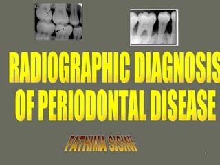
Radiographs are an essential tool for diagnosing periodontal disease by assessing bone loss. Early periodontitis appears on radiographs as localized bone erosions while advanced cases show generalized horizontal bone loss. Vertical bone defects can also be seen, appearing as widened ligament spaces or loss of cortical plates. Furcation involvement initially widens the ligament but may progress to deep vertical defects. Aggressive periodontitis in young people causes rapid, widespread bone destruction and early tooth loss. Follow-up radiographs after treatment can demonstrate bone fill-in and sharpening of bony contours.
![The Role of Radiology in Assessment of Periodontal Disease ,[object Object],[object Object],[object Object],[object Object],[object Object],[object Object],[object Object],[object Object]](data:image/gif;base64,R0lGODlhAQABAIAAAAAAAP///yH5BAEAAAAALAAAAAABAAEAAAIBRAA7)
More Related Content
What's hot
What's hot (20)
Viewers also liked
Viewers also liked (20)
Similar to radiographic diagnosis of periodontal disease
Similar to radiographic diagnosis of periodontal disease (20)
More from shabeel pn
More from shabeel pn (20)
Recently uploaded
Recently uploaded (20)
radiographic diagnosis of periodontal disease
- 1. RADIOGRAPHIC DIAGNOSIS OF PERIODONTAL DISEASE FATHIMA SISINI
- 39. thank you......