Title : Liver
Objectives : to
1. Describe liver cirrhosis.
2. Outline liver abscess.
3. Identify alcoholic liver diseases.
4. Study metabolic liver diseases.
Liver cirrhosis:
 End stage of many chronic liver diseases.
 Refers to diffuse hepatic fibrosis with replacement
of normal lobular architecture by parenchymal nodules
separated by fibrous tissue.
 suspected on clinical, laboratory, and imaging parameters,
but final diagnosis requires liver biopsy.
 Architectural changes best appreciated on reticulin stain.
 The morphologic classification of cirrhosis
is based on size of nodules:
o Micronodular : if nodules are less than 3 mm.
o Macronodular : if nodules are greater than 3 mm.
 The size of nodules can define etiology of cirrhosis ,
but this is relative since micronodular cirrhosis may
convert into macronodular type .
 Features indicating etiology of cirrhosis :-
• Biliary cirrhosis: garland-shaped nodules, and
clear halo in nodular periphery due to edema.
• Post hepatitic cirrhosis: necro inflammation.
• HBV : ground glass hepatocytes.
• HCV: lymphoid aggregates and lymphoid follicles.
• Autoimmune hepatitis: numerous plasma cells.
• Genetic hemochromatosis: severe parenchymal siderosis.
• a1-Antitrypsin deficiency: hepatocellular inclusions
which are eosinophilic, PAS positive.
 Hepatocellular carcinoma commonly arises in cirrhotic liver.
Its precancerous stages ( dysplastic foci , large and small
liver cell dysplasia) should be looked for.
Liver abscess :
 In developing countries: most liver abscesses
represent parasitic infections (amebic, echinococcal).
 In developed countries: most are pyogenic, the organism
reach liver by:
(1) portal vein.
(2) arterial supply.
(3) ascending cholangitis.
(4) direct invasion of liver from nearby source.
(5) penetrating injury.
Morphology :
 Liver abscesses can be solitary or multiple:
o Bacteremic spread: multiple small abscesses.
o Direct extension and trauma: solitary large abscesses.
o Biliary abscesses are usually multiple.
 Gross and microscopic features are those of any abscess.
 The causative organism can be identified in fungal or
parasitic abscesses .
The liver shows a small abscess filled with
many neutrophils.
Amoebic Liver abscess
amoebic Liver abscess- trophozoites
Alcoholic liver diseases:
Hepatic Steatosis (Fatty Liver):
 Fatty liver is large , soft , yellow and greasy.
 Small microvesicular lipid droplets in hepatocytes.
 With chronic intake of alcohol: large, clear macrovesicular.
 Initially is centrilobular, in severe cases involve entire lobule.
 Fatty change is completely reversible if there is abstention
from alcohol intake.
Alcoholic Steato Hepatitis- ASH:
 Grossly :
o liver is mottled red with bile-stained areas.
o it often contains visible nodules and fibrosis.
 Microscopically:
o Hepatocyte swelling and necrosis.
o Mallory bodies: cytokeratin intermediate filaments
visible as eosinophilic cytoplasmic inclusions.
o Neutrophilic reaction: around degenerating hepatocytes
particularly those with Mallory bodies.
o Fibrosis.
Common pathological findings in alcoholic steatohepatitis. A. Ballooning and Mallory
bodies in damaged hepatocytes . B. Steatosis and polymorphonuclear infiltration (arrow).
C. Centrilobular fibrosis. D. Bridging fibrosis in cases of advanced disease.
Alcoholic liver cirrhosis:
 Final and irreversible form of alcoholic liver disease,
usually evolves slowly.
 Initially cirrhotic liver is yellow-tan, fatty, and enlarged;
later on brown ,shrunken, non fatty organ.
 Uniformly sized micronodules , but with time scattered
larger nodules create a "hobnail" appearance.
 Mallory bodies rarely evident ,thus it resembles cirrhosis
developing from viral hepatitis and other causes.
Metabolic liver diseases:
Nonalcoholic fatty liver (NAFL) :
 Patients are not heavy drinkers.
 Men and women are equally affected.
 Strong association with obesity, dyslipidemia, and
insulin resistance (type 2) diabetes.
 Patients are largely asymptomatic, with abnormalities
only in lab. tests (elevated serum aminotransferases
and/or Ɣ- glutamyl transpeptidase ).
 Liver biopsy shows :
• Steatosis (Large and small vesicles of fat)
• No hepatic inflammation,No hepatocyte death, No fibrosis.
Nonalcoholic steatohepatitis, or NASH:
 Liver biopsy shows:
• Steatosis.
• multifocal parenchymal inflammation, Mallory body ,
hepatocyte death and sinusoidal fibrosis.
• Cirrhosis : may occur.
HEMOCHROMATOSIS :
 Excessive accumulation of body iron, deposited
in parenchymal organs such as liver and pancreas.
 Tow major types :
o Hereditary ( primary ) hemochromatosis :
• autosomal-recessive inherited disorder.
• gene located on chromosome 6 called HFE gene.
o Acquired ( secondary ) hemochromatosis: include:-
• Parenteral iron overload:
 Transfusions.
 Iron-dextran injections.
• Ineffective erythropoiesis:
 β-Thalassemia.
 Sideroblastic anemia.
• Increased oral intake of iron.
• Congenital atransferrinemia.
• Chronic liver disease:
 Chronic alcoholic liver disease.
 Porphyria cutanea tarda.
 Fully developed cases exhibit:
(1) micronodular cirrhosis in all patients.
(2) diabetes mellitus in 75% to 80% of patients.
(3) skin pigmentation in 75% to 80% of patients.
 Symptoms first appear in fifth to sixth decades of life.
 Males predominate (5 - 7:1).
 Death may result from cirrhosis or cardiac disease.
 Risk for hepatocellular carcinoma is 200-fold increased.
 Screening involves :
• very high levels of serum iron and serum ferritin.
• liver biopsy if indicated.
• identification of HFE gene for genetic screening.
 Treatment: regular phlebotomy.
Morphology:
 Liver is slightly larger than normal, dense,
and chocolate brown.
 Iron evident as golden-yellow hemosiderin granules
which stain blue with Prussian blue stain.
 Inflammation is absent.
 Fibrous septa develop slowly, leading ultimately
to micronodular type of cirrhosis .
The dark brown color
of liver, pancreas
(bottom center) and
lymph nodes
(bottom right) in
middle-aged man
with hereditary
hemochromatosis .
liver cells bear within them Hemosiderin
granules .
Iron(Prussian Blue ) staining: blue granules of hemosiderin
WILSON DISEASE :
 Autosomal-recessive disorder marked by
accumulation of toxic levels of copper in
many tissues and organs.
 The gene is ATP7B, located on chromosome 13.
 Morphology:
• Fatty change in hepatocyte.
• acute hepatitis.
• chronic hepatitis.
• with progression cirrhosis will develop.
• Excess copper deposition demonstrated by special stain:
rhodanine stain for copper.
Liver histology - rhodamine staining for copper
 Clinical Features:
• rarely manifests before 6 years of age.
• most common presentation is:
 acute or chronic liver disease.
 Neuropsychiatric manifestations (mild behavioral changes,
frank psychosis, or Parkinson disease-like syndrome).
 Eye lesions called Kayser-Fleischer rings:
green to brown deposits of copper in cornea.
 Diagnosis:
• decrease in serum ceruloplasmin.
• increase in hepatic copper content.
• increased urinary excretion of copper.
• Serum copper levels are of no diagnostic value,
since tmay be low, normal, or elevated, depending
on stage of evolution of disease.
 Treatment: copper chelation therapy (D-penicillamine).
Summary:
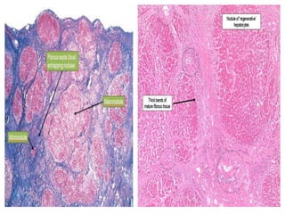
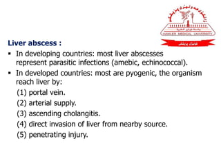
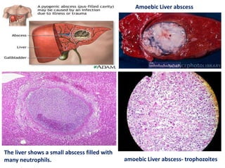
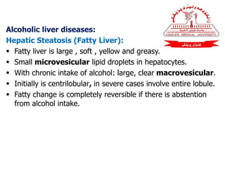
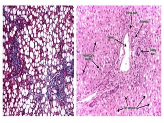
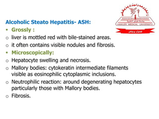
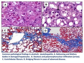
1. Liver cirrhosis is the end stage of many chronic
liver diseases.
2. Liver abscesses are either due to parasitic infection
or pyogenic.
3. Alcoholic liver diseases include steatosis , ASH ,
and alcoholic liver cirrhosis.
4. Metabolic liver diseases include NAFL , NASH,
hemochromatosis, and wilson disease.
Questions:
1. Enumerate alcoholic liver diseases, and write
short assay on one of them?
2. How you diagnose wilson disease?
THANK YOU

Liver 2

  • 1.
    Title : Liver Objectives: to 1. Describe liver cirrhosis. 2. Outline liver abscess. 3. Identify alcoholic liver diseases. 4. Study metabolic liver diseases.
  • 2.
    Liver cirrhosis:  Endstage of many chronic liver diseases.  Refers to diffuse hepatic fibrosis with replacement of normal lobular architecture by parenchymal nodules separated by fibrous tissue.  suspected on clinical, laboratory, and imaging parameters, but final diagnosis requires liver biopsy.  Architectural changes best appreciated on reticulin stain.
  • 3.
     The morphologicclassification of cirrhosis is based on size of nodules: o Micronodular : if nodules are less than 3 mm. o Macronodular : if nodules are greater than 3 mm.  The size of nodules can define etiology of cirrhosis , but this is relative since micronodular cirrhosis may convert into macronodular type .
  • 4.
     Features indicatingetiology of cirrhosis :- • Biliary cirrhosis: garland-shaped nodules, and clear halo in nodular periphery due to edema. • Post hepatitic cirrhosis: necro inflammation. • HBV : ground glass hepatocytes. • HCV: lymphoid aggregates and lymphoid follicles.
  • 5.
    • Autoimmune hepatitis:numerous plasma cells. • Genetic hemochromatosis: severe parenchymal siderosis. • a1-Antitrypsin deficiency: hepatocellular inclusions which are eosinophilic, PAS positive.  Hepatocellular carcinoma commonly arises in cirrhotic liver. Its precancerous stages ( dysplastic foci , large and small liver cell dysplasia) should be looked for.
  • 7.
    Liver abscess : In developing countries: most liver abscesses represent parasitic infections (amebic, echinococcal).  In developed countries: most are pyogenic, the organism reach liver by: (1) portal vein. (2) arterial supply. (3) ascending cholangitis. (4) direct invasion of liver from nearby source. (5) penetrating injury.
  • 8.
    Morphology :  Liverabscesses can be solitary or multiple: o Bacteremic spread: multiple small abscesses. o Direct extension and trauma: solitary large abscesses. o Biliary abscesses are usually multiple.  Gross and microscopic features are those of any abscess.  The causative organism can be identified in fungal or parasitic abscesses .
  • 9.
    The liver showsa small abscess filled with many neutrophils. Amoebic Liver abscess amoebic Liver abscess- trophozoites
  • 10.
    Alcoholic liver diseases: HepaticSteatosis (Fatty Liver):  Fatty liver is large , soft , yellow and greasy.  Small microvesicular lipid droplets in hepatocytes.  With chronic intake of alcohol: large, clear macrovesicular.  Initially is centrilobular, in severe cases involve entire lobule.  Fatty change is completely reversible if there is abstention from alcohol intake.
  • 12.
    Alcoholic Steato Hepatitis-ASH:  Grossly : o liver is mottled red with bile-stained areas. o it often contains visible nodules and fibrosis.  Microscopically: o Hepatocyte swelling and necrosis. o Mallory bodies: cytokeratin intermediate filaments visible as eosinophilic cytoplasmic inclusions. o Neutrophilic reaction: around degenerating hepatocytes particularly those with Mallory bodies. o Fibrosis.
  • 13.
    Common pathological findingsin alcoholic steatohepatitis. A. Ballooning and Mallory bodies in damaged hepatocytes . B. Steatosis and polymorphonuclear infiltration (arrow). C. Centrilobular fibrosis. D. Bridging fibrosis in cases of advanced disease.
  • 14.
    Alcoholic liver cirrhosis: Final and irreversible form of alcoholic liver disease, usually evolves slowly.  Initially cirrhotic liver is yellow-tan, fatty, and enlarged; later on brown ,shrunken, non fatty organ.  Uniformly sized micronodules , but with time scattered larger nodules create a "hobnail" appearance.  Mallory bodies rarely evident ,thus it resembles cirrhosis developing from viral hepatitis and other causes.
  • 15.
    Metabolic liver diseases: Nonalcoholicfatty liver (NAFL) :  Patients are not heavy drinkers.  Men and women are equally affected.  Strong association with obesity, dyslipidemia, and insulin resistance (type 2) diabetes.  Patients are largely asymptomatic, with abnormalities only in lab. tests (elevated serum aminotransferases and/or Ɣ- glutamyl transpeptidase ).
  • 16.
     Liver biopsyshows : • Steatosis (Large and small vesicles of fat) • No hepatic inflammation,No hepatocyte death, No fibrosis. Nonalcoholic steatohepatitis, or NASH:  Liver biopsy shows: • Steatosis. • multifocal parenchymal inflammation, Mallory body , hepatocyte death and sinusoidal fibrosis. • Cirrhosis : may occur.
  • 17.
    HEMOCHROMATOSIS :  Excessiveaccumulation of body iron, deposited in parenchymal organs such as liver and pancreas.  Tow major types : o Hereditary ( primary ) hemochromatosis : • autosomal-recessive inherited disorder. • gene located on chromosome 6 called HFE gene. o Acquired ( secondary ) hemochromatosis: include:-
  • 18.
    • Parenteral ironoverload:  Transfusions.  Iron-dextran injections. • Ineffective erythropoiesis:  β-Thalassemia.  Sideroblastic anemia. • Increased oral intake of iron. • Congenital atransferrinemia. • Chronic liver disease:  Chronic alcoholic liver disease.  Porphyria cutanea tarda.
  • 19.
     Fully developedcases exhibit: (1) micronodular cirrhosis in all patients. (2) diabetes mellitus in 75% to 80% of patients. (3) skin pigmentation in 75% to 80% of patients.  Symptoms first appear in fifth to sixth decades of life.  Males predominate (5 - 7:1).
  • 20.
     Death mayresult from cirrhosis or cardiac disease.  Risk for hepatocellular carcinoma is 200-fold increased.  Screening involves : • very high levels of serum iron and serum ferritin. • liver biopsy if indicated. • identification of HFE gene for genetic screening.  Treatment: regular phlebotomy.
  • 21.
    Morphology:  Liver isslightly larger than normal, dense, and chocolate brown.  Iron evident as golden-yellow hemosiderin granules which stain blue with Prussian blue stain.  Inflammation is absent.  Fibrous septa develop slowly, leading ultimately to micronodular type of cirrhosis .
  • 22.
    The dark browncolor of liver, pancreas (bottom center) and lymph nodes (bottom right) in middle-aged man with hereditary hemochromatosis . liver cells bear within them Hemosiderin granules . Iron(Prussian Blue ) staining: blue granules of hemosiderin
  • 23.
    WILSON DISEASE : Autosomal-recessive disorder marked by accumulation of toxic levels of copper in many tissues and organs.  The gene is ATP7B, located on chromosome 13.  Morphology: • Fatty change in hepatocyte. • acute hepatitis. • chronic hepatitis. • with progression cirrhosis will develop.
  • 24.
    • Excess copperdeposition demonstrated by special stain: rhodanine stain for copper. Liver histology - rhodamine staining for copper
  • 25.
     Clinical Features: •rarely manifests before 6 years of age. • most common presentation is:  acute or chronic liver disease.  Neuropsychiatric manifestations (mild behavioral changes, frank psychosis, or Parkinson disease-like syndrome).  Eye lesions called Kayser-Fleischer rings: green to brown deposits of copper in cornea.
  • 26.
     Diagnosis: • decreasein serum ceruloplasmin. • increase in hepatic copper content. • increased urinary excretion of copper. • Serum copper levels are of no diagnostic value, since tmay be low, normal, or elevated, depending on stage of evolution of disease.  Treatment: copper chelation therapy (D-penicillamine).
  • 27.
    Summary: 1. Liver cirrhosisis the end stage of many chronic liver diseases. 2. Liver abscesses are either due to parasitic infection or pyogenic. 3. Alcoholic liver diseases include steatosis , ASH , and alcoholic liver cirrhosis. 4. Metabolic liver diseases include NAFL , NASH, hemochromatosis, and wilson disease.
  • 28.
    Questions: 1. Enumerate alcoholicliver diseases, and write short assay on one of them? 2. How you diagnose wilson disease? THANK YOU