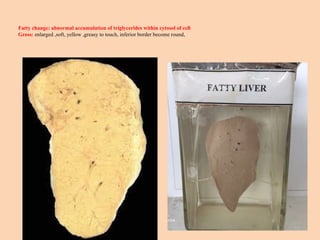
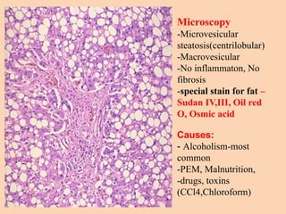
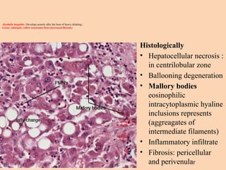
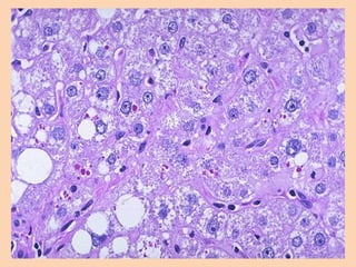
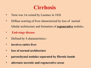
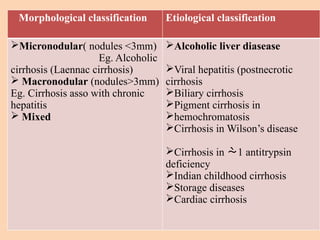
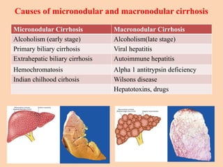
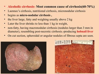
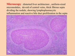
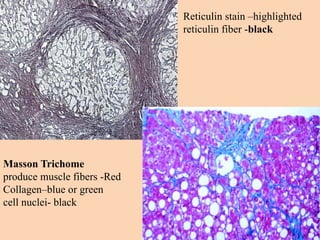
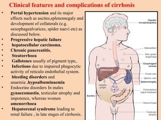
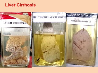
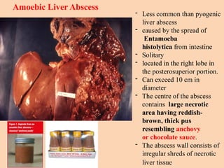
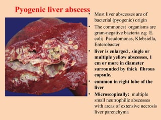
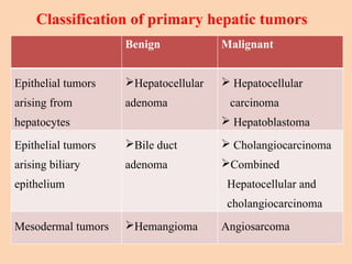
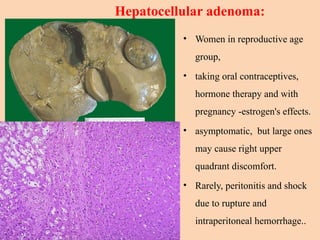
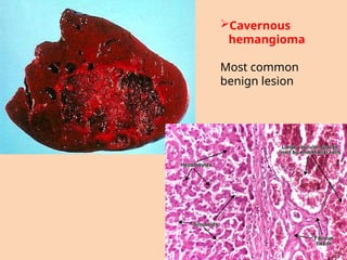
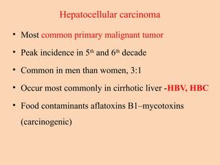
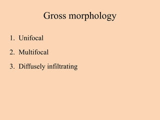
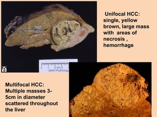
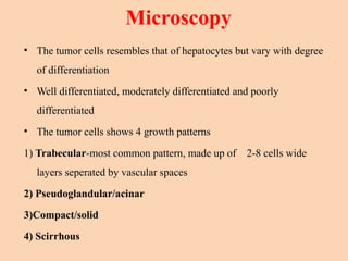
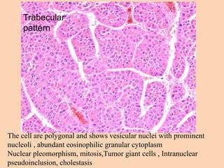
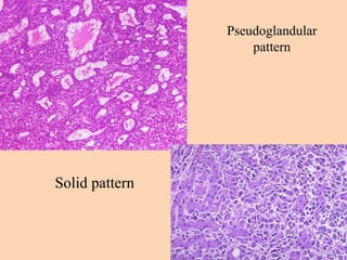
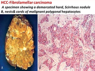
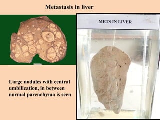
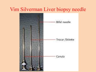
HEPATOBILLIARY
SYSTEM
TOPIC TO COVER
SPECIMENS
• CVC Liver
• Fatty Liver
• Cirrhosis
• Amoebic abscess
• Tumours of Liver,
Hepatocellular carcinoma
• Liver metastasis
SLIDES
• Fatty Liver
• Cirrhosis
• CHARTS -Jaundice
Normal liver histology
Chronic Venous Congestion (CVC)- Liver
Cut section of liver- alternate dark and light
areas are seen- known as “Nutmeg
appearance”
CVC liver histology
Hepatocytes around central
vein- necrosis (centrilobular
necrosis)- due to
deoxygeneated blood
Intermediate hepatocytes
undergo fatty change
Periportal hepatocytes are
normal due to adequate
perfusion.
Causes: Right sided heart failure-most common
Others –Constrictive pericarditis, tricuspid stenosis, obstruction of
inferior vena cava and hepatic vein
Alcoholic liver diseases and cirrhosis
• Spectrum of liver injury associated with acute and
chronic alcoholism
• Three stages
1) Alcoholic steatosis (fatty liver)
2) Alcoholic hepatitis
3) Alcoholic cirrhosis
Fatty change: abnormal accumulation of triglycerides within cytosol of cell
Gross: enlarged ,soft, yellow ,greasy to touch, inferior border become round,
Microscopy
-Microvesicular
steatosis(centrilobular)
-Macrovesicular
-No inflammaton, No
fibrosis
-special stain for fat –
Sudan IV,III, Oil red
O, Osmic acid
Causes:
- Alcoholism-most
common
-PEM, Malnutrition,
-drugs, toxins
(CCl4,Chloroform)
Alcoholic hepatitis :Develops acutely after the bout of heavy drinking ,
Gross: enlarged; yellow (steatosis) firm (increased fibrosis)
Histologically
• Hepatocellular necrosis :
in centrilobular zone
• Ballooning degeneration
• Mallory bodies
eosinophilic
intracytoplasmic hyaline
inclusions represents
(aggreagates of
intermediate filaments)
• Inflammatory infiltrate
• Fibrosis: pericellular
and perivenular
Cirrhosis
• Term was 1st coined by Laennec in 1826
• Diffuse scarring of liver characterized by loss of normal
lobular architecture and formation of regenerative nodules.
• End-stage disease
• Defined by 4 characteristics:-
• involves entire liver
• loss of normal architecture
• parenchymal nodules separated by fibrotic bands
• alternate necrotic and regenerative areas
Morphological classification Etiological classification
Micronodular( nodules <3mm)
Eg. Alcoholic
cirrhosis (Laennac cirrhosis)
 Macronodular (nodules>3mm)
Eg. Cirrhosis asso with chronic
hepatitis
 Mixed
Alcoholic liver diasease
Viral hepatitis (postnecrotic
cirrhosis
Biliary cirrhosis
Pigment cirrhosis in
hemochromatosis
Cirrhosis in Wilson’s disease
Cirrhosis in 1 antitrypsin
deficiency
Indian childhood cirrhosis
Storage diseases
Cardiac cirrhosis
Causes of micronodular and macronodular cirrhosis
Micronodular Cirrhosis Macronodular Cirrhosis
Alcoholism (early stage) Alcoholism(late stage)
Primary biliary cirrhosis Viral hepatitis
Extrahepatic biliary cirrhosis Autoimmune hepatitis
Hemochromatosis Alpha 1 antitrypsin deficiency
Indian chilhood cirhosis Wilsons disease
Hepatotoxins, drugs
• Alcoholic cirrhosis- Most common cause of cirrhosis(60-70%)
• Laennec’s cirrhosis, nutritional cirrhosis, micronodular cirrhosis
• begins as micro-nodular cirrhosis ,
• the liver large, fatty and weighing usually above 2 kg
• Later the liver shrinks to less than 1 kg in weight,
• non-fatty, having macronodular cirrhosis (nodules larger than 3 mm in
diameter), resembling post-necrotic cirrhosis.-producing hobnail liver
• On cut section, spheroidal or angular nodules of fibrous septa are seen.
Microscopy : distorted liver architecture , uniform-sized
micronodules, devoid of central veins, thick fibrous septa
dividing the nodule, showing lymphoplasmacytic
inflammation and reactive bile duct proliferation in the septa
Masson Trichome
produce muscle fibers -Red
Collagen–blue or green
cell nuclei- black
Reticulin stain –highlighted
reticulin fiber -black
• Portal hypertension and its major
effects such as ascites,splenomegaly and
development of collaterals (e.g.
oesophagealvarices, spider naevi etc) as
discussed below.
• Progressive hepatic failure
• hepatocellular carcinoma,
• Chronic pancreatitis,
• Steatorrhoea
• Gallstones usually of pigment type,.
• Infections due to impaired phagocytic
activity of reticulo endothelial system.
• bleeding disorders and
anaemia ,hypoalbuminaemia
• Endocrine disorders In males
gynaecomastia, testicular atrophy and
impotence, whereas women
amenorrhoea
• Hepatorenal syndrome leading to
renal failure , in late stages of cirrhosis.
Clinical features and complications of cirrhosis
Liver Cirrhosis
Amoebic Liver Abscess
- Less common than pyogenic
liver abscess
- caused by the spread of
Entamoeba
histolytica from intestine
- Solitary
- located in the right lobe in
the posterosuperior portion.
- Can exceed 10 cm in
diameter
- The centre of the abscess
contains large necrotic
area having reddish-
brown, thick pus
resembling anchovy
or chocolate sauce.
- The abscess wall consists of
irregular shreds of necrotic
liver tissue
Pyogenic liver abscess
• Most liver abscesses are of
bacterial (pyogenic) origin
• The commonest organisms are
gram-negative bacteria e.g E.
coli; Pseudomonas, Klebsiella,
Enterobacter
• liver is enlarged , single or
multiple yellow abscesses, 1
cm or more in diameter
surrounded by thick fibrous
capsule.
• common in right lobe of the
liver
• Microscopically: multiple
small neutrophilic abscesses
with areas of extensive necrosis
liver parenchyma
Benign Malignant
Epithelial tumors
arising from
hepatocytes
Hepatocellular
adenoma
 Hepatocellular
carcinoma
 Hepatoblastoma
Epithelial tumors
arising biliary
epithelium
Bile duct
adenoma
 Cholangiocarcinoma
Combined
Hepatocellular and
cholangiocarcinoma
Mesodermal tumors Hemangioma Angiosarcoma
Classification of primary hepatic tumors
Hepatocellular adenoma:
• Women in reproductive age
group,
• taking oral contraceptives,
hormone therapy and with
pregnancy -estrogen's effects.
• asymptomatic, but large ones
may cause right upper
quadrant discomfort.
• Rarely, peritonitis and shock
due to rupture and
intraperitoneal hemorrhage..
Cavernous
hemangioma
Most common
benign lesion
Hepatocellular carcinoma
• Most common primary malignant tumor
• Peak incidence in 5th
and 6th
decade
• Common in men than women, 3:1
• Occur most commonly in cirrhotic liver -HBV, HBC
• Food contaminants aflatoxins B1–mycotoxins
(carcinogenic)
Gross morphology
1. Unifocal
2. Multifocal
3. Diffusely infiltrating
Unifocal HCC:
single, yellow
brown, large mass
with areas of
necrosis ,
hemorrhage
Multifocal HCC:
Multiple masses 3-
5cm in diameter
scattered throughout
the liver
Microscopy
• The tumor cells resembles that of hepatocytes but vary with degree
of differentiation
• Well differentiated, moderately differentiated and poorly
differentiated
• The tumor cells shows 4 growth patterns
1) Trabecular-most common pattern, made up of 2-8 cells wide
layers seperated by vascular spaces
2) Pseudoglandular/acinar
3)Compact/solid
4) Scirrhous
Trabecular
pattern
The cell are polygonal and shows vesicular nuclei with prominent
nucleoli , abundant eosinophilic granular cytoplasm
Nuclear pleomorphism, mitosis,Tumor giant cells , Intranuclear
pseudoinclusion, cholestasis
Pseudoglandular
pattern
Solid pattern
Fibrolamellar hepatocellular carcinoma
• Clinicopathological variant
• Young Male and Female adults
• 20-40 years
• M:F = 1:1
• No association with HBV, cirrhosis.
• Better prognosis
• Single large hard scirrhous with fibrous bands.
Metastatic Tumours
• More common than primary tumors
• Stomach, Breast, lung, colon,oesaphagus, pancreas, leu/lym
• Sarcomas rarely metastases
• Noncirrhotic liver
• Gross: liver is enlarged and shows multiple, spherical,
nodular masses which shows characteristic central
umbilication due to central necrosis
Metastasis in liver
Large nodules with central
umbilication, in between
normal parenchyma is seen
NORMAL BILIRUBIN METABOLISM
JAUNDICE
• Yellowish discouloration of skin, sclera, and mucous membranes due to increased level
of serum bilirubin.
• Clinically evident when serum bilirubin level exceeds 2.0 mg/dl.
• Normal Serum bilirubin level-0-1 mg/dl
• Direct bilirubin level- 0.3 mg/dl
• Indirect bilirubin level- 0.7 mg/dl
• Jaundice classify :
1)According to type of bilirubin increased in plasma :
-unconjugated (Indirect) hyperbilirubinemia
-conjugated (direct) hyperbilirubinemia
2) According to etiology:
- Hemolytic
- Hepatocellular
- Obstructive
3)According to site of disease:
-Prehepatic
-Hepatic
-Posthepatic
• Pre hepatic jaundice:
- Haemolytic anaemia
- Ineffective erythropoiesis ( megaloblastic
anaemia, thalassemias)
- Resorption of large hematoma unconjugated
• Hepatic Jaundice
- Gilbert's syndrome
- Crigler–Najjar syndrome
- Physiological jaundice of newborn
- Hepatocellular disease: hepatitis, cirrhosis
- Intrahepatic cholestasis: Dubin–Johnson syndrome
- Primary biliary cirrhosis, primary sclerosing cholangitis,
biliary atresia
• Post hepatic jaundice: Conjugated
- Carcinoma head of pancreas,
- Ca ampulla of vater,
- Gallstone , sticture of common bile duct
Parameter Pre hepatic Hepatocellular Post hepatic
Basic mechanism haemolysis Deficient uptake,
conjugation or
excretion by
hepatocytes
Defective excretion
due to obstruction of
biliary tract
Type of serum
bilirubin raised
Mainly unconjugated unconjugated +
conjugated
Mainly conjugated
Urine bilirubin absent Present Present
Urine urobilinogen increased variable decreased
causes Haemolytic anaemia Viral hepatitis Common duct stone
Prothrombin time normal Abnormal that is not
corrected with
vitamin K
Abnormal that is
corrected with
vitamin K
Additional features Features of
haemolysis on blood
smear
Marked rise of serum
ALT and AST
Marked rise of ALP
Liver Function Test
Liver chemistry test Clinical implication of abnormality
ALT Hepatocellular damage
AST Hepatocellular damage
Bilirubin Cholestasis, impair conjugation, or biliary obstruction
ALP Cholestasis, infiltrative disease, or biliary obstruction
PT Synthetic function
Albumin Synthetic function
GGT Cholestasis or biliary obstruction
Bile acids Cholestasis or biliary obstruction
5`-nucleotidase Cholestasis or biliary obstruction
LDH Hepatocellular damage, not specific
LIVER BIOPSY
• Easy and safe, useful supplement to LFT.
• Indications:
1)hepatomegaly of unknown cause
2) distinguish -surgical and medical jaundice
3) diagnosis of primary /secondary neoplasms.
4) presence of systemic disease.
5) observe effectiveness of treatment
• contraindications: hydatid cyst,suspected haemangiomas, subphrenic abscess, bleeding diathesis
• disadvantages: small biopsy, bile duct peritonitis
• tests done: frozen section, routine HP, glycogen demonstration
Vim Silverman Liver biopsy needle
Hepatobiliary Practical for MBBS student

Hepatobiliary Practical for MBBS student

  • 1.
  • 2.
    TOPIC TO COVER SPECIMENS •CVC Liver • Fatty Liver • Cirrhosis • Amoebic abscess • Tumours of Liver, Hepatocellular carcinoma • Liver metastasis SLIDES • Fatty Liver • Cirrhosis • CHARTS -Jaundice
  • 3.
  • 4.
    Chronic Venous Congestion(CVC)- Liver Cut section of liver- alternate dark and light areas are seen- known as “Nutmeg appearance”
  • 5.
    CVC liver histology Hepatocytesaround central vein- necrosis (centrilobular necrosis)- due to deoxygeneated blood Intermediate hepatocytes undergo fatty change Periportal hepatocytes are normal due to adequate perfusion. Causes: Right sided heart failure-most common Others –Constrictive pericarditis, tricuspid stenosis, obstruction of inferior vena cava and hepatic vein
  • 6.
    Alcoholic liver diseasesand cirrhosis • Spectrum of liver injury associated with acute and chronic alcoholism • Three stages 1) Alcoholic steatosis (fatty liver) 2) Alcoholic hepatitis 3) Alcoholic cirrhosis
  • 7.
    Fatty change: abnormalaccumulation of triglycerides within cytosol of cell Gross: enlarged ,soft, yellow ,greasy to touch, inferior border become round,
  • 8.
    Microscopy -Microvesicular steatosis(centrilobular) -Macrovesicular -No inflammaton, No fibrosis -specialstain for fat – Sudan IV,III, Oil red O, Osmic acid Causes: - Alcoholism-most common -PEM, Malnutrition, -drugs, toxins (CCl4,Chloroform)
  • 9.
    Alcoholic hepatitis :Developsacutely after the bout of heavy drinking , Gross: enlarged; yellow (steatosis) firm (increased fibrosis) Histologically • Hepatocellular necrosis : in centrilobular zone • Ballooning degeneration • Mallory bodies eosinophilic intracytoplasmic hyaline inclusions represents (aggreagates of intermediate filaments) • Inflammatory infiltrate • Fibrosis: pericellular and perivenular
  • 11.
    Cirrhosis • Term was1st coined by Laennec in 1826 • Diffuse scarring of liver characterized by loss of normal lobular architecture and formation of regenerative nodules. • End-stage disease • Defined by 4 characteristics:- • involves entire liver • loss of normal architecture • parenchymal nodules separated by fibrotic bands • alternate necrotic and regenerative areas
  • 12.
    Morphological classification Etiologicalclassification Micronodular( nodules <3mm) Eg. Alcoholic cirrhosis (Laennac cirrhosis)  Macronodular (nodules>3mm) Eg. Cirrhosis asso with chronic hepatitis  Mixed Alcoholic liver diasease Viral hepatitis (postnecrotic cirrhosis Biliary cirrhosis Pigment cirrhosis in hemochromatosis Cirrhosis in Wilson’s disease Cirrhosis in 1 antitrypsin deficiency Indian childhood cirrhosis Storage diseases Cardiac cirrhosis
  • 13.
    Causes of micronodularand macronodular cirrhosis Micronodular Cirrhosis Macronodular Cirrhosis Alcoholism (early stage) Alcoholism(late stage) Primary biliary cirrhosis Viral hepatitis Extrahepatic biliary cirrhosis Autoimmune hepatitis Hemochromatosis Alpha 1 antitrypsin deficiency Indian chilhood cirhosis Wilsons disease Hepatotoxins, drugs
  • 14.
    • Alcoholic cirrhosis-Most common cause of cirrhosis(60-70%) • Laennec’s cirrhosis, nutritional cirrhosis, micronodular cirrhosis • begins as micro-nodular cirrhosis , • the liver large, fatty and weighing usually above 2 kg • Later the liver shrinks to less than 1 kg in weight, • non-fatty, having macronodular cirrhosis (nodules larger than 3 mm in diameter), resembling post-necrotic cirrhosis.-producing hobnail liver • On cut section, spheroidal or angular nodules of fibrous septa are seen.
  • 15.
    Microscopy : distortedliver architecture , uniform-sized micronodules, devoid of central veins, thick fibrous septa dividing the nodule, showing lymphoplasmacytic inflammation and reactive bile duct proliferation in the septa
  • 16.
    Masson Trichome produce musclefibers -Red Collagen–blue or green cell nuclei- black Reticulin stain –highlighted reticulin fiber -black
  • 17.
    • Portal hypertensionand its major effects such as ascites,splenomegaly and development of collaterals (e.g. oesophagealvarices, spider naevi etc) as discussed below. • Progressive hepatic failure • hepatocellular carcinoma, • Chronic pancreatitis, • Steatorrhoea • Gallstones usually of pigment type,. • Infections due to impaired phagocytic activity of reticulo endothelial system. • bleeding disorders and anaemia ,hypoalbuminaemia • Endocrine disorders In males gynaecomastia, testicular atrophy and impotence, whereas women amenorrhoea • Hepatorenal syndrome leading to renal failure , in late stages of cirrhosis. Clinical features and complications of cirrhosis
  • 18.
  • 19.
    Amoebic Liver Abscess -Less common than pyogenic liver abscess - caused by the spread of Entamoeba histolytica from intestine - Solitary - located in the right lobe in the posterosuperior portion. - Can exceed 10 cm in diameter - The centre of the abscess contains large necrotic area having reddish- brown, thick pus resembling anchovy or chocolate sauce. - The abscess wall consists of irregular shreds of necrotic liver tissue
  • 20.
    Pyogenic liver abscess •Most liver abscesses are of bacterial (pyogenic) origin • The commonest organisms are gram-negative bacteria e.g E. coli; Pseudomonas, Klebsiella, Enterobacter • liver is enlarged , single or multiple yellow abscesses, 1 cm or more in diameter surrounded by thick fibrous capsule. • common in right lobe of the liver • Microscopically: multiple small neutrophilic abscesses with areas of extensive necrosis liver parenchyma
  • 21.
    Benign Malignant Epithelial tumors arisingfrom hepatocytes Hepatocellular adenoma  Hepatocellular carcinoma  Hepatoblastoma Epithelial tumors arising biliary epithelium Bile duct adenoma  Cholangiocarcinoma Combined Hepatocellular and cholangiocarcinoma Mesodermal tumors Hemangioma Angiosarcoma Classification of primary hepatic tumors
  • 22.
    Hepatocellular adenoma: • Womenin reproductive age group, • taking oral contraceptives, hormone therapy and with pregnancy -estrogen's effects. • asymptomatic, but large ones may cause right upper quadrant discomfort. • Rarely, peritonitis and shock due to rupture and intraperitoneal hemorrhage..
  • 23.
  • 24.
    Hepatocellular carcinoma • Mostcommon primary malignant tumor • Peak incidence in 5th and 6th decade • Common in men than women, 3:1 • Occur most commonly in cirrhotic liver -HBV, HBC • Food contaminants aflatoxins B1–mycotoxins (carcinogenic)
  • 25.
    Gross morphology 1. Unifocal 2.Multifocal 3. Diffusely infiltrating
  • 26.
    Unifocal HCC: single, yellow brown,large mass with areas of necrosis , hemorrhage Multifocal HCC: Multiple masses 3- 5cm in diameter scattered throughout the liver
  • 27.
    Microscopy • The tumorcells resembles that of hepatocytes but vary with degree of differentiation • Well differentiated, moderately differentiated and poorly differentiated • The tumor cells shows 4 growth patterns 1) Trabecular-most common pattern, made up of 2-8 cells wide layers seperated by vascular spaces 2) Pseudoglandular/acinar 3)Compact/solid 4) Scirrhous
  • 28.
    Trabecular pattern The cell arepolygonal and shows vesicular nuclei with prominent nucleoli , abundant eosinophilic granular cytoplasm Nuclear pleomorphism, mitosis,Tumor giant cells , Intranuclear pseudoinclusion, cholestasis
  • 29.
  • 30.
    Fibrolamellar hepatocellular carcinoma •Clinicopathological variant • Young Male and Female adults • 20-40 years • M:F = 1:1 • No association with HBV, cirrhosis. • Better prognosis • Single large hard scirrhous with fibrous bands.
  • 32.
    Metastatic Tumours • Morecommon than primary tumors • Stomach, Breast, lung, colon,oesaphagus, pancreas, leu/lym • Sarcomas rarely metastases • Noncirrhotic liver • Gross: liver is enlarged and shows multiple, spherical, nodular masses which shows characteristic central umbilication due to central necrosis
  • 33.
    Metastasis in liver Largenodules with central umbilication, in between normal parenchyma is seen
  • 35.
  • 36.
    JAUNDICE • Yellowish discoulorationof skin, sclera, and mucous membranes due to increased level of serum bilirubin. • Clinically evident when serum bilirubin level exceeds 2.0 mg/dl. • Normal Serum bilirubin level-0-1 mg/dl • Direct bilirubin level- 0.3 mg/dl • Indirect bilirubin level- 0.7 mg/dl • Jaundice classify : 1)According to type of bilirubin increased in plasma : -unconjugated (Indirect) hyperbilirubinemia -conjugated (direct) hyperbilirubinemia 2) According to etiology: - Hemolytic - Hepatocellular - Obstructive 3)According to site of disease: -Prehepatic -Hepatic -Posthepatic
  • 37.
    • Pre hepaticjaundice: - Haemolytic anaemia - Ineffective erythropoiesis ( megaloblastic anaemia, thalassemias) - Resorption of large hematoma unconjugated • Hepatic Jaundice - Gilbert's syndrome - Crigler–Najjar syndrome - Physiological jaundice of newborn - Hepatocellular disease: hepatitis, cirrhosis - Intrahepatic cholestasis: Dubin–Johnson syndrome - Primary biliary cirrhosis, primary sclerosing cholangitis, biliary atresia • Post hepatic jaundice: Conjugated - Carcinoma head of pancreas, - Ca ampulla of vater, - Gallstone , sticture of common bile duct
  • 38.
    Parameter Pre hepaticHepatocellular Post hepatic Basic mechanism haemolysis Deficient uptake, conjugation or excretion by hepatocytes Defective excretion due to obstruction of biliary tract Type of serum bilirubin raised Mainly unconjugated unconjugated + conjugated Mainly conjugated Urine bilirubin absent Present Present Urine urobilinogen increased variable decreased causes Haemolytic anaemia Viral hepatitis Common duct stone Prothrombin time normal Abnormal that is not corrected with vitamin K Abnormal that is corrected with vitamin K Additional features Features of haemolysis on blood smear Marked rise of serum ALT and AST Marked rise of ALP
  • 39.
    Liver Function Test Liverchemistry test Clinical implication of abnormality ALT Hepatocellular damage AST Hepatocellular damage Bilirubin Cholestasis, impair conjugation, or biliary obstruction ALP Cholestasis, infiltrative disease, or biliary obstruction PT Synthetic function Albumin Synthetic function GGT Cholestasis or biliary obstruction Bile acids Cholestasis or biliary obstruction 5`-nucleotidase Cholestasis or biliary obstruction LDH Hepatocellular damage, not specific
  • 41.
    LIVER BIOPSY • Easyand safe, useful supplement to LFT. • Indications: 1)hepatomegaly of unknown cause 2) distinguish -surgical and medical jaundice 3) diagnosis of primary /secondary neoplasms. 4) presence of systemic disease. 5) observe effectiveness of treatment • contraindications: hydatid cyst,suspected haemangiomas, subphrenic abscess, bleeding diathesis • disadvantages: small biopsy, bile duct peritonitis • tests done: frozen section, routine HP, glycogen demonstration
  • 42.
    Vim Silverman Liverbiopsy needle

Editor's Notes

  • #39 no single test provide accurate global assessment of hepatic function