Title : Liver
Objectives: to
1. Describe anatomy and microarchitecture of liver.
2. Out line liver biopsy.
3. Study infectious disorders of liver .
4. Explain fulminant hepatitis.
Anatomy:
 Liver lies in right hypochondrium.
 Topographic division of liver into right, left, caudate,
and quadrate lobes.
 Physiologicaly or functionaly: divided in to right and
left lobes.
 Surgically: divided into eight segments.
 Adult liver weighs 1400 to 1600 gm,
representing 2.5% of body weight.
 Incoming blood arrives portal vein and hepatic artery
through hilum of liver (porta hepatis).
 Major bile ducts exit in porta hepatis.
 Blood from all sources is collected into hepatic vein
which exits into inferior vena cava.
Microarchitecture:
 Liver is divided into 1 to 2 mm hexagonal lobules.
 In lobule, parenchyma is divided into three zones:
o zone 1 being closest to portal tract (periportal).
o zone 2 being intermediate ( midzone ).
o zone 3 abutting central vein (centrilobular ).
o This zonation is important since many hepatic disorders
exhibit a zonal distribution.
Liver Biopsy
 There are four methods :
1. blind percutaneous needle biopsy.
2. peritoneoscopy .
3. open surgery .
4. transvenous (transjugular or less often transfemoral).
 C T or U/S guided Fine Needle Aspiration (FNA) :
for diagnosing focal hepatic lesions.
Infectious Disorders:
 Most hepatic infections are viral in origin.
 Others include: miliary tuberculosis, malaria,
staph , salmonella , candida, and amebiasis.
VIRAL HEPATITIS : caused by:
(1) infectious mononucleosis (Epstein-Barr virus).
(2) cytomegalovirus: newborn or immunosuppression.
(3) yellow fever: tropical countries.
(4) rubella, adenovirus, herpesvirus, or enterovirus :
children and immunosuppression.
(5) Hepatotropic viruses (most important).
Viral hepatitis caused by Hepatotropic viruses
VIRUS TYPE INFECTION ROUTE DISEASE
Hepatitis A (HAV) RNA Fecal–oral Acute
Hepatitis B (HBV) DNA Parenteral Acute, chronic
Hepatitis C (HCV) RNA Parenteral Acute, chronic
Hepatitis D (HDV) RNA
Pathogenic when
combined with HBV
Acute, chronic
Hepatitis E (HEV) RNA Fecal–oral Acute
Acute viral Hepatitis
Pathology:
Parenchymal changes:
• Hepatocyte injury: swelling (ballooning degeneration).
• Cholestasis (canalicular bile plugs).
• Hepatocyte necrosis: isolated cells or cell clusters.
• Cytolysis (rupture), or apoptosis (shrinkage) with
acidophil bodies (Councilman bodies) formation.
• If severe: bridging necrosis ( portal-portal,
central-central, portal-central ).
• Lobular disarray: loss of normal architecture.
Regenerative changes:
• Hepatocyte proliferation.
Sinusoidal cell reactive changes:
• phagocytosed cellular debris in Kupffer cells.
• Influx of mononuclear cells into sinusoids.
Portal tracts:
• Inflammation: predominantly mononuclear.
• Inflammatory spillover into adjacent parenchyma.
Chronic Hepatitis:
 Symptomatic, biochemical, or serologic
evidence of continuing or relapsing
hepatic disease for more than 6 months, with
histologically documented inflammation and necrosis.
 Hepatitis viruses (HBV, HCV, and HBV+HDV)
are responsible for most cases.
 Other causes include :
o chronic alcoholism.
o Wilson disease.
o α1-antitrypsin deficiency.
o drugs ( isoniazid, α-methyldopa, methotrexate ).
o autoimmunity.
Chronic viral Hepatitis:
Pathology:
Changes shared with acute hepatitis:
• Hepatocyte injury, necrosis, and regeneration.
• Sinusoidal cell reactive changes.
Portal tracts:
• Inflammation:
 Confined to portal tracts.
 Spill over into adjacent parenchyma, with necrosis of
hepatocytes (interface hepatitis originally termed
piecemeal necrosis).
 Bridging inflammation and necrosis.
Fibrosis:
 Portal deposition.
 Portal and periportal deposition.
 Formation of bridging fibrous septa.
HBV :
 ground-glass hepatocytes ( accumulations of viral
antigen in endoplasmic reticulum ).
 sanded nuclei ( nuclear viral inclusions ).
HCV :
 bile duct epithelial cell proliferation.
 lymphoid aggregate formation .
Cirrhosis : The end-stage outcome.
Chronic viral hepatitis
ground glass
hepatocytes in chronic HBV
Lymphoid follicles in portal tract of patient
with chronic hepatitis C.
Sanded nuclei (nuclear inclusions) are
seen in HBV-replicative hepatocytes.
Serologic Diagnosis in viral hepatitis
HAV: Incubation period
(2–6 wks).
IgM: appears in blood
at onset of symptoms,
constituting a reliable
Marker of acute infection .
Fecal shedding of virus ends
as IgM titer rises.
IgM begins to decline in few
months and is followed by
appearance of
IgG which persists for years,
perhaps for life, providing
Protective immunity against
Reinfection by all strains of
HAV, Hence HAV vaccine is
effective.
HBV: ( incubation period 6 to 8 wks)
HBsAg : appears before onset of
symptoms, peaks during overt
disease, declines in 3 to 6 months.
HBeAg: appear soon after HBsAg, and signify
active viral replication.
IgM anti-HBc : shortly before onset of
symptoms, concurrent with onset of elevation
of serum aminotransferases. Over months,
IgM antibody is replaced by IgG anti-HBc.
Anti-HBe : shortly after disappearance of
HBeAg
IgG anti-HBs : after disappearance of HBsAg.
and may persist for life, conferring protection.
The carrier state is defined by presence of
HBsAg in serum for 6 months or longer after
initial detection.
The persistence of circulating HBsAg, HBeAg ,
usually with anti-HBc and occasionally
anti-HBs indicate progressive liver
damage.
HCV : incubation period 6 and 12 wks .
HCV is detectable in blood for 1 to 3
weeks, coincident with elevations in
Serum transaminases (ALT) .
anti-HCV antibodies emerge after 3 to 6
weeks.
In chronic HCV infection, circulating HCV
persists in many patients despite
presence of neutralizing antibodies,
Hence, in patients with symptoms of
chronic hepatitis, HCV testing must be
performed to confirm diagnosis of
HCV infection.
A clinical feature that is quite
characteristic of chronic HCV infection
is episodic elevations in serum
aminotransferases, with intervening
normal or near-normal periods.
Fulminant hepatitis and hepatic failure:
Hepatic insufficiency progresses from onset
of symptoms to hepatic encephalopathy within
2 to 3 weeks.
Causes :
o HAV or HBV.
o Drug and chemical toxicity : acetaminophen , isoniazid ,
halothane, and methyldopa.
o Miscellaneous : mycotoxins of mushroom.
o Unknown : 18% of cases
Clinical features:
 Jaundice , encephalopathy, and fetor hepaticus.
 Mortality rate ranges from 25% to 90% in absence
of liver transplantation.
Morphology :
 Grossly: necrotic areas have a muddy red appearance .
 Microscopically : complete destruction of hepatocytes
in contiguous lobules leaves only a collapsed reticulin
framework and preserved portal tracts.
there may be surprisingly little inflammation.
Massive hepatic necrosis in fulminant hepatitis
Summary:
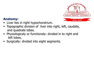
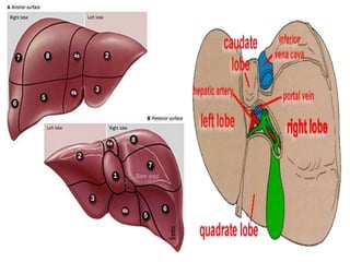
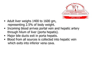
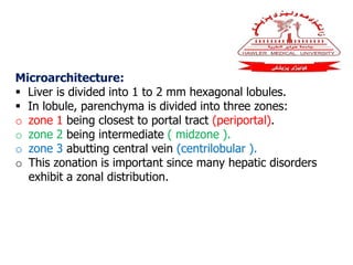
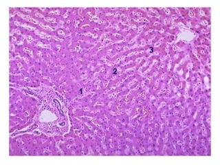
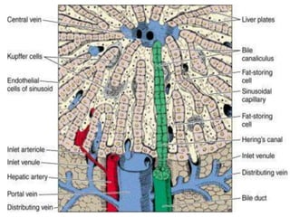
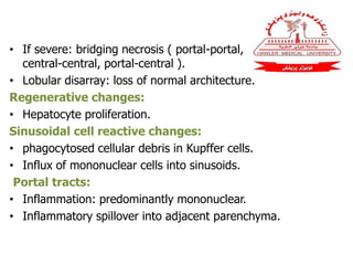
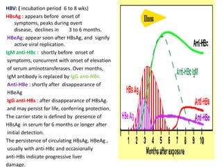
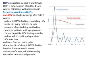
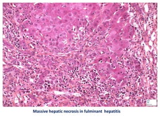
1. Liver lies in right hypochondrium and
divided in to right and left lobes.
2. Microarchitecture : liver is divided into
1 to 2 mm hexagonal lobules.
3. There are four methods for liver biopsy.
4. Most hepatic infections are viral in origin.
5. In fulminant hepatitis hepatic insufficiency
progresses from onset of symptoms to hepatic
encephalopathy within2 to 3 weeks.
Questions:
1. What are the pathological changes in chronic
viral hepatitis?
2. Draw a table for hepatotropic viruses including their
types, infectious routes, and diseases they cause?
THANK YOU

Liver 1

  • 1.
    Title : Liver Objectives:to 1. Describe anatomy and microarchitecture of liver. 2. Out line liver biopsy. 3. Study infectious disorders of liver . 4. Explain fulminant hepatitis.
  • 2.
    Anatomy:  Liver liesin right hypochondrium.  Topographic division of liver into right, left, caudate, and quadrate lobes.  Physiologicaly or functionaly: divided in to right and left lobes.  Surgically: divided into eight segments.
  • 4.
     Adult liverweighs 1400 to 1600 gm, representing 2.5% of body weight.  Incoming blood arrives portal vein and hepatic artery through hilum of liver (porta hepatis).  Major bile ducts exit in porta hepatis.  Blood from all sources is collected into hepatic vein which exits into inferior vena cava.
  • 5.
    Microarchitecture:  Liver isdivided into 1 to 2 mm hexagonal lobules.  In lobule, parenchyma is divided into three zones: o zone 1 being closest to portal tract (periportal). o zone 2 being intermediate ( midzone ). o zone 3 abutting central vein (centrilobular ). o This zonation is important since many hepatic disorders exhibit a zonal distribution.
  • 8.
    Liver Biopsy  Thereare four methods : 1. blind percutaneous needle biopsy. 2. peritoneoscopy . 3. open surgery . 4. transvenous (transjugular or less often transfemoral).  C T or U/S guided Fine Needle Aspiration (FNA) : for diagnosing focal hepatic lesions.
  • 9.
    Infectious Disorders:  Mosthepatic infections are viral in origin.  Others include: miliary tuberculosis, malaria, staph , salmonella , candida, and amebiasis. VIRAL HEPATITIS : caused by: (1) infectious mononucleosis (Epstein-Barr virus). (2) cytomegalovirus: newborn or immunosuppression. (3) yellow fever: tropical countries. (4) rubella, adenovirus, herpesvirus, or enterovirus : children and immunosuppression. (5) Hepatotropic viruses (most important).
  • 10.
    Viral hepatitis causedby Hepatotropic viruses VIRUS TYPE INFECTION ROUTE DISEASE Hepatitis A (HAV) RNA Fecal–oral Acute Hepatitis B (HBV) DNA Parenteral Acute, chronic Hepatitis C (HCV) RNA Parenteral Acute, chronic Hepatitis D (HDV) RNA Pathogenic when combined with HBV Acute, chronic Hepatitis E (HEV) RNA Fecal–oral Acute
  • 11.
    Acute viral Hepatitis Pathology: Parenchymalchanges: • Hepatocyte injury: swelling (ballooning degeneration). • Cholestasis (canalicular bile plugs). • Hepatocyte necrosis: isolated cells or cell clusters. • Cytolysis (rupture), or apoptosis (shrinkage) with acidophil bodies (Councilman bodies) formation.
  • 12.
    • If severe:bridging necrosis ( portal-portal, central-central, portal-central ). • Lobular disarray: loss of normal architecture. Regenerative changes: • Hepatocyte proliferation. Sinusoidal cell reactive changes: • phagocytosed cellular debris in Kupffer cells. • Influx of mononuclear cells into sinusoids. Portal tracts: • Inflammation: predominantly mononuclear. • Inflammatory spillover into adjacent parenchyma.
  • 14.
    Chronic Hepatitis:  Symptomatic,biochemical, or serologic evidence of continuing or relapsing hepatic disease for more than 6 months, with histologically documented inflammation and necrosis.  Hepatitis viruses (HBV, HCV, and HBV+HDV) are responsible for most cases.  Other causes include : o chronic alcoholism. o Wilson disease. o α1-antitrypsin deficiency. o drugs ( isoniazid, α-methyldopa, methotrexate ). o autoimmunity.
  • 15.
    Chronic viral Hepatitis: Pathology: Changesshared with acute hepatitis: • Hepatocyte injury, necrosis, and regeneration. • Sinusoidal cell reactive changes. Portal tracts: • Inflammation:  Confined to portal tracts.  Spill over into adjacent parenchyma, with necrosis of hepatocytes (interface hepatitis originally termed piecemeal necrosis).  Bridging inflammation and necrosis.
  • 16.
    Fibrosis:  Portal deposition. Portal and periportal deposition.  Formation of bridging fibrous septa. HBV :  ground-glass hepatocytes ( accumulations of viral antigen in endoplasmic reticulum ).  sanded nuclei ( nuclear viral inclusions ). HCV :  bile duct epithelial cell proliferation.  lymphoid aggregate formation . Cirrhosis : The end-stage outcome.
  • 17.
    Chronic viral hepatitis groundglass hepatocytes in chronic HBV
  • 18.
    Lymphoid follicles inportal tract of patient with chronic hepatitis C. Sanded nuclei (nuclear inclusions) are seen in HBV-replicative hepatocytes.
  • 19.
    Serologic Diagnosis inviral hepatitis HAV: Incubation period (2–6 wks). IgM: appears in blood at onset of symptoms, constituting a reliable Marker of acute infection . Fecal shedding of virus ends as IgM titer rises. IgM begins to decline in few months and is followed by appearance of IgG which persists for years, perhaps for life, providing Protective immunity against Reinfection by all strains of HAV, Hence HAV vaccine is effective.
  • 20.
    HBV: ( incubationperiod 6 to 8 wks) HBsAg : appears before onset of symptoms, peaks during overt disease, declines in 3 to 6 months. HBeAg: appear soon after HBsAg, and signify active viral replication. IgM anti-HBc : shortly before onset of symptoms, concurrent with onset of elevation of serum aminotransferases. Over months, IgM antibody is replaced by IgG anti-HBc. Anti-HBe : shortly after disappearance of HBeAg IgG anti-HBs : after disappearance of HBsAg. and may persist for life, conferring protection. The carrier state is defined by presence of HBsAg in serum for 6 months or longer after initial detection. The persistence of circulating HBsAg, HBeAg , usually with anti-HBc and occasionally anti-HBs indicate progressive liver damage.
  • 21.
    HCV : incubationperiod 6 and 12 wks . HCV is detectable in blood for 1 to 3 weeks, coincident with elevations in Serum transaminases (ALT) . anti-HCV antibodies emerge after 3 to 6 weeks. In chronic HCV infection, circulating HCV persists in many patients despite presence of neutralizing antibodies, Hence, in patients with symptoms of chronic hepatitis, HCV testing must be performed to confirm diagnosis of HCV infection. A clinical feature that is quite characteristic of chronic HCV infection is episodic elevations in serum aminotransferases, with intervening normal or near-normal periods.
  • 22.
    Fulminant hepatitis andhepatic failure: Hepatic insufficiency progresses from onset of symptoms to hepatic encephalopathy within 2 to 3 weeks. Causes : o HAV or HBV. o Drug and chemical toxicity : acetaminophen , isoniazid , halothane, and methyldopa. o Miscellaneous : mycotoxins of mushroom. o Unknown : 18% of cases
  • 23.
    Clinical features:  Jaundice, encephalopathy, and fetor hepaticus.  Mortality rate ranges from 25% to 90% in absence of liver transplantation. Morphology :  Grossly: necrotic areas have a muddy red appearance .  Microscopically : complete destruction of hepatocytes in contiguous lobules leaves only a collapsed reticulin framework and preserved portal tracts. there may be surprisingly little inflammation.
  • 24.
    Massive hepatic necrosisin fulminant hepatitis
  • 25.
    Summary: 1. Liver liesin right hypochondrium and divided in to right and left lobes. 2. Microarchitecture : liver is divided into 1 to 2 mm hexagonal lobules. 3. There are four methods for liver biopsy. 4. Most hepatic infections are viral in origin. 5. In fulminant hepatitis hepatic insufficiency progresses from onset of symptoms to hepatic encephalopathy within2 to 3 weeks.
  • 26.
    Questions: 1. What arethe pathological changes in chronic viral hepatitis? 2. Draw a table for hepatotropic viruses including their types, infectious routes, and diseases they cause? THANK YOU