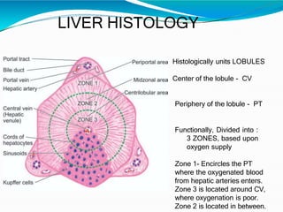
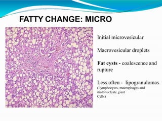
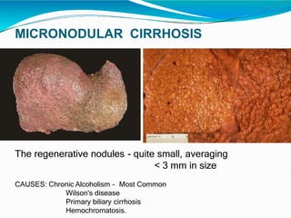
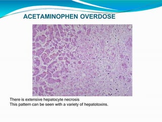
SlideShare a Scribd company logo
LIVER PATHOLOGY
Contents
•Introdution
•Normal anatomy & Histology
•Biopsy
•Viral hepatitis
•Steatosis and steatohepatitis
•Cirrhosis
•Vascular disorders
•Pigmentary disorders
•Drug/ toxin mediated injury
•Benign Epithelial tumors
•Malignant Epithelial tumors
•Mesenchymal tumors
•Metastatic tumors
LIVER
⚫Largest internal organ
⚫1.4–1.6 kg (3.1–3.5 lb)
⚫Reddish brown soft organ with four lobes of unequal
size and shape
Histologically units LOBULES
Center of the lobule - CV
Periphery of the lobule - PT
Functionally, Divided into :
3 ZONES, based upon
oxygen supply
Zone 1- Encircles the PT
where the oxygenated blood
from hepatic arteries enters.
Zone 3 is located around CV,
where oxygenation is poor.
Zone 2 is located in between.
LIVER HISTOLOGY
CV
PT
Liver Biopsy
•Liver histopathology remains a mainstay in the
diagnostic work-up of most liver diseases
•Biopsy remains the gold standard for the diagnosis
of cellular rejection
•Adequacy of biopsy,
•Site of biopsy (is it from liver?),
•Architecture on low power;
•Assess inflammation (degree, type),
•Necrosis, fibrosis, tumor cells
Pattern Recognition
• Acute Hepatitis
• Chronic hepatitis
• Fatty liver disease
• Biliary Pathology
• Vascular Pathology
• Metabolic liver disease
• Architecture and fibrosis
STEATOSIS
⚫This liver is slightly
enlarged
⚫Tense capsule
⚫Pale yellow seen
both on external
and cut surface
FATTY CHANGE: GROSS
Initial microvesicular
Macrovesicular droplets
Fat cysts - coalescence and
rupture
Less often - lipogranulomas
(Lymphocytes, macrophages and
multinucleate giant
Cells)
FATTY CHANGE: MICRO
VIRAL HEPATITIS
Microscopy :
Ballooning degeneration
Spotty necrosis
Predominantly sinusoidal and
lobular mononuclear cell infiltrate
(occasional neutrophils and
eosinophils)
Kupffer cell hyperplasia
Scattered apoptotic bodies
Canalicular cholestasis
Hepatocellular regeneration
Microscopy of CVH
• Predominantly portal tract inflammation
• Interface hepatitis
• Lobular acidophil bodies
•Mononuclear inflammatory cells, +/-plasma
cells
•Portal-based lymphoid aggregates and
lymphoid follicle formation
• Bile ductular proliferation
• Progressive fibrosis with eventual cirrhosis
VIRAL HEPATITIS: TRICHROME
This trichrome stain demonstrates the collapse of the liver parenchyma with
viral hepatitis. The blue-staining areas are the connective tissue of many portal
tracts that have collapsed together.
Hepatitis A - mononuclear infiltrate
rich in plasma cells
Hepatitis B - “ground-glass”
hepatocytes - cells with
endoplasmic
reticulum swollen by HBsAg
Hepatitis C - shows lymphoid
aggregates or fully formed
lymphoid follicles
ALCOHOLIC
HEPATITIS
- Mallory's hyaline, neutrophils, necrosis of hepatocytes,
collagen deposition, and fatty change
MALLORY'S HYALINE
- Rope like red hyaline material within hepatocytes
- also known as "alcoholic" hyaline, Mallory- Denk Bodies - chronic
alcoholism
- aggregates of intermediate filaments in the cytoplasm resulting from
hepatocyte injury.
CIRRHOSIS
CIRRHOSIS
⚫Diffuse nodulation of liver due to fibrous bands
subdividing liver into regenerative nodules
⚫Nodules : >3 mm - macronodular
< less than 3 mm - Micronodular
MICRONODULAR CIRRHOSIS
The regenerative nodules - quite small, averaging
< 3 mm in size
CAUSES: Chronic Alcoholism – Most Common
Wilson's disease
Primary biliary cirrhosis
Hemochromatosis.
MACRONODULAR
CIRRHOSIS
CAUSES: Viral hepatitis (B or C) - most common cause
Wilson's disease
Alpha-1-antitrypsin deficiency
CIRRHOSIS: micro
Regenerative nodules of hepatocytes are surrounded by fibrous
connective tissue that bridges between portal tracts.
Within this collagenous tissue are scattered lymphocytes as well as a
proliferation of bile ducts.
PRIMARY BILIARY CIRRHOSIS
A rare autoimmune disease
(mostly of middle-aged women)
Characterized by destruction of
bile ductules within the triads of the
liver.
Intense chronic inflammatory
infiltrate
Loss of bile ductules.
Micronodular cirrhosis
PIGMENTARY DISORDERS
OF THE LIVER
HEMOSIDEROSIS
Hemosiderosis - accumulation of iron.
The hepatocytes and Kupffer cells here
are full of granular brown deposits of
hemosiderin from accumulation of
excess iron in the liver.
Hemochromatosis - used when organ
dysfunction occurs.
The iron accumulation may lead to a
micronodular cirrhosis - "pigment"
cirrhosis).
Iron stain highlights the periportal predominance of iron
deposition seen in hemochromatosis.
There are larger dark blue granules in the hepatocytes closer
to the portal tracts.
HEREDITARY HEMOCHROMATOSIS (HHC):
GROSS
- Dark brown - liver, the pancreas and lymph nodes -due to
extensive iron deposition
- HHC results from a mutation involving the hemochromatosis gene (HFE)
that leads to increased iron absorption from the gut.
LIPOFUSCIN PIGMENT
-Pale golden brown finely granular pigment in nearly all hepatocytes is
lipochrome (lipofuscin).
-This is a "wear and tear" pigment from the accumulation of
autophagolysosomes over time.
- This pigment is of no real pathologic importance
CHOLESTASIS: MICRO
- accumulations of pigment - bile
- often this is due to extrahepatic biliary tract obstruction
- Bile may also accumulate in liver (called cholestasis) when there is
hepatocyte injury.
INTRAHEPATIC LITHIASIS
- Small stone in an intrahepatic bile duct
- Produce a localized cholestasis, but the serum bilirubin is NOT
increased, because there is plenty of non-obstructed liver to
clear the bilirubin from the blood
- Serum Alkaline Phos is increased with biliary tract obstruction at any
level
VASCULAR
DISORDERS
CHRONIC PASSIVE CONGESTION
"nutmeg" liver - chronic passive congestion of the liver.
the dark red congested regions that represent accumulation of
RBC's in centrilobular regions.
This is usually due to a "RIGHT SIDED" heart failure.
CENTRILOBULAR NECROSIS
If the passive congestion is pronounced, then there can be centrilobular
necrosis, because the oxygenation in zone 3 of the hepatic lobule is
decreased.
The light brown pigment in the necrotic hepatocytes around the central vein is
lipochrome.
CPC - CARDIAC CIRRHOSIS
"cardiac cirrhosis" - fibrosis bridging between central zonal regions,
The portal tracts appear to be in the center of the reorganized lobule.
Unlike a true cirrhosis, there is minimal nodular regeneration.
INFARCTION OF THE LIVER
Infarcts are uncommon because the liver has two blood supplies-portal
venous system and hepatic arterial system.
The infarcts seen here are yellow, with geographic borders and surrounding
hyperemia.
About half of liver infarcts occur with arteritis, and the remaining half are due
to a variety of causes.
ACETAMINOPHEN OVERDOSE
There is extensive hepatocyte necrosis
This pattern can be seen with a variety of hepatotoxins.
POLYCYSTIC KIDNEY DISEASE
(PCKD) - POLYCYSTIC LIVER
- Manifest with renal failure beginning in
middle age
- Sometimes the liver can be affected as
well by polycystic change.
- Less commonly the pancreas is involved.
- These patients with DPKD can also have
berry aneurysms in the cerebral arteries.
EXTRAHEPATIC BILIARY ATRESIA
•Inflammation with stricture of
hepatic or common bile ducts.
•This leads to marked cholestasis with
intrahepatic bile duct proliferation,
fibrosis, and cirrhosis.
•The liver is rock hard.
•The dark green color comes from
formalin acting on bile pigments in the
liver from marked cholestasis, turning
bilrubin to biliverdin.
EXTRAHEPATIC BILIARY ATRESIA
Microscopy:
•Numerous brown-green bile plugs,
•Bile duct proliferation
•Extensive fibrosis.
α-1-ANTITRYPSIN DEFICIENCY
AAT deficiency - chronic obstructive
pulmonary disease with panlobular
emphysema.
The periportal red hyaline globules seen
with periodic acid-Schiff (PAS) stain
The globules are collections of alpha-1-
antitrypsin not being excreted from
hepatocytes.
This may eventually lead to chronic
hepatitis and cirrhosis.
TUMORS OF THE
LIVER &
INTRAHEPATIC DUCTS
BENIGN EPITHELIAL
TUMORS
FOCAL NODULAR HYPERPLASIA
(FNH)
⚫Benign nonneoplastic hepatic lesion,
⚫Arising in a noncirrhotic liver parenchyma
⚫Most common in young adults
⚫May represent hyperplastic response to arterial
malformation or other vascular anomaly
Gross :
•Well demarcated,
•Unencapsulated
•Solitary lesion
•Lighter in color than the surrounding liver parenchyma
•Subcapsular region
•Measure < 5 cm
•Cut surface :
•Central scar
•Radiating fibrous septa
Micro :
•Bland hepatocytes
•Surrounded by fibrous septa
•Artery branches
•Bile ductular reaction
•Variable amount of mixed
inflammatory infiltrate
•Portal tracts are absent except
at periphery
•Hepatocytes - similar to
those in the surrounding
liver
•1 - 2 cells thick
•Supported by an intact
reticulin framework
FNH
⚫Positive stains: alpha-1-antitrypsin
⚫Negative stains: p53, CD143 (angiotensin I-converting
enzyme: reduced expression
⚫DD:
⚫ Budd-Chiari syndrome or cirrhosis (adjacent liver is not
normal)
⚫ Fibrolamellar hepatocellular carcinoma (marked atypia of
hepatocytes)
⚫ Hepatocellular adenoma (encapsulated, monoclonal)
⚫ Peritumoral hyperplasia
⚫ Mesenchymal hamartoma
GROSS: LIVER CELL ADENOMA
⚫Solitary (70 %), pale, yellow-tan
⚫bile-stained nodules,
⚫Often subcapsular, 10-30 cm,
⚫Sharply demarcated
⚫Usually right lobe,
⚫Adjacent liver is noncirrhotic
LIVER CELL ADENOMA: MICRO
⚫Sheets and cords 1-3 cells thick of normal appearing hepatocytes
with variable glycogen
⚫Prominent “free floating” arterial vessels and draining veins
throughout the tumor
⚫Intact reticulin framework
⚫No/rare mitotic figures
⚫No portal tracts, no central
veins or connection with biliary
system
LIVER CELL ADENOMA
⚫Positive stains: ER, PR
⚫Negative stains: p53
⚫DD:
⚫HEPATOCELLULAR CARCINOMA (mitotic activity,
atypia, trabecular growth, cell plates > 2 cells thick,
vascular invasion, infiltrative, often different clinical
features)
⚫ FOCAL NODULAR HYPERPLASIA (central stellate
scar and radiating fibrous septa)
BILE DUCT ADENOMA (Cholangioma)
⚫30% - incidental finding
⚫GROSS:
Small, well-circumscribed but unencapsulated,
firm, gray-white/ tan,
subcapsular nodule;
85% solitary;
usually 5 mm or less
BDA: MICRO:
⚫Small tubules set in a fibrous
stroma with lymphocytes
⚫Single layer of cuboidal cells;
⚫possible mucin secretion;
⚫no bile in the lumen
⚫Normal portal tracts often included
Biliary Hamartoma
⚫ Within cirrhotic livers, most commonly
seen chronic hepatitis C and alcohol-
related liver disease
⚫ single or multiple
Micro :
•composed of bile duct
structures that grow in an
irregular inter-anastomosing
fashion.
•duct structures have open
lumens, associated with either a
myxoid or fibrotic stroma
Intraductal
papillary
neoplasm of the
bile ducts
GROSS:
⚫Inner surface of ducts has velvety friable papillary
growths / excrescences with masses filling dilated major
bile ducts
⚫Masses are soft, friable, white-red-tan
Micro:
⚫Multiple papillary tumors
composed of fibrovascular
cores
⚫Lined by columnar,
pseudostratified, biliary-type
cells with numerous
cytoplasmic mucin vacuoles
⚫Ovarian stroma is absent
⚫Varying cytologic atypia and
mitotic activity
⚫May have associated tubular
adenocarcinoma with invasion
Differential diagnosis
Mucinous cystic neoplasm
Also a cystic hepatic lesion
Lined by biliary type epithelium
Has ovarian type stroma and
Does not communicate with biliary lumen
Cholangiocarcinoma
May show areas of cystic degeneration
Does not communicate with biliary lumen
Simple cyst, simple hepatic cyst, Simple biliary cyst, bile
duct cyst
Lacks papillary projections
MALIGNANT TUMORS
Hepatocellular Carcinoma
GROSS
⚫Unifocal, multifocal or diffusely infiltrative soft tumor,
⚫Paler than normal tissue, may be green due to bile
⚫Extensive intrahepatic metastases are common
⚫Snakelike masses of tumor may involve the portal
vein (35-80%), hepatic vein (20%) or inferior vena
cava
⚫Hemorrhage and necrosis are common
⚫Occasionally tumor is pedunculated
⚫Liver usually cirrhotic, often enlarged
HEPATOCELLULAR CARCINOMA
HCC: MICRO
Patterns:
⚫ Trabecular (most common) - 4+ cells surrounded by
layer of flattened endothelial cells
⚫ Pseudoglandular (acinar with proteinaceous material or bile in
lumina)
⚫ Solid and macrotrabecular
Cells
⚫ Polygonal with distinct cell membranes
⚫ Higher N/C ratio
⚫ Abundant granular eosinophilic cytoplasm
⚫ Round nuclei with coarse chromatin and thickened
nuclear membrane
⚫ Prominent nucleoli
⚫Sinusoidal vessels surrounding tumor cells is important
diagnostic feature
⚫Scanty stroma, from well differentiated to bizarre (often
within same tumor)
HEPATOCELLULAR CARCINOMA
Differential diagnosis
1. Hepatocellular adenoma:
Neoplastic hepatocytes are not expanded on reticulin stain (no loss)
Noncirrhotic background
2. Intrahepatic cholangiocarcinoma:
Discrete gland formation surrounded by desmoplastic stroma
Negative for hepatocellular markers; positive for CK7
3. Dysplastic nodule in cirrhosis:
~ 1 cm lesion
Preserved portal tracts
4. Metastatic neuroendocrine neoplasms:
Positive staining for neuroendocrine markers while negative for hepatocellular markers
5. Metastatic carcinomas
Possible clinical history of prior malignancy
Differing immunohistochemical profile
CLEAR CELL VARIANT - HCC
⚫Predominant appearance in
5-16% of cases, but some
clear cells present in 20-40%
of cases
⚫Tumor cells have prominent
clear cytoplasm due to
cytoplasmic fat or glycogen
⚫May need to hunt for
typical HCC to rule out
metastatic tumor
⚫Similar prognosis to classic
tumor
FIBROLAMELLAR VARIANT- HCC
GROSS:
⚫Single (75%)
⚫Large (mean 13 cm)
⚫Hard
⚫Scirrhous
⚫Well-circumscribed
⚫Bulging
⚫White-brown tumor with fibrous bands throughout and
central stellate scar
⚫Most cases involve left lobe, but may involve both lobes
⚫Variable bile staining, hemorrhage and necrosis
FIBROLAMELLAR VARIANT - HCC
Micro:
⚫ Nests, sheets or cords of well
differentiated oncocytic cells in
⚫ Background of dense, acellular
collagen bundles
⚫ That may contain small, thick-
walled vessels
⚫ Fibrotic tissue coalesces into
central scar
Differential diagnosis
1. Cholangiocarcinoma:
Truly glandular, often conspicuous pleomorphism
2. Focal nodular hyperplasia:
Ductular reaction around central scar
Lesional cells are not characteristically oncocytic
Does not have the nuclear features of fibrolamellar carcinoma
3. Hepatocellular carcinoma, sclerosing variant:
No oncocytes, smaller tumor cells, pseudoglandular pattern common
4. Metastatic carcinoma with sclerotic stroma:
Conspicuous pleomorphism
5. Neuroendocrine tumors:
Nuclear features are often not typical of fibrolamellar carcinoma
Positive for neuroendocrine marker
ONCOCYTIC VARIANT -HCC
⚫Oncocytes are present in fibrolamellar variant and
⚫Occasionally in classic hepatocellular carcinoma
⚫Rarely these cells predominate without fibrous
stroma of fibrolamellar variant
⚫Cytoplasm is intensely eosinophilic with coarse
granules
PLEOMORPHIC (GIANT CELL) VARIANT
⚫<1% of all hepatocellular
carcinomas,
⚫Although 15% have some
tumor giant cells
⚫Multinucleated tumor giant
cells predominate,
⚫marked loss of cell cohesion
SARCOMATOID VARIANT - HCC
⚫1-9% of all HCC have prominent
sarcomatoid pattern
⚫Diffuse collection of spindle cells
resembling fibrosarcoma
⚫Classic HCC is also present;
⚫May have pleomorphic and
osteoclast-like giant cells
Differential diagnosis
1. Undifferentiated pleomorphic sarcoma and fibrosarcoma
No area of classic HCC
Immunohistochemistry (entirely negative for pancytokeratin)
2. Metastatic sarcomatoid carcinoma:
Will not have area of classic HCC
Usually multiple nodules
Immunohistochemistry (pancytokeratin+)
Negative for hepatocellular markers,
SCLEROSING: HCC
Micro:
⚫Fibrous septa separate trabecular cell plates but no
lamellar fibrosis
⚫Cell plates 3 or more cells thick
⚫Tumor cells may have pseudoglandular (acinar)
features compared to fibrolamellar variant
⚫Tumor cells are smaller
⚫Lack vesicular nuclei and prominent nucleoli
⚫Have less abundant and granular cytoplasm
⚫No apparent endothelial sinusoidal cells
CHOLANGIOCARCINOMA
Gross:
Large, nonencapsulated, well
demarcated,
Firm, white-tan to gray
Nodular intrahepatic mass
More frequent in the right lobe of the
liver
Satellite nodules in 30%
Noncirrhotic background liver in
most cases
Grossly - 3 growth patterns
1. Mass forming: hepatic parenchymal solid mass
2. Periductal infiltrating: infiltrates along the portal
tracts, causing bile duct strictures
3. Intraductal growth: papillary or polypoid growth
inside a dilated bile duct
Micro:
⚫Moderate to well differentiated adeno
ca
⚫Glandular and tubular structures,
⚫Mucin production and dense
desmoplasia
⚫Epithelial cells are anaplastic,
cuboidal to columnar with
eosinophilic cytoplasm and round
central nuclei,
⚫Tumor cells are heterogeneous even
within the same gland but resemble
bile duct cells, not hepatocytes
⚫Multicentricity and perineural
invasion are common
HEPATOBLASTOMA
•Malignant primary hepatic blastomatous tumor
•Most frequent liver tumor in children
•1 - 1.5 cases per million
•80 - 90% between 5 months to 6 years old
•Slight male predominance
Gross :
•Single or multinodular
•Well defined boundaries
•Tan-brown cut surface
•Undifferentiated subtypes have a variegated appearance
•Necrosis and hemorrhage are present in posttreatment
specimens
•Osteoid may be present- firm and gritty
Fetal pattern
•Small to medium sized cells
•Thin trabeculae or nests
•Resembling hepatocytes of
fetal liver
•Clear or finely granular
cytoplasm
•Small round nucleus with
indistinct nucleolus
•Foci of extramedullary
hematopoiesis are usually
present
•Low mitotic activity,
•Referred to as well
differentiated hepatoblastoma
Embryonal pattern
•Resembles liver at 6 - 8 weeks of gestation
•Solid nests or glandular / acinar morphology
•Papillae and pseudorosettes
•Dark and granular cytoplasm without glycogen or lipids
•Enlarged nuclei with coarse chromatin,
•Resembling blastemal cells
•Extramedullary hematopoiesis- absent
•Increased mitotic activity
Small cell undifferentiated
pattern
•Small, round blue tumor
•Solid sheets of
discohesive small cells
•Abundant mitoses,
apoptosis and necrosis
Cholangioblastic pattern
Small ducts within or around hepatocellular components
Mesenchymal pattern
•Mature and immature fibrous tissue
•Osteoid or osteoid-like tissue
•Hyaline cartilage
•Small subset may exhibit teratoid features:
endodermal, neuroectodermal (neuronal cells, glial
tissue, melanin producing cells) and complex
tissue (striated muscle)
MESENCHYMAL
TUMORS
HEMANGIOMA: GROSS
Gross:
⚫solitary (70-90%), usually 2-
4 cm, although tumors up to
20 cm are overrepresented
in studies of excisions
⚫Soft, red-purple, well
circumscribed
⚫Subcapsular or deep
⚫Collapse when sectioned as
blood oozes out
HEMANGIOMA: MICRO
Micro:
⚫Variably sized vascular
spaces
⚫Lined by flat endothelial
cells and
⚫Fibrous stroma
Epithelioid hemangioendothelioma
Low grade Malignant endothelial neoplasm
GROSS :
Poorly circumscribed, firm, white-tan mass
Variable size, up to 18 cm
MICROSCOPY
oCords, strands or small nests of large endothelial cells with
abundant eosinophilic cytoplasm
oEmbedded in a myxohyaline stroma
oTumor cells have vesicular, round to oval, sometimes indented
nuclei
oIntracytoplasmic, round,
clear vacuoles representing
small vascular lumina,
oMay contain erythrocytes
Peliosis Hepatis
Peliosis hepatis represents blood-fi lled cysts
in the hepatic parenchyma
certain drugs,particularly anabolic steroids,
high-dose oral contraceptive,
or azathioprine in patients with solid organ
transplants,TB,IMMUNO COMPRO MISED
Most cases ofpeliosis are detected
incidentally,
but rupture,intraperitoneal hemorrhage,and
death
have been reported.Peliosis may regress on
stopping hormone
or drug causing the lesion.
M/E blood-filled cystic spaces,separated by
cords of normal or compressed hepatocytes,
distributed randomly in liver parenchyma
METASTASES TO THE LIVER
Numerous mass lesions that are of variable size
Larger ones demonstrate central necrosis
METASTATIC CARCINOMA
Microscopically, metastatic infiltrating ductal carcinoma from breast is seen
on the right, with normal liver parenchyma on the left.
REFERNCES :
1. Mills SE , Carter D ,Sternberg diagnostic pathology , 4th
edition , Lipincott Williams and wilkins.
2. Rosai J,Rosai and Ackerman surgical pathology, 9th edition
3. Differential diagnosis in surgicalpathology ,3rd edition
4. Robbins and cotran, Pathologic basis of disease.
5. Internet sources
THANK YOU
liver pathology.pptx

More Related Content

What's hot

Non neoplastic lesions of lymph node
Non neoplastic lesions of lymph nodeNon neoplastic lesions of lymph node
Non neoplastic lesions of lymph node
Manoj Madakshira Gopal
 
approach to lymph node cytology part 1
approach to lymph node cytology part 1approach to lymph node cytology part 1
approach to lymph node cytology part 1
Kamalesh Lenka
 
Glomerular diseases
Glomerular diseases Glomerular diseases
Glomerular diseases
Mohammad Manzoor
 
Diseases of the kidney
Diseases of the kidneyDiseases of the kidney
Diseases of the kidney
Dr. Varughese George
 
Gastric polyps
Gastric polypsGastric polyps
Endothelium in health and diseases
Endothelium in health and diseasesEndothelium in health and diseases
Endothelium in health and diseases
Meghana P
 
Unusual type and site of Lymphoma(Extranodal Lymphoma) Dr.Argha Baruah
Unusual type and site of Lymphoma(Extranodal Lymphoma) Dr.Argha BaruahUnusual type and site of Lymphoma(Extranodal Lymphoma) Dr.Argha Baruah
Unusual type and site of Lymphoma(Extranodal Lymphoma) Dr.Argha Baruah
Argha Baruah
 
cytology of the breast
cytology of the breastcytology of the breast
cytology of the breast
Hayelom kassaye
 
Intestinal polyps
Intestinal polypsIntestinal polyps
Neoplasm of hematopoietic tissue
Neoplasm of hematopoietic tissueNeoplasm of hematopoietic tissue
Neoplasm of hematopoietic tissue
Emmanuel Oppong
 
High Grade Papillary Serous Cystadenocarcinoma of Bilateral Ovaries with Caps...
High Grade Papillary Serous Cystadenocarcinoma of Bilateral Ovaries with Caps...High Grade Papillary Serous Cystadenocarcinoma of Bilateral Ovaries with Caps...
High Grade Papillary Serous Cystadenocarcinoma of Bilateral Ovaries with Caps...
Dr. Varughese George
 
Eosinophils in lymph node
Eosinophils in lymph nodeEosinophils in lymph node
Eosinophils in lymph node
Monika Nema
 
ADA
ADAADA
Renal Pathology Lectures_Ppt Series (4 in 1)
Renal Pathology Lectures_Ppt Series (4 in 1)Renal Pathology Lectures_Ppt Series (4 in 1)
Renal Pathology Lectures_Ppt Series (4 in 1)
Imhotep Virtual Medical School
 
Cytology of bone lesions
Cytology of bone lesionsCytology of bone lesions
Interpretation of endoscopic intestinal biopsy
Interpretation of endoscopic intestinal biopsyInterpretation of endoscopic intestinal biopsy
Interpretation of endoscopic intestinal biopsy
Appy Akshay Agarwal
 
Chronic myeloid Leukemia
Chronic myeloid LeukemiaChronic myeloid Leukemia
Chronic myeloid Leukemia
MLT LECTURES BY TANVEER TARA
 
Ulcers of the intestine
Ulcers of the intestineUlcers of the intestine
Ulcers of the intestine
Manoj Madakshira Gopal
 
Paroxysmal nocturnal hemoglobinuria (PNH)
Paroxysmal nocturnal hemoglobinuria (PNH)Paroxysmal nocturnal hemoglobinuria (PNH)
Paroxysmal nocturnal hemoglobinuria (PNH)
DrAbdulrazzaqAlagbar
 
Non hodgkin lymphoma
Non hodgkin lymphomaNon hodgkin lymphoma
Non hodgkin lymphoma
tashagarwal
 

What's hot (20)

Non neoplastic lesions of lymph node
Non neoplastic lesions of lymph nodeNon neoplastic lesions of lymph node
Non neoplastic lesions of lymph node
 
approach to lymph node cytology part 1
approach to lymph node cytology part 1approach to lymph node cytology part 1
approach to lymph node cytology part 1
 
Glomerular diseases
Glomerular diseases Glomerular diseases
Glomerular diseases
 
Diseases of the kidney
Diseases of the kidneyDiseases of the kidney
Diseases of the kidney
 
Gastric polyps
Gastric polypsGastric polyps
Gastric polyps
 
Endothelium in health and diseases
Endothelium in health and diseasesEndothelium in health and diseases
Endothelium in health and diseases
 
Unusual type and site of Lymphoma(Extranodal Lymphoma) Dr.Argha Baruah
Unusual type and site of Lymphoma(Extranodal Lymphoma) Dr.Argha BaruahUnusual type and site of Lymphoma(Extranodal Lymphoma) Dr.Argha Baruah
Unusual type and site of Lymphoma(Extranodal Lymphoma) Dr.Argha Baruah
 
cytology of the breast
cytology of the breastcytology of the breast
cytology of the breast
 
Intestinal polyps
Intestinal polypsIntestinal polyps
Intestinal polyps
 
Neoplasm of hematopoietic tissue
Neoplasm of hematopoietic tissueNeoplasm of hematopoietic tissue
Neoplasm of hematopoietic tissue
 
High Grade Papillary Serous Cystadenocarcinoma of Bilateral Ovaries with Caps...
High Grade Papillary Serous Cystadenocarcinoma of Bilateral Ovaries with Caps...High Grade Papillary Serous Cystadenocarcinoma of Bilateral Ovaries with Caps...
High Grade Papillary Serous Cystadenocarcinoma of Bilateral Ovaries with Caps...
 
Eosinophils in lymph node
Eosinophils in lymph nodeEosinophils in lymph node
Eosinophils in lymph node
 
ADA
ADAADA
ADA
 
Renal Pathology Lectures_Ppt Series (4 in 1)
Renal Pathology Lectures_Ppt Series (4 in 1)Renal Pathology Lectures_Ppt Series (4 in 1)
Renal Pathology Lectures_Ppt Series (4 in 1)
 
Cytology of bone lesions
Cytology of bone lesionsCytology of bone lesions
Cytology of bone lesions
 
Interpretation of endoscopic intestinal biopsy
Interpretation of endoscopic intestinal biopsyInterpretation of endoscopic intestinal biopsy
Interpretation of endoscopic intestinal biopsy
 
Chronic myeloid Leukemia
Chronic myeloid LeukemiaChronic myeloid Leukemia
Chronic myeloid Leukemia
 
Ulcers of the intestine
Ulcers of the intestineUlcers of the intestine
Ulcers of the intestine
 
Paroxysmal nocturnal hemoglobinuria (PNH)
Paroxysmal nocturnal hemoglobinuria (PNH)Paroxysmal nocturnal hemoglobinuria (PNH)
Paroxysmal nocturnal hemoglobinuria (PNH)
 
Non hodgkin lymphoma
Non hodgkin lymphomaNon hodgkin lymphoma
Non hodgkin lymphoma
 

Similar to liver pathology.pptx

liver pathgology.pptx
liver pathgology.pptxliver pathgology.pptx
liver pathgology.pptx
gimspathcme2022
 
Liver surgical pathology
Liver surgical pathologyLiver surgical pathology
Liver surgical pathology
specialclass
 
LIVER HISTOPATHOLOGY (Budd chiari syndrome, Cirrohosis, Chlangiocarcinoma)
LIVER HISTOPATHOLOGY (Budd chiari syndrome, Cirrohosis, Chlangiocarcinoma)LIVER HISTOPATHOLOGY (Budd chiari syndrome, Cirrohosis, Chlangiocarcinoma)
LIVER HISTOPATHOLOGY (Budd chiari syndrome, Cirrohosis, Chlangiocarcinoma)
Dr. Shubhi Saxena
 
Liver 2
Liver 2Liver 2
Lecture 1-8th-nov-07
Lecture 1-8th-nov-07Lecture 1-8th-nov-07
Lecture 1-8th-nov-07
Waleed Rafiq
 
Renal pathology iv
Renal pathology ivRenal pathology iv
Renal pathology iv
med_students0
 
Bleeding and thrombosis final
Bleeding and thrombosis final Bleeding and thrombosis final
Bleeding and thrombosis final
DR MANOJ PRABHAKARAN
 
The diseases of the hepatobiliary system
The diseases of the hepatobiliary systemThe diseases of the hepatobiliary system
The diseases of the hepatobiliary system
Dr. Varughese George
 
Inherited tubular disorders
Inherited tubular disorders Inherited tubular disorders
Inherited tubular disorders
Chetan Ganteppanavar
 
Liver & liver diseases
Liver & liver diseasesLiver & liver diseases
Liver & liver diseases
HIRANGER
 
01 jfailcirr
01 jfailcirr01 jfailcirr
01 jfailcirr
Hotimah Masdan Salim
 
Aubf lec block b
Aubf lec block bAubf lec block b
Aubf lec block b
Hazel Acosta
 
Liver abscess
Liver abscessLiver abscess
Liver abscess
pukar kc
 
Cirrhosis of the Liver.pptx..............
Cirrhosis of the Liver.pptx..............Cirrhosis of the Liver.pptx..............
Cirrhosis of the Liver.pptx..............
MadhuSM4
 
Liver & its diseases
Liver & its diseasesLiver & its diseases
Liver & its diseases
RAJESH EAPEN
 
Vascular disordersof hepatobiliary
Vascular disordersof hepatobiliaryVascular disordersof hepatobiliary
Vascular disordersof hepatobiliary
Rashed Hassen
 
IMAGING IN PORTAL HYPERTENSION.pptx
IMAGING IN PORTAL HYPERTENSION.pptxIMAGING IN PORTAL HYPERTENSION.pptx
IMAGING IN PORTAL HYPERTENSION.pptx
aasrithakotha2
 
Pathology of Blood and Urine
Pathology of Blood and UrinePathology of Blood and Urine
Pathology of Blood and Urine
Monika P. Maske
 
Spleen
SpleenSpleen
parvati- hemo- final.pptx
parvati- hemo- final.pptxparvati- hemo- final.pptx
parvati- hemo- final.pptx
DarshanS239776
 

Similar to liver pathology.pptx (20)

liver pathgology.pptx
liver pathgology.pptxliver pathgology.pptx
liver pathgology.pptx
 
Liver surgical pathology
Liver surgical pathologyLiver surgical pathology
Liver surgical pathology
 
LIVER HISTOPATHOLOGY (Budd chiari syndrome, Cirrohosis, Chlangiocarcinoma)
LIVER HISTOPATHOLOGY (Budd chiari syndrome, Cirrohosis, Chlangiocarcinoma)LIVER HISTOPATHOLOGY (Budd chiari syndrome, Cirrohosis, Chlangiocarcinoma)
LIVER HISTOPATHOLOGY (Budd chiari syndrome, Cirrohosis, Chlangiocarcinoma)
 
Liver 2
Liver 2Liver 2
Liver 2
 
Lecture 1-8th-nov-07
Lecture 1-8th-nov-07Lecture 1-8th-nov-07
Lecture 1-8th-nov-07
 
Renal pathology iv
Renal pathology ivRenal pathology iv
Renal pathology iv
 
Bleeding and thrombosis final
Bleeding and thrombosis final Bleeding and thrombosis final
Bleeding and thrombosis final
 
The diseases of the hepatobiliary system
The diseases of the hepatobiliary systemThe diseases of the hepatobiliary system
The diseases of the hepatobiliary system
 
Inherited tubular disorders
Inherited tubular disorders Inherited tubular disorders
Inherited tubular disorders
 
Liver & liver diseases
Liver & liver diseasesLiver & liver diseases
Liver & liver diseases
 
01 jfailcirr
01 jfailcirr01 jfailcirr
01 jfailcirr
 
Aubf lec block b
Aubf lec block bAubf lec block b
Aubf lec block b
 
Liver abscess
Liver abscessLiver abscess
Liver abscess
 
Cirrhosis of the Liver.pptx..............
Cirrhosis of the Liver.pptx..............Cirrhosis of the Liver.pptx..............
Cirrhosis of the Liver.pptx..............
 
Liver & its diseases
Liver & its diseasesLiver & its diseases
Liver & its diseases
 
Vascular disordersof hepatobiliary
Vascular disordersof hepatobiliaryVascular disordersof hepatobiliary
Vascular disordersof hepatobiliary
 
IMAGING IN PORTAL HYPERTENSION.pptx
IMAGING IN PORTAL HYPERTENSION.pptxIMAGING IN PORTAL HYPERTENSION.pptx
IMAGING IN PORTAL HYPERTENSION.pptx
 
Pathology of Blood and Urine
Pathology of Blood and UrinePathology of Blood and Urine
Pathology of Blood and Urine
 
Spleen
SpleenSpleen
Spleen
 
parvati- hemo- final.pptx
parvati- hemo- final.pptxparvati- hemo- final.pptx
parvati- hemo- final.pptx
 

More from gimspathcme2022

Massive-transfusion.pptx
Massive-transfusion.pptxMassive-transfusion.pptx
Massive-transfusion.pptx
gimspathcme2022
 
nucleicacidamplificationtechnologynattest-190404045749.pptx
nucleicacidamplificationtechnologynattest-190404045749.pptxnucleicacidamplificationtechnologynattest-190404045749.pptx
nucleicacidamplificationtechnologynattest-190404045749.pptx
gimspathcme2022
 
Angiogenesis in health and disease.pptx
Angiogenesis in health and disease.pptxAngiogenesis in health and disease.pptx
Angiogenesis in health and disease.pptx
gimspathcme2022
 
vivek autopsy 2.pptx
vivek autopsy 2.pptxvivek autopsy 2.pptx
vivek autopsy 2.pptx
gimspathcme2022
 
fetalautopsy-1.pptx
fetalautopsy-1.pptxfetalautopsy-1.pptx
fetalautopsy-1.pptx
gimspathcme2022
 
urine basics.pptx
urine basics.pptxurine basics.pptx
urine basics.pptx
gimspathcme2022
 
heartfailure.pptx
heartfailure.pptxheartfailure.pptx
heartfailure.pptx
gimspathcme2022
 
Actinomycetes
ActinomycetesActinomycetes
Actinomycetes
gimspathcme2022
 
Neoplasia
NeoplasiaNeoplasia
Neoplasia
gimspathcme2022
 
Hypersensitivity
HypersensitivityHypersensitivity
Hypersensitivity
gimspathcme2022
 
Immune.pptx
Immune.pptxImmune.pptx
Immune.pptx
gimspathcme2022
 
EDEMA,TYPES.pptx
EDEMA,TYPES.pptxEDEMA,TYPES.pptx
EDEMA,TYPES.pptx
gimspathcme2022
 
csf.pptx
csf.pptxcsf.pptx
csf.pptx
gimspathcme2022
 
12- Blood Groups and Blood Transfusion 2018-converted.pptx
12- Blood Groups and Blood Transfusion 2018-converted.pptx12- Blood Groups and Blood Transfusion 2018-converted.pptx
12- Blood Groups and Blood Transfusion 2018-converted.pptx
gimspathcme2022
 
Pediatric autopsy.pptx
Pediatric autopsy.pptxPediatric autopsy.pptx
Pediatric autopsy.pptx
gimspathcme2022
 
Kidney failure.pptx
Kidney failure.pptxKidney failure.pptx
Kidney failure.pptx
gimspathcme2022
 
special stain
special stainspecial stain
special stain
gimspathcme2022
 
NEOPLASIA.pptx
NEOPLASIA.pptxNEOPLASIA.pptx
NEOPLASIA.pptx
gimspathcme2022
 
Autopsy techniques.ppt
Autopsy techniques.pptAutopsy techniques.ppt
Autopsy techniques.ppt
gimspathcme2022
 
Bilirubin &jaundice.pptx
Bilirubin &jaundice.pptxBilirubin &jaundice.pptx
Bilirubin &jaundice.pptx
gimspathcme2022
 

More from gimspathcme2022 (20)

Massive-transfusion.pptx
Massive-transfusion.pptxMassive-transfusion.pptx
Massive-transfusion.pptx
 
nucleicacidamplificationtechnologynattest-190404045749.pptx
nucleicacidamplificationtechnologynattest-190404045749.pptxnucleicacidamplificationtechnologynattest-190404045749.pptx
nucleicacidamplificationtechnologynattest-190404045749.pptx
 
Angiogenesis in health and disease.pptx
Angiogenesis in health and disease.pptxAngiogenesis in health and disease.pptx
Angiogenesis in health and disease.pptx
 
vivek autopsy 2.pptx
vivek autopsy 2.pptxvivek autopsy 2.pptx
vivek autopsy 2.pptx
 
fetalautopsy-1.pptx
fetalautopsy-1.pptxfetalautopsy-1.pptx
fetalautopsy-1.pptx
 
urine basics.pptx
urine basics.pptxurine basics.pptx
urine basics.pptx
 
heartfailure.pptx
heartfailure.pptxheartfailure.pptx
heartfailure.pptx
 
Actinomycetes
ActinomycetesActinomycetes
Actinomycetes
 
Neoplasia
NeoplasiaNeoplasia
Neoplasia
 
Hypersensitivity
HypersensitivityHypersensitivity
Hypersensitivity
 
Immune.pptx
Immune.pptxImmune.pptx
Immune.pptx
 
EDEMA,TYPES.pptx
EDEMA,TYPES.pptxEDEMA,TYPES.pptx
EDEMA,TYPES.pptx
 
csf.pptx
csf.pptxcsf.pptx
csf.pptx
 
12- Blood Groups and Blood Transfusion 2018-converted.pptx
12- Blood Groups and Blood Transfusion 2018-converted.pptx12- Blood Groups and Blood Transfusion 2018-converted.pptx
12- Blood Groups and Blood Transfusion 2018-converted.pptx
 
Pediatric autopsy.pptx
Pediatric autopsy.pptxPediatric autopsy.pptx
Pediatric autopsy.pptx
 
Kidney failure.pptx
Kidney failure.pptxKidney failure.pptx
Kidney failure.pptx
 
special stain
special stainspecial stain
special stain
 
NEOPLASIA.pptx
NEOPLASIA.pptxNEOPLASIA.pptx
NEOPLASIA.pptx
 
Autopsy techniques.ppt
Autopsy techniques.pptAutopsy techniques.ppt
Autopsy techniques.ppt
 
Bilirubin &jaundice.pptx
Bilirubin &jaundice.pptxBilirubin &jaundice.pptx
Bilirubin &jaundice.pptx
 

Recently uploaded

NARCOTICS- POLICY AND PROCEDURES FOR ITS USE
NARCOTICS- POLICY AND PROCEDURES FOR ITS USENARCOTICS- POLICY AND PROCEDURES FOR ITS USE
NARCOTICS- POLICY AND PROCEDURES FOR ITS USE
Dr. Ahana Haroon
 
pharmacy exam preparation for undergradute students.pptx
pharmacy exam preparation for undergradute students.pptxpharmacy exam preparation for undergradute students.pptx
pharmacy exam preparation for undergradute students.pptx
AdugnaWari
 
5 Effective Homeopathic Medicines for Irregular Periods
5 Effective Homeopathic Medicines for Irregular Periods5 Effective Homeopathic Medicines for Irregular Periods
5 Effective Homeopathic Medicines for Irregular Periods
Dr. Deepika's Homeopathy - Gaur City
 
“Psychiatry and the Humanities”: An Innovative Course at the University of Mo...
“Psychiatry and the Humanities”: An Innovative Course at the University of Mo...“Psychiatry and the Humanities”: An Innovative Course at the University of Mo...
“Psychiatry and the Humanities”: An Innovative Course at the University of Mo...
Université de Montréal
 
How to Control Your Asthma Tips by gokuldas hospital.
How to Control Your Asthma Tips by gokuldas hospital.How to Control Your Asthma Tips by gokuldas hospital.
How to Control Your Asthma Tips by gokuldas hospital.
Gokuldas Hospital
 
Call Girls In Mumbai +91-7426014248 High Profile Call Girl Mumbai
Call Girls In Mumbai +91-7426014248 High Profile Call Girl MumbaiCall Girls In Mumbai +91-7426014248 High Profile Call Girl Mumbai
Call Girls In Mumbai +91-7426014248 High Profile Call Girl Mumbai
Mobile Problem
 
Cervical Disc Arthroplasty ORSI 2024.pptx
Cervical Disc Arthroplasty ORSI 2024.pptxCervical Disc Arthroplasty ORSI 2024.pptx
Cervical Disc Arthroplasty ORSI 2024.pptx
LEFLOT Jean-Louis
 
District Residency Programme (DRP) for PGs in India.pptx
District Residency Programme (DRP) for PGs in India.pptxDistrict Residency Programme (DRP) for PGs in India.pptx
District Residency Programme (DRP) for PGs in India.pptx
CommunityMedicine46
 
Giloy in Ayurveda - Classical Categorization and Synonyms
Giloy in Ayurveda - Classical Categorization and SynonymsGiloy in Ayurveda - Classical Categorization and Synonyms
Giloy in Ayurveda - Classical Categorization and Synonyms
Planet Ayurveda
 
Nano-gold for Cancer Therapy chemistry investigatory project
Nano-gold for Cancer Therapy chemistry investigatory projectNano-gold for Cancer Therapy chemistry investigatory project
Nano-gold for Cancer Therapy chemistry investigatory project
SIVAVINAYAKPK
 
Medical Quiz ( Online Quiz for API Meet 2024 ).pdf
Medical Quiz ( Online Quiz for API Meet 2024 ).pdfMedical Quiz ( Online Quiz for API Meet 2024 ).pdf
Medical Quiz ( Online Quiz for API Meet 2024 ).pdf
Jim Jacob Roy
 
RESPIRATORY DISEASES by bhavya kelavadiya
RESPIRATORY DISEASES by bhavya kelavadiyaRESPIRATORY DISEASES by bhavya kelavadiya
RESPIRATORY DISEASES by bhavya kelavadiya
Bhavyakelawadiya
 
Foundation of Yoga, YCB Level-3, Unit-1
Foundation of Yoga, YCB Level-3, Unit-1 Foundation of Yoga, YCB Level-3, Unit-1
Foundation of Yoga, YCB Level-3, Unit-1
Jyoti Bhaghasra
 
Physical demands in sports - WCSPT Oslo 2024
Physical demands in sports - WCSPT Oslo 2024Physical demands in sports - WCSPT Oslo 2024
Physical demands in sports - WCSPT Oslo 2024
Torstein Dalen-Lorentsen
 
Ophthalmic drugs latest. Xxxxxxzxxxxxx.pdf
Ophthalmic drugs latest. Xxxxxxzxxxxxx.pdfOphthalmic drugs latest. Xxxxxxzxxxxxx.pdf
Ophthalmic drugs latest. Xxxxxxzxxxxxx.pdf
MuhammadMuneer49
 
Ageing, the Elderly, Gerontology and Public Health
Ageing, the Elderly, Gerontology and Public HealthAgeing, the Elderly, Gerontology and Public Health
Ageing, the Elderly, Gerontology and Public Health
phuakl
 
13. PROM premature rupture of membranes
13.  PROM premature rupture of membranes13.  PROM premature rupture of membranes
13. PROM premature rupture of membranes
TigistuMelak
 
Travel Clinic Cardiff: Health Advice for International Travelers
Travel Clinic Cardiff: Health Advice for International TravelersTravel Clinic Cardiff: Health Advice for International Travelers
Travel Clinic Cardiff: Health Advice for International Travelers
NX Healthcare
 
PGx Analysis in VarSeq: A User’s Perspective
PGx Analysis in VarSeq: A User’s PerspectivePGx Analysis in VarSeq: A User’s Perspective
PGx Analysis in VarSeq: A User’s Perspective
Golden Helix
 
Public Health Lecture 4 Social Sciences and Public Health
Public Health Lecture 4 Social Sciences and Public HealthPublic Health Lecture 4 Social Sciences and Public Health
Public Health Lecture 4 Social Sciences and Public Health
phuakl
 

Recently uploaded (20)

NARCOTICS- POLICY AND PROCEDURES FOR ITS USE
NARCOTICS- POLICY AND PROCEDURES FOR ITS USENARCOTICS- POLICY AND PROCEDURES FOR ITS USE
NARCOTICS- POLICY AND PROCEDURES FOR ITS USE
 
pharmacy exam preparation for undergradute students.pptx
pharmacy exam preparation for undergradute students.pptxpharmacy exam preparation for undergradute students.pptx
pharmacy exam preparation for undergradute students.pptx
 
5 Effective Homeopathic Medicines for Irregular Periods
5 Effective Homeopathic Medicines for Irregular Periods5 Effective Homeopathic Medicines for Irregular Periods
5 Effective Homeopathic Medicines for Irregular Periods
 
“Psychiatry and the Humanities”: An Innovative Course at the University of Mo...
“Psychiatry and the Humanities”: An Innovative Course at the University of Mo...“Psychiatry and the Humanities”: An Innovative Course at the University of Mo...
“Psychiatry and the Humanities”: An Innovative Course at the University of Mo...
 
How to Control Your Asthma Tips by gokuldas hospital.
How to Control Your Asthma Tips by gokuldas hospital.How to Control Your Asthma Tips by gokuldas hospital.
How to Control Your Asthma Tips by gokuldas hospital.
 
Call Girls In Mumbai +91-7426014248 High Profile Call Girl Mumbai
Call Girls In Mumbai +91-7426014248 High Profile Call Girl MumbaiCall Girls In Mumbai +91-7426014248 High Profile Call Girl Mumbai
Call Girls In Mumbai +91-7426014248 High Profile Call Girl Mumbai
 
Cervical Disc Arthroplasty ORSI 2024.pptx
Cervical Disc Arthroplasty ORSI 2024.pptxCervical Disc Arthroplasty ORSI 2024.pptx
Cervical Disc Arthroplasty ORSI 2024.pptx
 
District Residency Programme (DRP) for PGs in India.pptx
District Residency Programme (DRP) for PGs in India.pptxDistrict Residency Programme (DRP) for PGs in India.pptx
District Residency Programme (DRP) for PGs in India.pptx
 
Giloy in Ayurveda - Classical Categorization and Synonyms
Giloy in Ayurveda - Classical Categorization and SynonymsGiloy in Ayurveda - Classical Categorization and Synonyms
Giloy in Ayurveda - Classical Categorization and Synonyms
 
Nano-gold for Cancer Therapy chemistry investigatory project
Nano-gold for Cancer Therapy chemistry investigatory projectNano-gold for Cancer Therapy chemistry investigatory project
Nano-gold for Cancer Therapy chemistry investigatory project
 
Medical Quiz ( Online Quiz for API Meet 2024 ).pdf
Medical Quiz ( Online Quiz for API Meet 2024 ).pdfMedical Quiz ( Online Quiz for API Meet 2024 ).pdf
Medical Quiz ( Online Quiz for API Meet 2024 ).pdf
 
RESPIRATORY DISEASES by bhavya kelavadiya
RESPIRATORY DISEASES by bhavya kelavadiyaRESPIRATORY DISEASES by bhavya kelavadiya
RESPIRATORY DISEASES by bhavya kelavadiya
 
Foundation of Yoga, YCB Level-3, Unit-1
Foundation of Yoga, YCB Level-3, Unit-1 Foundation of Yoga, YCB Level-3, Unit-1
Foundation of Yoga, YCB Level-3, Unit-1
 
Physical demands in sports - WCSPT Oslo 2024
Physical demands in sports - WCSPT Oslo 2024Physical demands in sports - WCSPT Oslo 2024
Physical demands in sports - WCSPT Oslo 2024
 
Ophthalmic drugs latest. Xxxxxxzxxxxxx.pdf
Ophthalmic drugs latest. Xxxxxxzxxxxxx.pdfOphthalmic drugs latest. Xxxxxxzxxxxxx.pdf
Ophthalmic drugs latest. Xxxxxxzxxxxxx.pdf
 
Ageing, the Elderly, Gerontology and Public Health
Ageing, the Elderly, Gerontology and Public HealthAgeing, the Elderly, Gerontology and Public Health
Ageing, the Elderly, Gerontology and Public Health
 
13. PROM premature rupture of membranes
13.  PROM premature rupture of membranes13.  PROM premature rupture of membranes
13. PROM premature rupture of membranes
 
Travel Clinic Cardiff: Health Advice for International Travelers
Travel Clinic Cardiff: Health Advice for International TravelersTravel Clinic Cardiff: Health Advice for International Travelers
Travel Clinic Cardiff: Health Advice for International Travelers
 
PGx Analysis in VarSeq: A User’s Perspective
PGx Analysis in VarSeq: A User’s PerspectivePGx Analysis in VarSeq: A User’s Perspective
PGx Analysis in VarSeq: A User’s Perspective
 
Public Health Lecture 4 Social Sciences and Public Health
Public Health Lecture 4 Social Sciences and Public HealthPublic Health Lecture 4 Social Sciences and Public Health
Public Health Lecture 4 Social Sciences and Public Health
 

liver pathology.pptx

  • 2. Contents •Introdution •Normal anatomy & Histology •Biopsy •Viral hepatitis •Steatosis and steatohepatitis •Cirrhosis •Vascular disorders •Pigmentary disorders •Drug/ toxin mediated injury •Benign Epithelial tumors •Malignant Epithelial tumors •Mesenchymal tumors •Metastatic tumors
  • 3. LIVER ⚫Largest internal organ ⚫1.4–1.6 kg (3.1–3.5 lb) ⚫Reddish brown soft organ with four lobes of unequal size and shape
  • 4. Histologically units LOBULES Center of the lobule - CV Periphery of the lobule - PT Functionally, Divided into : 3 ZONES, based upon oxygen supply Zone 1- Encircles the PT where the oxygenated blood from hepatic arteries enters. Zone 3 is located around CV, where oxygenation is poor. Zone 2 is located in between. LIVER HISTOLOGY CV PT
  • 5.
  • 6.
  • 7. Liver Biopsy •Liver histopathology remains a mainstay in the diagnostic work-up of most liver diseases •Biopsy remains the gold standard for the diagnosis of cellular rejection •Adequacy of biopsy, •Site of biopsy (is it from liver?), •Architecture on low power; •Assess inflammation (degree, type), •Necrosis, fibrosis, tumor cells
  • 8. Pattern Recognition • Acute Hepatitis • Chronic hepatitis • Fatty liver disease • Biliary Pathology • Vascular Pathology • Metabolic liver disease • Architecture and fibrosis
  • 10. ⚫This liver is slightly enlarged ⚫Tense capsule ⚫Pale yellow seen both on external and cut surface FATTY CHANGE: GROSS
  • 11. Initial microvesicular Macrovesicular droplets Fat cysts - coalescence and rupture Less often - lipogranulomas (Lymphocytes, macrophages and multinucleate giant Cells) FATTY CHANGE: MICRO
  • 13. Microscopy : Ballooning degeneration Spotty necrosis Predominantly sinusoidal and lobular mononuclear cell infiltrate (occasional neutrophils and eosinophils) Kupffer cell hyperplasia Scattered apoptotic bodies Canalicular cholestasis Hepatocellular regeneration
  • 14. Microscopy of CVH • Predominantly portal tract inflammation • Interface hepatitis • Lobular acidophil bodies •Mononuclear inflammatory cells, +/-plasma cells •Portal-based lymphoid aggregates and lymphoid follicle formation • Bile ductular proliferation • Progressive fibrosis with eventual cirrhosis
  • 15.
  • 16. VIRAL HEPATITIS: TRICHROME This trichrome stain demonstrates the collapse of the liver parenchyma with viral hepatitis. The blue-staining areas are the connective tissue of many portal tracts that have collapsed together.
  • 17. Hepatitis A - mononuclear infiltrate rich in plasma cells Hepatitis B - “ground-glass” hepatocytes - cells with endoplasmic reticulum swollen by HBsAg Hepatitis C - shows lymphoid aggregates or fully formed lymphoid follicles
  • 18. ALCOHOLIC HEPATITIS - Mallory's hyaline, neutrophils, necrosis of hepatocytes, collagen deposition, and fatty change
  • 19. MALLORY'S HYALINE - Rope like red hyaline material within hepatocytes - also known as "alcoholic" hyaline, Mallory- Denk Bodies - chronic alcoholism - aggregates of intermediate filaments in the cytoplasm resulting from hepatocyte injury.
  • 21. CIRRHOSIS ⚫Diffuse nodulation of liver due to fibrous bands subdividing liver into regenerative nodules ⚫Nodules : >3 mm - macronodular < less than 3 mm - Micronodular
  • 22. MICRONODULAR CIRRHOSIS The regenerative nodules - quite small, averaging < 3 mm in size CAUSES: Chronic Alcoholism – Most Common Wilson's disease Primary biliary cirrhosis Hemochromatosis.
  • 23. MACRONODULAR CIRRHOSIS CAUSES: Viral hepatitis (B or C) - most common cause Wilson's disease Alpha-1-antitrypsin deficiency
  • 24. CIRRHOSIS: micro Regenerative nodules of hepatocytes are surrounded by fibrous connective tissue that bridges between portal tracts. Within this collagenous tissue are scattered lymphocytes as well as a proliferation of bile ducts.
  • 25. PRIMARY BILIARY CIRRHOSIS A rare autoimmune disease (mostly of middle-aged women) Characterized by destruction of bile ductules within the triads of the liver. Intense chronic inflammatory infiltrate Loss of bile ductules. Micronodular cirrhosis
  • 27. HEMOSIDEROSIS Hemosiderosis - accumulation of iron. The hepatocytes and Kupffer cells here are full of granular brown deposits of hemosiderin from accumulation of excess iron in the liver. Hemochromatosis - used when organ dysfunction occurs. The iron accumulation may lead to a micronodular cirrhosis - "pigment" cirrhosis).
  • 28. Iron stain highlights the periportal predominance of iron deposition seen in hemochromatosis. There are larger dark blue granules in the hepatocytes closer to the portal tracts.
  • 29. HEREDITARY HEMOCHROMATOSIS (HHC): GROSS - Dark brown - liver, the pancreas and lymph nodes -due to extensive iron deposition - HHC results from a mutation involving the hemochromatosis gene (HFE) that leads to increased iron absorption from the gut.
  • 30. LIPOFUSCIN PIGMENT -Pale golden brown finely granular pigment in nearly all hepatocytes is lipochrome (lipofuscin). -This is a "wear and tear" pigment from the accumulation of autophagolysosomes over time. - This pigment is of no real pathologic importance
  • 31. CHOLESTASIS: MICRO - accumulations of pigment - bile - often this is due to extrahepatic biliary tract obstruction - Bile may also accumulate in liver (called cholestasis) when there is hepatocyte injury.
  • 32. INTRAHEPATIC LITHIASIS - Small stone in an intrahepatic bile duct - Produce a localized cholestasis, but the serum bilirubin is NOT increased, because there is plenty of non-obstructed liver to clear the bilirubin from the blood - Serum Alkaline Phos is increased with biliary tract obstruction at any level
  • 34. CHRONIC PASSIVE CONGESTION "nutmeg" liver - chronic passive congestion of the liver. the dark red congested regions that represent accumulation of RBC's in centrilobular regions. This is usually due to a "RIGHT SIDED" heart failure.
  • 35. CENTRILOBULAR NECROSIS If the passive congestion is pronounced, then there can be centrilobular necrosis, because the oxygenation in zone 3 of the hepatic lobule is decreased. The light brown pigment in the necrotic hepatocytes around the central vein is lipochrome.
  • 36. CPC - CARDIAC CIRRHOSIS "cardiac cirrhosis" - fibrosis bridging between central zonal regions, The portal tracts appear to be in the center of the reorganized lobule. Unlike a true cirrhosis, there is minimal nodular regeneration.
  • 37. INFARCTION OF THE LIVER Infarcts are uncommon because the liver has two blood supplies-portal venous system and hepatic arterial system. The infarcts seen here are yellow, with geographic borders and surrounding hyperemia. About half of liver infarcts occur with arteritis, and the remaining half are due to a variety of causes.
  • 38. ACETAMINOPHEN OVERDOSE There is extensive hepatocyte necrosis This pattern can be seen with a variety of hepatotoxins.
  • 39. POLYCYSTIC KIDNEY DISEASE (PCKD) - POLYCYSTIC LIVER - Manifest with renal failure beginning in middle age - Sometimes the liver can be affected as well by polycystic change. - Less commonly the pancreas is involved. - These patients with DPKD can also have berry aneurysms in the cerebral arteries.
  • 40. EXTRAHEPATIC BILIARY ATRESIA •Inflammation with stricture of hepatic or common bile ducts. •This leads to marked cholestasis with intrahepatic bile duct proliferation, fibrosis, and cirrhosis. •The liver is rock hard. •The dark green color comes from formalin acting on bile pigments in the liver from marked cholestasis, turning bilrubin to biliverdin.
  • 41. EXTRAHEPATIC BILIARY ATRESIA Microscopy: •Numerous brown-green bile plugs, •Bile duct proliferation •Extensive fibrosis.
  • 42. α-1-ANTITRYPSIN DEFICIENCY AAT deficiency - chronic obstructive pulmonary disease with panlobular emphysema. The periportal red hyaline globules seen with periodic acid-Schiff (PAS) stain The globules are collections of alpha-1- antitrypsin not being excreted from hepatocytes. This may eventually lead to chronic hepatitis and cirrhosis.
  • 43. TUMORS OF THE LIVER & INTRAHEPATIC DUCTS
  • 45.
  • 46. FOCAL NODULAR HYPERPLASIA (FNH) ⚫Benign nonneoplastic hepatic lesion, ⚫Arising in a noncirrhotic liver parenchyma ⚫Most common in young adults ⚫May represent hyperplastic response to arterial malformation or other vascular anomaly
  • 47. Gross : •Well demarcated, •Unencapsulated •Solitary lesion •Lighter in color than the surrounding liver parenchyma •Subcapsular region •Measure < 5 cm •Cut surface : •Central scar •Radiating fibrous septa
  • 48. Micro : •Bland hepatocytes •Surrounded by fibrous septa •Artery branches •Bile ductular reaction •Variable amount of mixed inflammatory infiltrate •Portal tracts are absent except at periphery
  • 49. •Hepatocytes - similar to those in the surrounding liver •1 - 2 cells thick •Supported by an intact reticulin framework
  • 50. FNH ⚫Positive stains: alpha-1-antitrypsin ⚫Negative stains: p53, CD143 (angiotensin I-converting enzyme: reduced expression ⚫DD: ⚫ Budd-Chiari syndrome or cirrhosis (adjacent liver is not normal) ⚫ Fibrolamellar hepatocellular carcinoma (marked atypia of hepatocytes) ⚫ Hepatocellular adenoma (encapsulated, monoclonal) ⚫ Peritumoral hyperplasia ⚫ Mesenchymal hamartoma
  • 51. GROSS: LIVER CELL ADENOMA ⚫Solitary (70 %), pale, yellow-tan ⚫bile-stained nodules, ⚫Often subcapsular, 10-30 cm, ⚫Sharply demarcated ⚫Usually right lobe, ⚫Adjacent liver is noncirrhotic
  • 52. LIVER CELL ADENOMA: MICRO ⚫Sheets and cords 1-3 cells thick of normal appearing hepatocytes with variable glycogen ⚫Prominent “free floating” arterial vessels and draining veins throughout the tumor ⚫Intact reticulin framework ⚫No/rare mitotic figures ⚫No portal tracts, no central veins or connection with biliary system
  • 53. LIVER CELL ADENOMA ⚫Positive stains: ER, PR ⚫Negative stains: p53 ⚫DD: ⚫HEPATOCELLULAR CARCINOMA (mitotic activity, atypia, trabecular growth, cell plates > 2 cells thick, vascular invasion, infiltrative, often different clinical features) ⚫ FOCAL NODULAR HYPERPLASIA (central stellate scar and radiating fibrous septa)
  • 54. BILE DUCT ADENOMA (Cholangioma) ⚫30% - incidental finding ⚫GROSS: Small, well-circumscribed but unencapsulated, firm, gray-white/ tan, subcapsular nodule; 85% solitary; usually 5 mm or less
  • 55. BDA: MICRO: ⚫Small tubules set in a fibrous stroma with lymphocytes ⚫Single layer of cuboidal cells; ⚫possible mucin secretion; ⚫no bile in the lumen ⚫Normal portal tracts often included
  • 56. Biliary Hamartoma ⚫ Within cirrhotic livers, most commonly seen chronic hepatitis C and alcohol- related liver disease ⚫ single or multiple Micro : •composed of bile duct structures that grow in an irregular inter-anastomosing fashion. •duct structures have open lumens, associated with either a myxoid or fibrotic stroma
  • 57. Intraductal papillary neoplasm of the bile ducts GROSS: ⚫Inner surface of ducts has velvety friable papillary growths / excrescences with masses filling dilated major bile ducts ⚫Masses are soft, friable, white-red-tan
  • 58. Micro: ⚫Multiple papillary tumors composed of fibrovascular cores ⚫Lined by columnar, pseudostratified, biliary-type cells with numerous cytoplasmic mucin vacuoles ⚫Ovarian stroma is absent ⚫Varying cytologic atypia and mitotic activity ⚫May have associated tubular adenocarcinoma with invasion
  • 59. Differential diagnosis Mucinous cystic neoplasm Also a cystic hepatic lesion Lined by biliary type epithelium Has ovarian type stroma and Does not communicate with biliary lumen Cholangiocarcinoma May show areas of cystic degeneration Does not communicate with biliary lumen Simple cyst, simple hepatic cyst, Simple biliary cyst, bile duct cyst Lacks papillary projections
  • 61. Hepatocellular Carcinoma GROSS ⚫Unifocal, multifocal or diffusely infiltrative soft tumor, ⚫Paler than normal tissue, may be green due to bile ⚫Extensive intrahepatic metastases are common ⚫Snakelike masses of tumor may involve the portal vein (35-80%), hepatic vein (20%) or inferior vena cava ⚫Hemorrhage and necrosis are common ⚫Occasionally tumor is pedunculated ⚫Liver usually cirrhotic, often enlarged
  • 63. HCC: MICRO Patterns: ⚫ Trabecular (most common) - 4+ cells surrounded by layer of flattened endothelial cells ⚫ Pseudoglandular (acinar with proteinaceous material or bile in lumina) ⚫ Solid and macrotrabecular Cells ⚫ Polygonal with distinct cell membranes ⚫ Higher N/C ratio ⚫ Abundant granular eosinophilic cytoplasm ⚫ Round nuclei with coarse chromatin and thickened nuclear membrane ⚫ Prominent nucleoli
  • 64. ⚫Sinusoidal vessels surrounding tumor cells is important diagnostic feature ⚫Scanty stroma, from well differentiated to bizarre (often within same tumor)
  • 66. Differential diagnosis 1. Hepatocellular adenoma: Neoplastic hepatocytes are not expanded on reticulin stain (no loss) Noncirrhotic background 2. Intrahepatic cholangiocarcinoma: Discrete gland formation surrounded by desmoplastic stroma Negative for hepatocellular markers; positive for CK7 3. Dysplastic nodule in cirrhosis: ~ 1 cm lesion Preserved portal tracts 4. Metastatic neuroendocrine neoplasms: Positive staining for neuroendocrine markers while negative for hepatocellular markers 5. Metastatic carcinomas Possible clinical history of prior malignancy Differing immunohistochemical profile
  • 67. CLEAR CELL VARIANT - HCC ⚫Predominant appearance in 5-16% of cases, but some clear cells present in 20-40% of cases ⚫Tumor cells have prominent clear cytoplasm due to cytoplasmic fat or glycogen ⚫May need to hunt for typical HCC to rule out metastatic tumor ⚫Similar prognosis to classic tumor
  • 68. FIBROLAMELLAR VARIANT- HCC GROSS: ⚫Single (75%) ⚫Large (mean 13 cm) ⚫Hard ⚫Scirrhous ⚫Well-circumscribed ⚫Bulging ⚫White-brown tumor with fibrous bands throughout and central stellate scar ⚫Most cases involve left lobe, but may involve both lobes ⚫Variable bile staining, hemorrhage and necrosis
  • 69. FIBROLAMELLAR VARIANT - HCC Micro: ⚫ Nests, sheets or cords of well differentiated oncocytic cells in ⚫ Background of dense, acellular collagen bundles ⚫ That may contain small, thick- walled vessels ⚫ Fibrotic tissue coalesces into central scar
  • 70. Differential diagnosis 1. Cholangiocarcinoma: Truly glandular, often conspicuous pleomorphism 2. Focal nodular hyperplasia: Ductular reaction around central scar Lesional cells are not characteristically oncocytic Does not have the nuclear features of fibrolamellar carcinoma 3. Hepatocellular carcinoma, sclerosing variant: No oncocytes, smaller tumor cells, pseudoglandular pattern common 4. Metastatic carcinoma with sclerotic stroma: Conspicuous pleomorphism 5. Neuroendocrine tumors: Nuclear features are often not typical of fibrolamellar carcinoma Positive for neuroendocrine marker
  • 71. ONCOCYTIC VARIANT -HCC ⚫Oncocytes are present in fibrolamellar variant and ⚫Occasionally in classic hepatocellular carcinoma ⚫Rarely these cells predominate without fibrous stroma of fibrolamellar variant ⚫Cytoplasm is intensely eosinophilic with coarse granules
  • 72. PLEOMORPHIC (GIANT CELL) VARIANT ⚫<1% of all hepatocellular carcinomas, ⚫Although 15% have some tumor giant cells ⚫Multinucleated tumor giant cells predominate, ⚫marked loss of cell cohesion
  • 73. SARCOMATOID VARIANT - HCC ⚫1-9% of all HCC have prominent sarcomatoid pattern ⚫Diffuse collection of spindle cells resembling fibrosarcoma ⚫Classic HCC is also present; ⚫May have pleomorphic and osteoclast-like giant cells
  • 74. Differential diagnosis 1. Undifferentiated pleomorphic sarcoma and fibrosarcoma No area of classic HCC Immunohistochemistry (entirely negative for pancytokeratin) 2. Metastatic sarcomatoid carcinoma: Will not have area of classic HCC Usually multiple nodules Immunohistochemistry (pancytokeratin+) Negative for hepatocellular markers,
  • 75. SCLEROSING: HCC Micro: ⚫Fibrous septa separate trabecular cell plates but no lamellar fibrosis ⚫Cell plates 3 or more cells thick ⚫Tumor cells may have pseudoglandular (acinar) features compared to fibrolamellar variant ⚫Tumor cells are smaller ⚫Lack vesicular nuclei and prominent nucleoli ⚫Have less abundant and granular cytoplasm ⚫No apparent endothelial sinusoidal cells
  • 76. CHOLANGIOCARCINOMA Gross: Large, nonencapsulated, well demarcated, Firm, white-tan to gray Nodular intrahepatic mass More frequent in the right lobe of the liver Satellite nodules in 30% Noncirrhotic background liver in most cases
  • 77. Grossly - 3 growth patterns 1. Mass forming: hepatic parenchymal solid mass 2. Periductal infiltrating: infiltrates along the portal tracts, causing bile duct strictures 3. Intraductal growth: papillary or polypoid growth inside a dilated bile duct
  • 78. Micro: ⚫Moderate to well differentiated adeno ca ⚫Glandular and tubular structures, ⚫Mucin production and dense desmoplasia ⚫Epithelial cells are anaplastic, cuboidal to columnar with eosinophilic cytoplasm and round central nuclei, ⚫Tumor cells are heterogeneous even within the same gland but resemble bile duct cells, not hepatocytes ⚫Multicentricity and perineural invasion are common
  • 79. HEPATOBLASTOMA •Malignant primary hepatic blastomatous tumor •Most frequent liver tumor in children •1 - 1.5 cases per million •80 - 90% between 5 months to 6 years old •Slight male predominance
  • 80. Gross : •Single or multinodular •Well defined boundaries •Tan-brown cut surface •Undifferentiated subtypes have a variegated appearance •Necrosis and hemorrhage are present in posttreatment specimens •Osteoid may be present- firm and gritty
  • 81.
  • 82. Fetal pattern •Small to medium sized cells •Thin trabeculae or nests •Resembling hepatocytes of fetal liver •Clear or finely granular cytoplasm •Small round nucleus with indistinct nucleolus •Foci of extramedullary hematopoiesis are usually present •Low mitotic activity, •Referred to as well differentiated hepatoblastoma
  • 83. Embryonal pattern •Resembles liver at 6 - 8 weeks of gestation •Solid nests or glandular / acinar morphology •Papillae and pseudorosettes •Dark and granular cytoplasm without glycogen or lipids •Enlarged nuclei with coarse chromatin, •Resembling blastemal cells •Extramedullary hematopoiesis- absent •Increased mitotic activity
  • 84. Small cell undifferentiated pattern •Small, round blue tumor •Solid sheets of discohesive small cells •Abundant mitoses, apoptosis and necrosis
  • 85. Cholangioblastic pattern Small ducts within or around hepatocellular components Mesenchymal pattern •Mature and immature fibrous tissue •Osteoid or osteoid-like tissue •Hyaline cartilage •Small subset may exhibit teratoid features: endodermal, neuroectodermal (neuronal cells, glial tissue, melanin producing cells) and complex tissue (striated muscle)
  • 87. HEMANGIOMA: GROSS Gross: ⚫solitary (70-90%), usually 2- 4 cm, although tumors up to 20 cm are overrepresented in studies of excisions ⚫Soft, red-purple, well circumscribed ⚫Subcapsular or deep ⚫Collapse when sectioned as blood oozes out
  • 88. HEMANGIOMA: MICRO Micro: ⚫Variably sized vascular spaces ⚫Lined by flat endothelial cells and ⚫Fibrous stroma
  • 89. Epithelioid hemangioendothelioma Low grade Malignant endothelial neoplasm GROSS : Poorly circumscribed, firm, white-tan mass Variable size, up to 18 cm
  • 90. MICROSCOPY oCords, strands or small nests of large endothelial cells with abundant eosinophilic cytoplasm oEmbedded in a myxohyaline stroma oTumor cells have vesicular, round to oval, sometimes indented nuclei oIntracytoplasmic, round, clear vacuoles representing small vascular lumina, oMay contain erythrocytes
  • 91. Peliosis Hepatis Peliosis hepatis represents blood-fi lled cysts in the hepatic parenchyma certain drugs,particularly anabolic steroids, high-dose oral contraceptive, or azathioprine in patients with solid organ transplants,TB,IMMUNO COMPRO MISED Most cases ofpeliosis are detected incidentally, but rupture,intraperitoneal hemorrhage,and death have been reported.Peliosis may regress on stopping hormone or drug causing the lesion. M/E blood-filled cystic spaces,separated by cords of normal or compressed hepatocytes, distributed randomly in liver parenchyma
  • 92. METASTASES TO THE LIVER Numerous mass lesions that are of variable size Larger ones demonstrate central necrosis
  • 93.
  • 94. METASTATIC CARCINOMA Microscopically, metastatic infiltrating ductal carcinoma from breast is seen on the right, with normal liver parenchyma on the left.
  • 95. REFERNCES : 1. Mills SE , Carter D ,Sternberg diagnostic pathology , 4th edition , Lipincott Williams and wilkins. 2. Rosai J,Rosai and Ackerman surgical pathology, 9th edition 3. Differential diagnosis in surgicalpathology ,3rd edition 4. Robbins and cotran, Pathologic basis of disease. 5. Internet sources

Editor's Notes

  1. Liver acini – suplied by one hepatic arteriole and one portal venule.
  2. The defining histologic feature of chronic viral hepatitis is mononuclear portal infiltration
  3. recognized feature of Wilson's disease (25%), primary biliary cirrhosis (24%), non-alcoholic cirrhosis (24%), hepatocellular carcinoma (23%) and morbid obesity (8%),
  4. The hepatocytes at the right are dead, and those at the left are dying.
  5. Liver disease is more likely to occur in children with AAT deficiency, while lung disease occurs in adults.
  6. Characteristic peripherally located atypical ductules composed of flattened duct epithelium often with no distinct lumen
  7. anabolic steroid related more often multiple Numerous nodules (>5) scattered in both lobes can also be seen (adenomatosis)
  8. but 7% are larger than 1 cm;
  9. Incidentally encountered by surgeons while doing abdominal Surgery to rule out metastatic disease.
  10. Arise in the setting of cirrhosis Worldwide, viral hepatitis is the most common cause, but in the U.S., chronic alcoholism is the most common cause. The satellite nodules - represent either intrahepatic spread of the tumor or multicentric origin of the tumor.
  11. Hepatocellular markers : Arginase, Hep par, Glypican 3, AFP
  12. Uncommon yet distinctive primary hepatocellular malignancy Typically affects young individuals from teens to 40 years
  13. Aka BILE DUCT CARCINOMA 10% of primary liver cancers Adenoca arising from intrahepatic bile duct epithelial cells