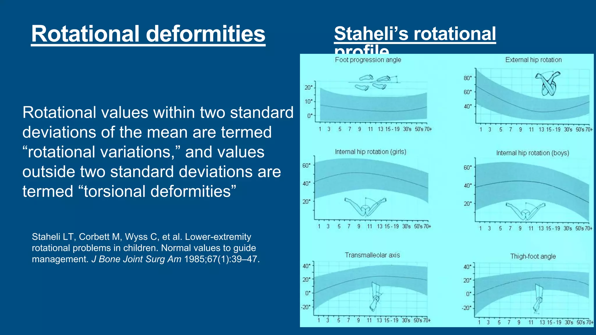
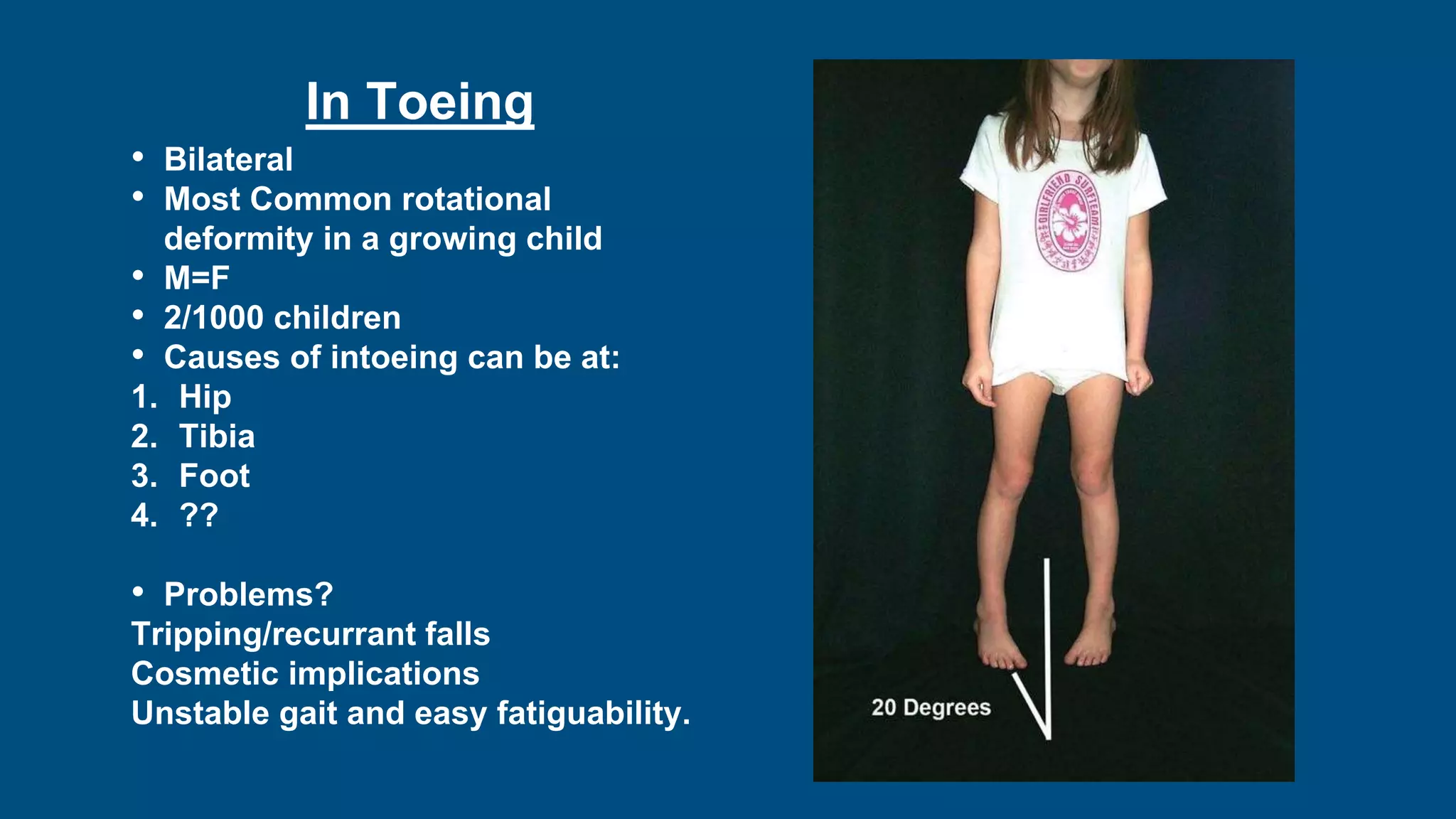
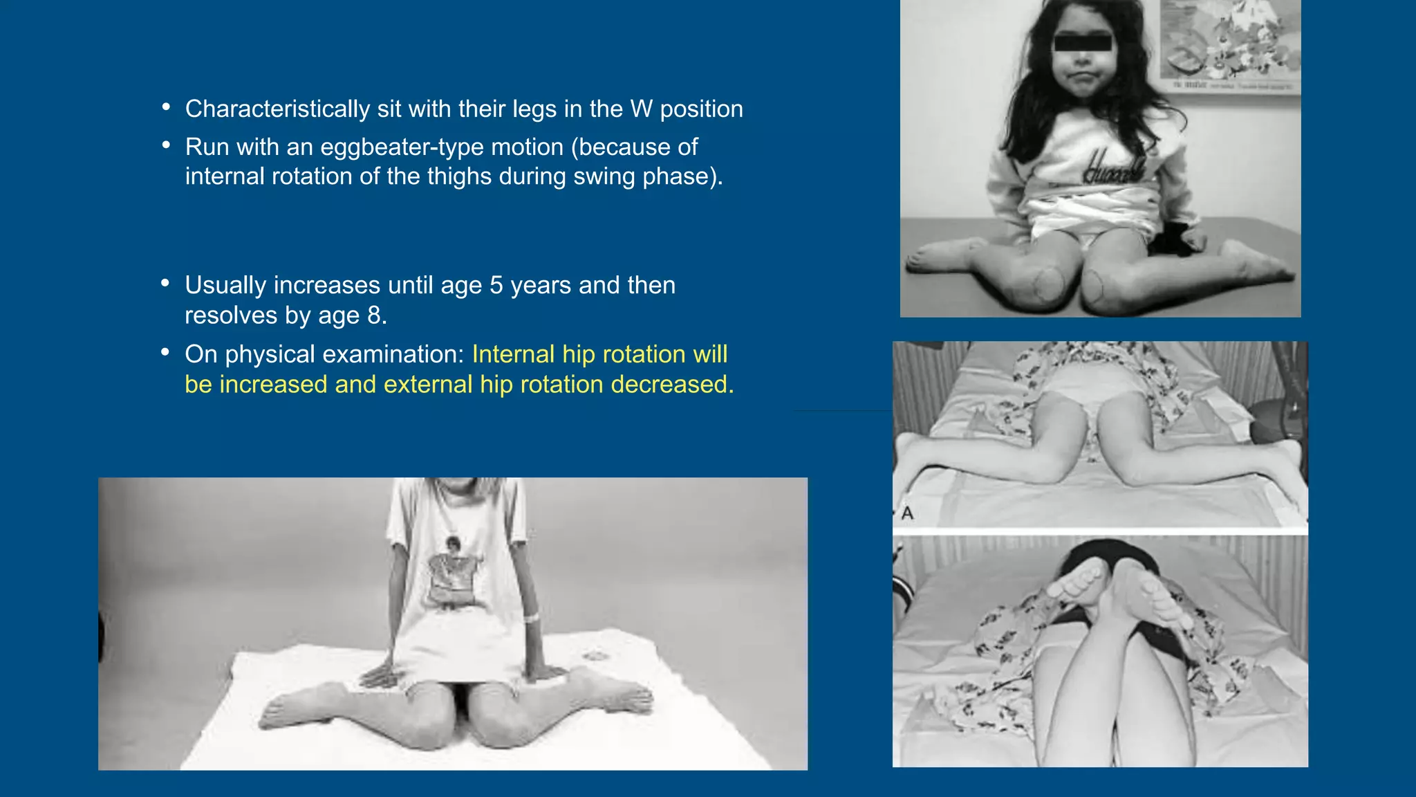
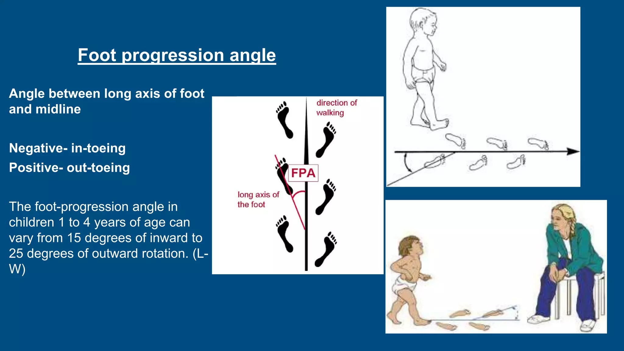
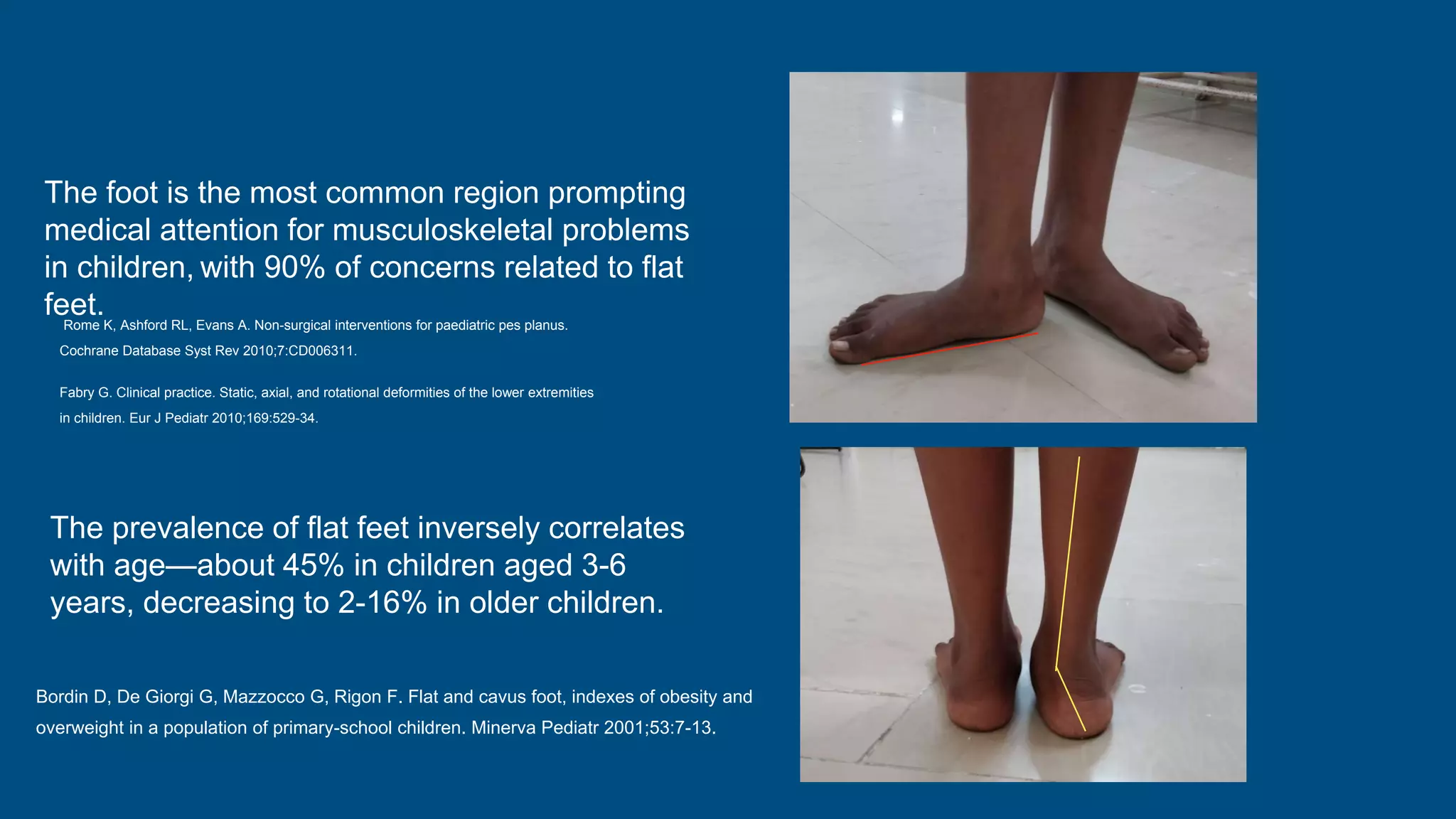
- Physiological variations in children such as intoeing, out-toeing, bowing of the legs, and flat feet are common and usually resolve on their own without intervention. Conditions that suggest an abnormality include asymmetry, limitation of movement, or progressive deformity.
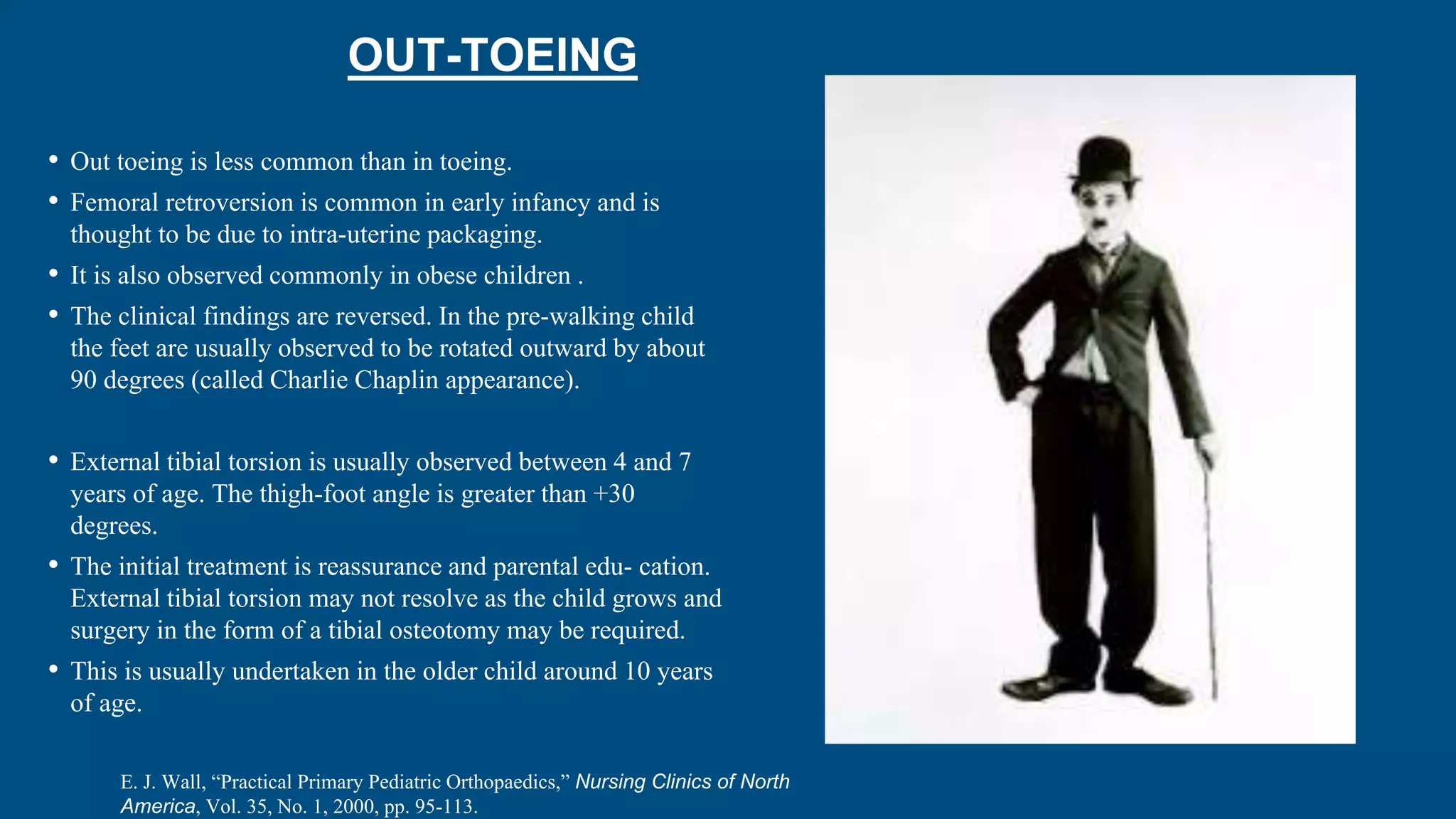
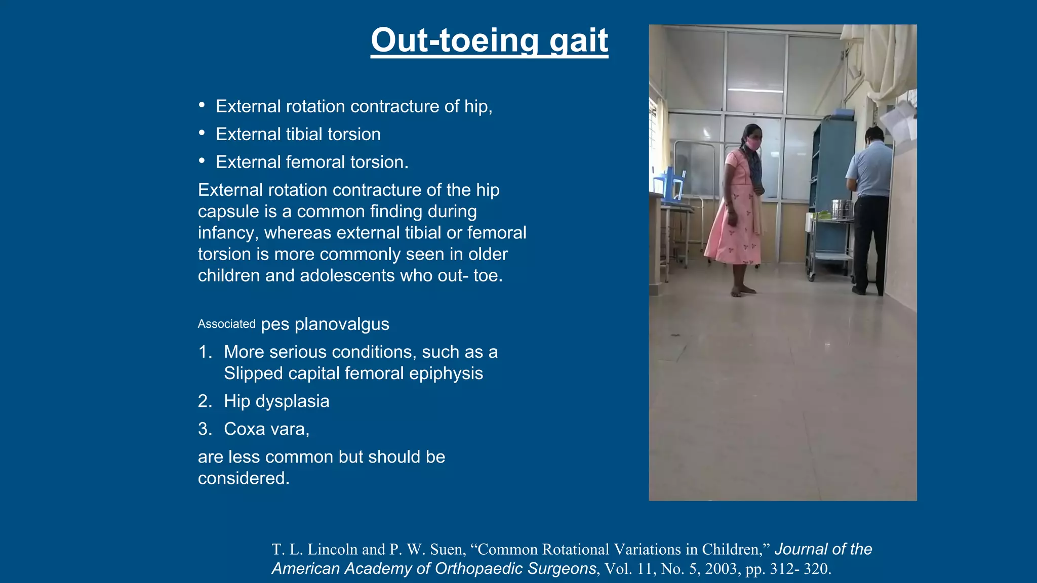
- Intoeing is most commonly caused by femoral anteversion or tibial torsion in young children and resolves by age 8 in 95% of cases. Out-toeing can be due to femoral retroversion or external tibial torsion.
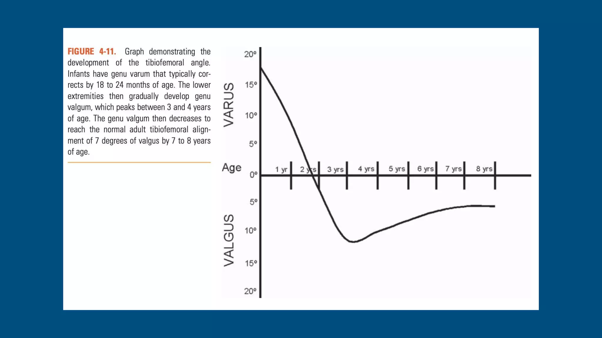
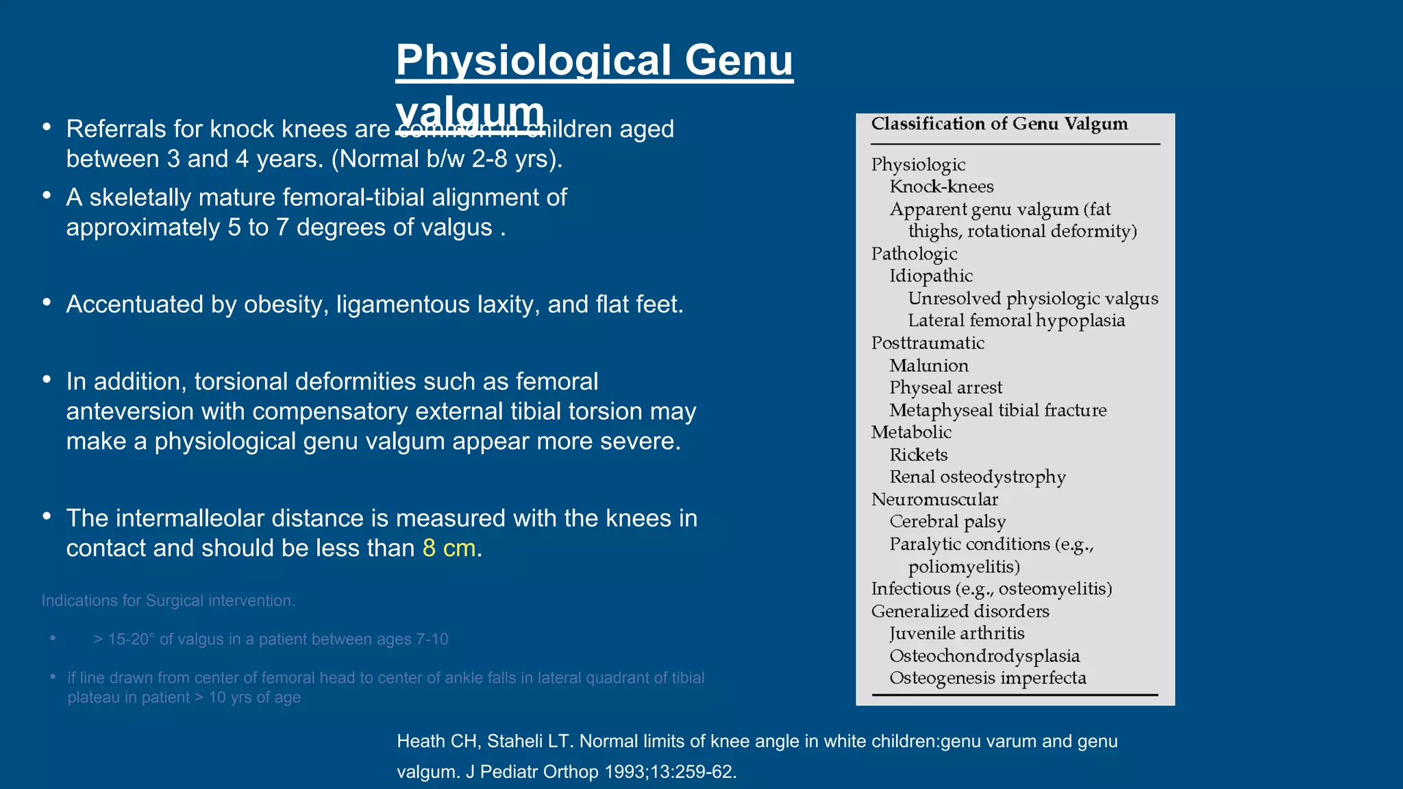
- Genu varum is normal in infants under 18 months while genu valgum peaks between 3-4 years and also typically resolves by age 7 without intervention.
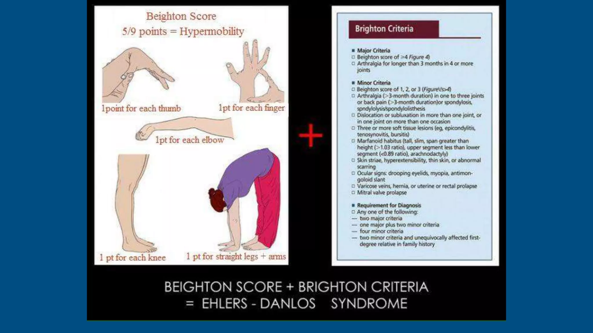
Jennifer Wambui, the Chief Exec. Director, CaCAI has a superb presentation about the Ponseti Method of Clubfoot treatment. The whole presentation is based on her own experience with Clubfoot and how she has triumphed over it.
Congenital disorders are commonly screened by pediatricians and certain disorders like club foot needs early intervention to get satisfactory results .I have tried to present common disorders in neonates for early diagnosis.
Leading the Way in Nephrology: Dr. David Greene's Work with Stem Cells for Ki...Dr. David Greene Arizona
As we watch Dr. Greene's continued efforts and research in Arizona, it's clear that stem cell therapy holds a promising key to unlocking new doors in the treatment of kidney disease. With each study and trial, we step closer to a world where kidney disease is no longer a life sentence but a treatable condition, thanks to pioneers like Dr. David Greene.
The Importance of Community Nursing Care.pdfAD Healthcare
NDIS and Community 24/7 Nursing Care is a specific type of support that may be provided under the NDIS for individuals with complex medical needs who require ongoing nursing care in a community setting, such as their home or a supported accommodation facility.
Global launch of the Healthy Ageing and Prevention Index 2nd wave – alongside...ILC- UK
The Healthy Ageing and Prevention Index is an online tool created by ILC that ranks countries on six metrics including, life span, health span, work span, income, environmental performance, and happiness. The Index helps us understand how well countries have adapted to longevity and inform decision makers on what must be done to maximise the economic benefits that comes with living well for longer.
Alongside the 77th World Health Assembly in Geneva on 28 May 2024, we launched the second version of our Index, allowing us to track progress and give new insights into what needs to be done to keep populations healthier for longer.
The speakers included:
Professor Orazio Schillaci, Minister of Health, Italy
Dr Hans Groth, Chairman of the Board, World Demographic & Ageing Forum
Professor Ilona Kickbusch, Founder and Chair, Global Health Centre, Geneva Graduate Institute and co-chair, World Health Summit Council
Dr Natasha Azzopardi Muscat, Director, Country Health Policies and Systems Division, World Health Organisation EURO
Dr Marta Lomazzi, Executive Manager, World Federation of Public Health Associations
Dr Shyam Bishen, Head, Centre for Health and Healthcare and Member of the Executive Committee, World Economic Forum
Dr Karin Tegmark Wisell, Director General, Public Health Agency of Sweden
Empowering ACOs: Leveraging Quality Management Tools for MIPS and BeyondHealth Catalyst
Join us as we delve into the crucial realm of quality reporting for MSSP (Medicare Shared Savings Program) Accountable Care Organizations (ACOs).
In this session, we will explore how a robust quality management solution can empower your organization to meet regulatory requirements and improve processes for MIPS reporting and internal quality programs. Learn how our MeasureAble application enables compliance and fosters continuous improvement.
Defecation
Normal defecation begins with movement in the left colon, moving stool toward the anus. When stool reaches the rectum, the distention causes relaxation of the internal sphincter and an awareness of the need to defecate. At the time of defecation, the external sphincter relaxes, and abdominal muscles contract, increasing intrarectal pressure and forcing the stool out
The Valsalva maneuver exerts pressure to expel faeces through a voluntary contraction of the abdominal muscles while maintaining forced expiration against a closed airway. Patients with cardiovascular disease, glaucoma, increased intracranial pressure, or a new surgical wound are at greater risk for cardiac dysrhythmias and elevated blood pressure with the Valsalva maneuver and need to avoid straining to pass the stool.
Normal defecation is painless, resulting in passage of soft, formed stool
CONSTIPATION
Constipation is a symptom, not a disease. Improper diet, reduced fluid intake, lack of exercise, and certain medications can cause constipation. For example, patients receiving opiates for pain after surgery often require a stool softener or laxative to prevent constipation. The signs of constipation include infrequent bowel movements (less than every 3 days), difficulty passing stools, excessive straining, inability to defecate at will, and hard feaces
IMPACTION
Fecal impaction results from unrelieved constipation. It is a collection of hardened feces wedged in the rectum that a person cannot expel. In cases of severe impaction the mass extends up into the sigmoid colon.
DIARRHEA
Diarrhea is an increase in the number of stools and the passage of liquid, unformed feces. It is associated with disorders affecting digestion, absorption, and secretion in the GI tract. Intestinal contents pass through the small and large intestine too quickly to allow for the usual absorption of fluid and nutrients. Irritation within the colon results in increased mucus secretion. As a result, feces become watery, and the patient is unable to control the urge to defecate. Normally an anal bag is safe and effective in long-term treatment of patients with fecal incontinence at home, in hospice, or in the hospital. Fecal incontinence is expensive and a potentially dangerous condition in terms of contamination and risk of skin ulceration
HEMORRHOIDS
Hemorrhoids are dilated, engorged veins in the lining of the rectum. They are either external or internal.
FLATULENCE
As gas accumulates in the lumen of the intestines, the bowel wall stretches and distends (flatulence). It is a common cause of abdominal fullness, pain, and cramping. Normally intestinal gas escapes through the mouth (belching) or the anus (passing of flatus)
FECAL INCONTINENCE
Fecal incontinence is the inability to control passage of feces and gas from the anus. Incontinence harms a patient’s body image
PREPARATION AND GIVING OF LAXATIVESACCORDING TO POTTER AND PERRY,
An enema is the instillation of a solution into the rectum and sig
CHAPTER 1 SEMESTER V - ROLE OF PEADIATRIC NURSE.pdfSachin Sharma
Pediatric nurses play a vital role in the health and well-being of children. Their responsibilities are wide-ranging, and their objectives can be categorized into several key areas:
1. Direct Patient Care:
Objective: Provide comprehensive and compassionate care to infants, children, and adolescents in various healthcare settings (hospitals, clinics, etc.).
This includes tasks like:
Monitoring vital signs and physical condition.
Administering medications and treatments.
Performing procedures as directed by doctors.
Assisting with daily living activities (bathing, feeding).
Providing emotional support and pain management.
2. Health Promotion and Education:
Objective: Promote healthy behaviors and educate children, families, and communities about preventive healthcare.
This includes tasks like:
Administering vaccinations.
Providing education on nutrition, hygiene, and development.
Offering breastfeeding and childbirth support.
Counseling families on safety and injury prevention.
3. Collaboration and Advocacy:
Objective: Collaborate effectively with doctors, social workers, therapists, and other healthcare professionals to ensure coordinated care for children.
Objective: Advocate for the rights and best interests of their patients, especially when children cannot speak for themselves.
This includes tasks like:
Communicating effectively with healthcare teams.
Identifying and addressing potential risks to child welfare.
Educating families about their child's condition and treatment options.
4. Professional Development and Research:
Objective: Stay up-to-date on the latest advancements in pediatric healthcare through continuing education and research.
Objective: Contribute to improving the quality of care for children by participating in research initiatives.
This includes tasks like:
Attending workshops and conferences on pediatric nursing.
Participating in clinical trials related to child health.
Implementing evidence-based practices into their daily routines.
By fulfilling these objectives, pediatric nurses play a crucial role in ensuring the optimal health and well-being of children throughout all stages of their development.
Antibiotic Stewardship by Anushri Srivastava.pptxAnushriSrivastav
Stewardship is the act of taking good care of something.
Antimicrobial stewardship is a coordinated program that promotes the appropriate use of antimicrobials (including antibiotics), improves patient outcomes, reduces microbial resistance, and decreases the spread of infections caused by multidrug-resistant organisms.
WHO launched the Global Antimicrobial Resistance and Use Surveillance System (GLASS) in 2015 to fill knowledge gaps and inform strategies at all levels.
ACCORDING TO apic.org,
Antimicrobial stewardship is a coordinated program that promotes the appropriate use of antimicrobials (including antibiotics), improves patient outcomes, reduces microbial resistance, and decreases the spread of infections caused by multidrug-resistant organisms.
ACCORDING TO pewtrusts.org,
Antibiotic stewardship refers to efforts in doctors’ offices, hospitals, long term care facilities, and other health care settings to ensure that antibiotics are used only when necessary and appropriate
According to WHO,
Antimicrobial stewardship is a systematic approach to educate and support health care professionals to follow evidence-based guidelines for prescribing and administering antimicrobials
In 1996, John McGowan and Dale Gerding first applied the term antimicrobial stewardship, where they suggested a causal association between antimicrobial agent use and resistance. They also focused on the urgency of large-scale controlled trials of antimicrobial-use regulation employing sophisticated epidemiologic methods, molecular typing, and precise resistance mechanism analysis.
Antimicrobial Stewardship(AMS) refers to the optimal selection, dosing, and duration of antimicrobial treatment resulting in the best clinical outcome with minimal side effects to the patients and minimal impact on subsequent resistance.
According to the 2019 report, in the US, more than 2.8 million antibiotic-resistant infections occur each year, and more than 35000 people die. In addition to this, it also mentioned that 223,900 cases of Clostridoides difficile occurred in 2017, of which 12800 people died. The report did not include viruses or parasites
VISION
Being proactive
Supporting optimal animal and human health
Exploring ways to reduce overall use of antimicrobials
Using the drugs that prevent and treat disease by killing microscopic organisms in a responsible way
GOAL
to prevent the generation and spread of antimicrobial resistance (AMR). Doing so will preserve the effectiveness of these drugs in animals and humans for years to come.
being to preserve human and animal health and the effectiveness of antimicrobial medications.
to implement a multidisciplinary approach in assembling a stewardship team to include an infectious disease physician, a clinical pharmacist with infectious diseases training, infection preventionist, and a close collaboration with the staff in the clinical microbiology laboratory
to prevent antimicrobial overuse, misuse and abuse.
to minimize the developme
1. Dr Puneeth K Pai
Physiological variations in
children
2. • Common cause of parental concern.
Referral by pediatrician, General
Orthopedist, General practitioner.
• Half of all new referrals to pediatric
orthopedic clinic were children with
normal variants of lower limb
development in a study by Molony
et al.
D. Molony, G. Hefferman, et al., “Normal Variants in the Pediatric Orthopedic
Population,” Irish Medical Journal, Vol. 99, No. 1, 2006, pp. 13-14.
3. The following features suggest a pathological
condition and not a normal variant :
• Abnormal perinatal history.
• Significant family history.
• Abnormal facies .
• Abnormal height for age.
• Asymmetry of limb findings
• Limitation of joint movements
• Leg length discrepancy/hypoplasia/hypertrophy
• Progressive deformity
• NOT normal for age
• Localised/Anglular deformities
What is not normal?
L. T. Staheli, “Lower Limb-Fundamentals of Pediatric Orthopedics,” 4th Edition, Lippincott Williams and Wi- lkins, Philadelphia, 2008.
4. Lower Limb
Rotational deformities
• Intoeing
• Out-toeing
Coronal deformities
• Genu varum
• Genu valgum
• Metatarsus Primus Varus
• Positional foot deformities- Pes supinates and pes
talus, Calcaneovalgus.
• Accessory bones/Sesamoids
Limb length discrepancy
What is not abnormal?
Upper limb
Carrying angle
General
Hyperlaxity
5. Rotational deformities
Rotational values within two standard
deviations of the mean are termed
“rotational variations,” and values
outside two standard deviations are
termed “torsional deformities”
Staheli LT, Corbett M, Wyss C, et al. Lower-extremity
rotational problems in children. Normal values to guide
management. J Bone Joint Surg Am 1985;67(1):39–47.
Staheli’s rotational
profile
6. • Bilateral
• Most Common rotational
deformity in a growing child
• M=F
• 2/1000 children
• Causes of intoeing can be at:
1. Hip
2. Tibia
3. Foot
4. ??
• Problems?
Tripping/recurrant falls
Cosmetic implications
Unstable gait and easy fatiguability.
In Toeing
7.
8. 95 % of all intoeing resolves by the age of 8 years
9. Femoral anteversion
1.5 degrees of correction per year;
more than 80 % of affected children, usually by age 10 years
most pronounced between ages 4 to 6 years
At birth, neonates have an average of 40° of femoral
anteversion.
By age 8 years, average anteversion decreases to the typical
adult value of 15°
10. • Characteristically sit with their legs in the W position
• Run with an eggbeater-type motion (because of
internal rotation of the thighs during swing phase).
• Usually increases until age 5 years and then
resolves by age 8.
• On physical examination: Internal hip rotation will
be increased and external hip rotation decreased.
11. No association between increased femoral ante-version
and degenerative joint disease has been proved, some
association with knee pain has been suggested.
Reikerås O: Patellofemoral characteristics in patients with increased femoral
anteversion. Skeletal Radiol 1992;21:311-313.
Knee pain may be particularly prevalent in children
with concomitantly increased femoral anteversion
and external tibial torsion (so-called miserable
malalignment syndrome).
Delgado ED, Schoenecker PL, Rich MM, Capelli AM: Treatment of severe
torsional malalignment syndrome. J Pediatr Orthop 1996;16:484-488.
12.
13.
14.
15. Foot progression angle
Angle between long axis of foot
and midline
Negative- in-toeing
Positive- out-toeing
The foot-progression angle in
children 1 to 4 years of age can
vary from 15 degrees of inward to
25 degrees of outward rotation. (L-
W)
16. Tibial intorsion
• Internal tibial torsion is the most common
cause of in-toeing from ages 1 to 3 years.
• 2/3 rd bilateral
• Left>Right
•Intrauterine positioning
•Expectant observation
•Most resolve by 4 years of age
•Disability due to persistant IR is rare
•No risk of degenerative arthritis
Fuchs R, Staheli LT: Sprinting and in- toeing. J Pediatr Orthop 1996;16:489-491
17. In a retrospective review of all intoeing
referrals to a Scottish paediatric
orthopaedic unit, no children required
surgery for their condition, with a 85%
being discharged on their first visit.
Similarly, in a large American series
reviewing 720 intoeing referrals in a
year, only one child required surgery.
Blackmur JP, Murray AW. Do children who in-toe need to be referred to an
orthopaedic clinic? J Pediatr Orthop B 2010;19:415-7.
Karol LA. Rotational deformities in the lower extremities. Curr Opin Pediatr 1997;9:77-80.
18. Surgical management reserved for :
• Children older than 8 years with
marked functional
• Cosmetic deformity
• Thigh-foot angle greater than
three standard deviations beyond
the mean (eg, thigh-foot angle
>15°).
19. OUT-TOEING
• Out toeing is less common than in toeing.
• Femoral retroversion is common in early infancy and is
thought to be due to intra-uterine packaging.
• It is also observed commonly in obese children .
• The clinical findings are reversed. In the pre-walking child
the feet are usually observed to be rotated outward by about
90 degrees (called Charlie Chaplin appearance).
• External tibial torsion is usually observed between 4 and 7
years of age. The thigh-foot angle is greater than +30
degrees.
• The initial treatment is reassurance and parental edu- cation.
External tibial torsion may not resolve as the child grows and
surgery in the form of a tibial osteotomy may be required.
• This is usually undertaken in the older child around 10 years
of age.
E. J. Wall, “Practical Primary Pediatric Orthopaedics,” Nursing Clinics of North
America, Vol. 35, No. 1, 2000, pp. 95-113.
20. Out-toeing gait
• External rotation contracture of hip,
• External tibial torsion
• External femoral torsion.
External rotation contracture of the hip
capsule is a common finding during
infancy, whereas external tibial or femoral
torsion is more commonly seen in older
children and adolescents who out- toe.
Associated pes planovalgus
1. More serious conditions, such as a
Slipped capital femoral epiphysis
2. Hip dysplasia
3. Coxa vara,
are less common but should be
considered.
T. L. Lincoln and P. W. Suen, “Common Rotational Variations in Children,” Journal of the
American Academy of Orthopaedic Surgeons, Vol. 11, No. 5, 2003, pp. 312- 320.
21. Coronal plane deformities
Angular alignment refers to the tibiofemoral
angle, which can be clinically assessed by the
intermalleolar and intercondylar distances
Salenius P, Vankka E. The development of the tibiofemoral angle in children. J Bone Joint
Surg Am 1975;57:259-61.
Salenius and Vankka
Landmark study of tibiofemoral angles in 1500 normal
children
Up to the age of 18 months present with genu varum (bow
legs; mean of 15°)
Genu valgum (knock knees; mean of 12°) deformity
ensues, which subsequently corrects itself to the normal
value in adults (7-8° valgus) by the age of 7 years.
22.
23.
24.
25. Physiological Genu
varum
Heath CH, Staheli LT. Normal limits of knee angle in white
children:genu varum and genu
valgum. J Pediatr Orthop 1993;13:259-62.
Physiologic genu varum is defined by a tibiofemoral angle of at least 10
degrees of varus, a radiographically normal physis, and apex lateral
bowing of the proximal end of the tibia and often the distal end of the
femur.
• Physiological genu varum is thought to relate to
intrauterine positioning
• Which leads to the contracture of the medial knee
joint capsule.
• This, in addition to the internal tibial torsion
common in this age group, accentuates the
deformity when children weight bear.
• Therefore, referrals for bow legs are common for
children aged between 10 and 14 months, the
average age at which children start to stand and
ambulate.
• The intercondylar distance is measured with the
medial malleoli in contact and should be less than
6 cm.
26. Physiological Genu
valgum
• Referrals for knock knees are common in children aged
between 3 and 4 years. (Normal b/w 2-8 yrs).
• A skeletally mature femoral-tibial alignment of
approximately 5 to 7 degrees of valgus .
• Accentuated by obesity, ligamentous laxity, and flat feet.
• In addition, torsional deformities such as femoral
anteversion with compensatory external tibial torsion may
make a physiological genu valgum appear more severe.
• The intermalleolar distance is measured with the knees in
contact and should be less than 8 cm.
Indications for Surgical intervention.
• > 15-20° of valgus in a patient between ages 7-10
• if line drawn from center of femoral head to center of ankle falls in lateral quadrant of tibial
plateau in patient > 10 yrs of age
Heath CH, Staheli LT. Normal limits of knee angle in white children:genu varum and genu
valgum. J Pediatr Orthop 1993;13:259-62.
27.
28.
29. Metatarsus adducts
• occurs in approximately 1 in 1,000 births
• equal frequency in males and females
• bilateral approximately 50% of cases
Associated conditions
• DDH (15-20%)
• Torticollis
Most common congenital foot deformity
30.
31. Metatarsus Primus Varus
• Metatarsus primus varus is an isolated
adducted first metatarsal.
• In contrast with simple metatarsus ad- ductus,
in metatarsus primus varus the lateral border
of the foot has a normal alignment, and there is
often a deepened vertical skin crease on the
medial border of the foot at the tar- some
tatarsal joint.
• In general, meta- tarsus primus varus is a
more rigid deformity than simple metatarsus
ad- ductus, and early casting is recommended.
• Persistent deformity in childhood is associated
with progressive hallux valgus.
• Opening medial cuneiform osteotomy has
been described for selective use in children
with a severe deformity. Lynch FR: Applications of the opening wedge cuneiform osteotomy in the
surgical repair of juvenile hallux abducto valgus. J Foot Ankle Surg
1995;34:103-123.
33. Os trigonum/trigonal process/the
Stieda process/ posterior process
The os trigonum is formed from the lateral portion of the groove in
the posterior aspect of the talus, through which passes the flexor
hallucis longus .
Between 8 and 11 years old, medial and lateral centers of
ossification appear
• Immobilization
• Steroid injections around the os trigonum.
• Open or arthroscopic excision should be reserved for those in
whom conservative therapy fails
• Marotta and Micheli reported improvement after excision of
the ossicle in a series of ballet dancers in whom conservative
treatment failed.
• Abramowitz and colleagues noted worse results after
resection in patients who had symptoms for longer than 2
years when compared with those who had symptoms of a
shorter duration.
• Wredmark and associates released the flexor hallucis sheath
if thickened at the time of os trigonum removal
34. Prehallux, Accessory scaphoid, Os tibiale externum, Os naviculare secundarium, Navicular secundum.
Accessory navicular
14% to 26%
Three types of accessory navicular bones have been
described.
Type I (os tibiale externum) is a small ossicle within the
substance of the tibialis tendon.
Type II is an 8- to 12-mm ossicle extending medially and
plantarward from the navicular bone and connected to the
navicular by a cartilaginous synchondrosis.
Type III is a cornuate navicular remaining after fusion of the
accessory navicular with the primary navicular bone.
35. Positional Foot deformitis
Pes Supinatus
• It is the main differential diagnosis of clubfoot.
• A supination is observed, but with no equinus or adduction of
the forefoot .
• It is also distinguished from clubfoot by its total reductibility.
Pes Talus
• The plantar surface of the foot is against the wall of the uterus,
which forces the foot into dorsiflexion due to intrauterine
constraints.
• The result is excessive dorsiflexion of the foot, which allows its
dorsum to come into contact with the anterior aspect of the lower
leg.
• It is sometimes associated with a valgus mal- position and is also
characterized by total reducibility.
Brasseur‐Daudruy, Marie; Abu Amara, Saad;
Ickowicz‐Onnient, Valentine; Touleimat, Salma;
Verspyck, Eric (2019). Clubfoot Versus Positional
Foot Deformities on Prenatal Ultrasound Imaging.
Journal of Ultrasound in Medicine, (), jum.15136–.
doi:10.1002/jum.15136
36. • Calcaneovalgus foot is a condition in infants where the
foot is pushed up against the front of the leg.
• It’s caused by a baby being crowded or growing in an
unusual position in the uterus.
• Risk factors
1. First born babies
2. Babies with more birth weight.
3. Oligohydramnios
Positional Calcaneovalgus
37. Flexible pes planus (flat feet)
Incidence -20% to 25%
generalized ligamentous laxity is common
25% are associated with gastrocnemius-soleus contracture
Differential diagnosis
• Tarsal coalition
• Congenital vertical talus
• Accessory navicular
38. The foot is the most common region prompting
medical attention for musculoskeletal problems
in children, with 90% of concerns related to flat
feet.
Rome K, Ashford RL, Evans A. Non-surgical interventions for paediatric pes planus.
Cochrane Database Syst Rev 2010;7:CD006311.
Fabry G. Clinical practice. Static, axial, and rotational deformities of the lower extremities
in children. Eur J Pediatr 2010;169:529-34.
The prevalence of flat feet inversely correlates
with age—about 45% in children aged 3-6
years, decreasing to 2-16% in older children.
Bordin D, De Giorgi G, Mazzocco G, Rigon F. Flat and cavus foot, indexes of obesity and
overweight in a population of primary-school children. Minerva Pediatr 2001;53:7-13.
39. Talonavicular joint coverage is a good radiological
criterion for discriminating between symptomatic
and asymptomatic flatfoot
Moraleda and Mubarak reported a mean loss of
talonavicular coverage of 25 ± 8◦ in asymptomatic
patients, 36 ± 9◦ in symptomatic patients managed
non-operatively and 39 ± 11◦ in symptomatic
patients managed surgically
40.
41. Limb length discrepancy
Approximately 15% of the adult population has a
leg length discrepancy (LLD) measuring greater
than 1 cm.
Most LLDs < 2 cm are idiopathic, due to normal
anatomic variation (asymmetry) of the human
body.
Rush WA, Steiner HA. A study of lower extremity length inequality. Am
J Roentgenol. 1946;56:616-23.
42. Hypermobility
Hypermobility syndrome (HMS) is a dominant
inherited connective tissue disorder described as
“generalized articular hypermobility, with or without
subluxation or dislocation.”
Ratio of type I to type III collagen is decreased in
skin.
Larsen, Beals, or Ehlers-Danlos syndrome
43.
44. prevalence of hypermobility in children as a phenomenon [as
opposed to joint hypermobility syndrome (JHS), i.e. symptomatic
hypermobility] depending on the age or ethnicity of the study
population or the inclusion criteria, has been reported to be
between 2.3 and 30%
Numerous extra-articular manifestations of JHS
have been similarly reported in children,
including
• chronic constipation and encopresis, enuresis
and urinary tract infections (UTI)
• higher skin extensibility
• lower systemic blood pressure
• lower bone quantitative ultrasound
measurements
• chronic fatigue syndrome
• temporomandibular joint disease
• fibromyalgia
• gross motor developmental delay
45.
46. The carrying angle is defined as the angle between the
long axis of the ulna and the long axis of the humerus
Carrying angle
F>M
Angle increases with age
Age-related increase from birth through
adolescence in carrying angle that was most
likely related more to the osseous development
of the elbow joint
Balasubramanian P, Madhuri V, Muliyil J. Carrying angle in children: a
normative study. J Pediatr Orthop B 2006; 15:37–40.