1) The document discusses limb amputation in animals, including the definition, indications, surgical anatomy, blood and nerve supply, site of operation, anesthesia procedure, surgical procedure, and postoperative care.
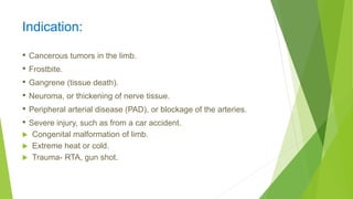
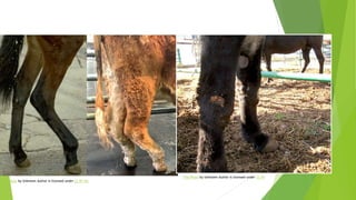
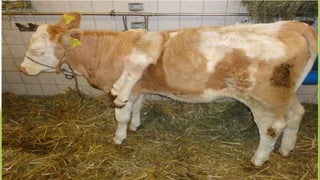
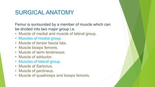
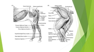
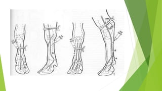
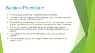
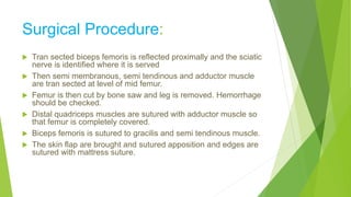
2) Key points covered include that amputation involves removing all or part of a limb through dividing one or more bones, indications for amputation include various medical conditions and injuries, and the surgical procedure described is for amputation of the hind limb in animals at the middle third of the femur above the stifle joint.
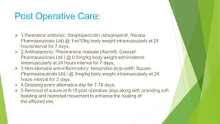
3) Postoperative care involves administering antibiotics, antihistamines, anti-inflammatories, dressing changes, and suture removal to aid healing after the amputation surgery.


![AMPUTATIONS OF LOWER LIMBS [Autosaved] copy.pptx](https://cdn.slidesharecdn.com/ss_thumbnails/amputationsoflowerlimbsautosavedcopy-240504162057-afe262d2-thumbnail.jpg?width=640&height=640&fit=bounds)


