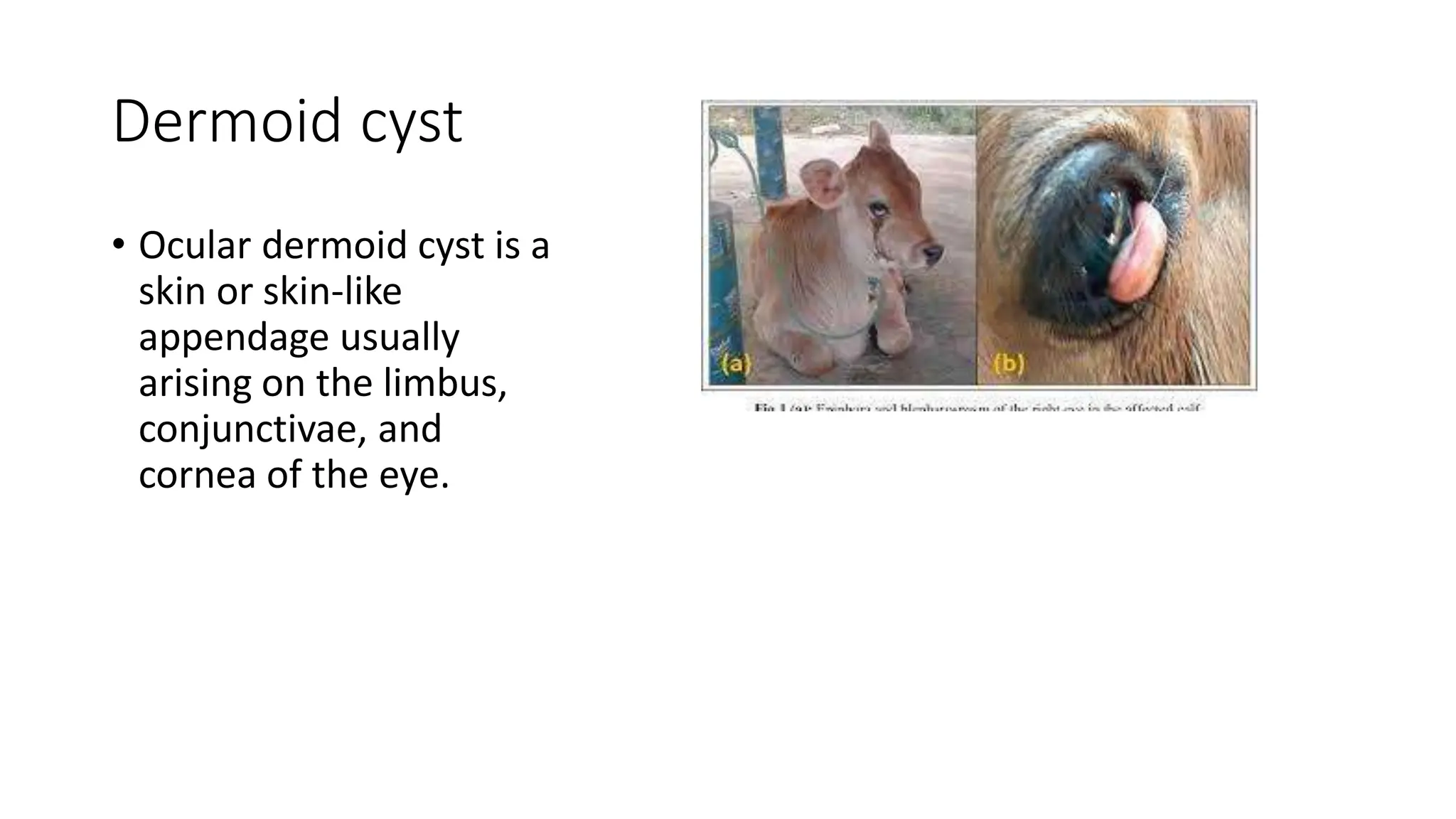
dusjagr & nano talk on open tools for agriculture research and learning
Ocular dermoid cyst.pdf
1. Dermoid cyst
• Ocular dermoid cyst is a
skin or skin-like
appendage usually
arising on the limbus,
conjunctivae, and
cornea of the eye.
2. Cause
• It forms due to heritable autosomal recessive and polygenic traits.
Clinical signs
• It can be unilateral or bilateral
• It may be associated with other ocular manifestation or with other
malformations.
• Hair from the lesions is predominantly responsible for the associated
irritation resulting in conjunctis and keratitis
• The condition may cause visual impairment.
• Dermoids may affect the eyelids, conjunctiva, nictitans, sclera and cornea.
• Bilateral ocular dermoids have been reported in cattle , sheep and a camel.
3. Treatment
• Sterilize the surgical equipment.
• Restrain the calf in lateral
recumbency
• Sedate the calf with diazepam 0.5
mg/kg body weight
• Infiltrate the cyst stalk with 1 ml
0.2% Lignocaine HCl to achieve
anaesthesia
• Excise the mass of dermoid by
grasping with forceps gently and the
tissue mass remove completely by
surgical blade.
4. • The dermoids are excised by superficial lamellar keratectomy while
attached to the cornea
• Apply Topical 1% atropine sulfate ointment twice daily to both eyes
for 5 days and
• Apply Bacitracin-neomycin-polymixin ophthalmic ointment twice daily
to both eyes for 2 weeks postoperatively
• Meloxicam and chloropheniramine chloride were given i/mly at the
dose of 2mg/kg body weight for 3 days.