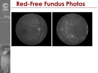
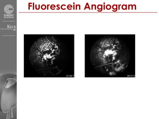
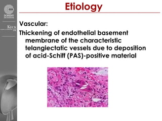
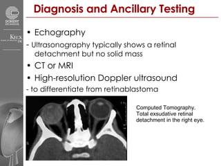
Doheny Eye Institute Grand Rounds Presenter Irina Bykhovskaya, MD Discussant Dr. SriniVas Sadda 3/9/2007
Patient Presentation HPI: 16 yo HM from juvenile hall c/o “black cloud” OS x 8 months POHx: h/o “fluid in the eye” and laser tx OS at age 8 yo PMH / FHx – unremarkable SLE: LLL/S/C/K/AC/I/L – wnl OU
Fundus Photos lipid-rich, yellow subretinal exudate  in the posterior pole OS
Fundus Photos OS
Red-Free Fundus Photos
Fluorescein Angiogram
Coats’ Disease Idiopathic condition  Telangiectatic and aneurismal vessels with intraretinal and subretinal exudates  1 st  described by Coats in 1908
Etiology Vascular: Thickening of endothelial basement membrane of the characteristic telangiectatic vessels due to deposition of acid-Schiff (PAS)-positive material
Clinical Presentation Painless Males : Females = 3:1 80% unilateral 2/3 cases present before age 10 Diagnosed at a median age of 5 years  No predilection for race or laterality  Poor vision, strabismus, leukocoria Visual acuity at presentation 20/200 to no light perception No genetic transmission No systemic associations
Clinical Picture Localized, lipid-rich, yellow subretinal exudate  Associated vascular anomalies: sheathing, telangiectasia, tortuosity, aneurismal dilatations, zones of capillary drop out, and occasionally neovascularization
Clinical Course Variable but generally progressive with acute exacerbations and quiescent stages Complications:  subretinal CMV serous RD hemorrhagic macrocysts secondary iridocyclitis Cataract neovascular glaucoma
FA Reveals localized anomalies in retinal vasculature Telangiectasia, aneurisms, beading of vessel walls, and vascular communication channels  Breakdown of blood-retinal barrier     Early and persistent leakage  Microvascular anomalies: Diffuse loss of capillary bed or areas of complete capillary nonperfusion surrounded by areas of arteriolar and venular anomalies
Histopathology Loss of vascular endothelium and pericytes  All levels of the retinal vascular circuit are involved  Proteinaceous subretinal exudates with prominent cholesterol clefts  and prominent histiocytes Total exudative retinal detachment
Histopathology cont’d Subretinal fluid with cholesterol clefts  and lipid-laden macrophages Telangiectasic retinal vessels  Intraretinal cholesterol deposition  triggering a giant cell reaction  foreign-body type
Differential Diagnosis Retinoblastoma Retinal detachment Persistentt hyperplastic primary vitreous Congenital cataract Norrie’s disease Familial exudative vitreoretinopathy  Eales’ disease Vasculitis  Tumor accompanied by exudation Diabetic vasculopathies BRVO Idiopathic juxtafoveal telangiectasis
Diagnosis and Ancillary Testing Echography -  Ultrasonography typically shows a retinal detachment but no solid mass CT or MRI High-resolution Doppler ultrasound - to differentiate from retinablastoma Computed Tomography.  Total exsudative retinal  detachment in the right eye.
Treatment Focal laser photocoagulation  to close areas of vascular leakage to allow resorption of exudate Near yellow wavelength 100 to 500 microns Cryotherapy if retina is exudatively detached Subretinal fluid drainage and vasoablation +/- scleral buckle
References  Shields JA, Shields CL, Honavar SG, Demirci H. Clinical variations and complications of Coats disease in 150 cases: the 2000 Sanford Gifford Memorial Lecture. Am J Ophthalmol. 2001;131:561–571. Shields JA, Shields CL. Review: coats disease: the 2001 LuEsther T. Mertz lecture. Retina. 2002;22:80–91.  Shields JA, Parsons HM, Shields CL, Shah P. Lesions simulating retinoblastoma. J Pediatr Ophthalmol Strabismus. 1991;28:338–340.  Bruno F Fernandes, Alexandre N Odashiro, Shawn Maloney, Moyses E Zajdenweber, Andressa G Lopes, and Miguel N Burnier, Jr. Clinical-histopathological correlation in a case of Coats' disease. Diagn Pathol. 2006; 1: 24.  L. Smithen, G. Brown, A. Brucker, L. Yannuzzi, C. Klais, R. Spaide. Coats’ Disease Diagnosed in Adulthood. Ophthalmology, Volume 112, Issue 6, Pages 1072-1078  Edward DP, Mafee MF, Garcia-Valenzuela E, Weiss RA. Coats' disease and persistent hyperplastic primary vitreous: role of MR imaging and CT.  Radiol Clin North Am  1998;36:1119-1131

Coats Disease

  • 1.
    Doheny Eye InstituteGrand Rounds Presenter Irina Bykhovskaya, MD Discussant Dr. SriniVas Sadda 3/9/2007
  • 2.
    Patient Presentation HPI:16 yo HM from juvenile hall c/o “black cloud” OS x 8 months POHx: h/o “fluid in the eye” and laser tx OS at age 8 yo PMH / FHx – unremarkable SLE: LLL/S/C/K/AC/I/L – wnl OU
  • 3.
    Fundus Photos lipid-rich,yellow subretinal exudate in the posterior pole OS
  • 4.
  • 5.
  • 6.
  • 7.
  • 8.
    Coats’ Disease Idiopathiccondition Telangiectatic and aneurismal vessels with intraretinal and subretinal exudates 1 st described by Coats in 1908
  • 9.
    Etiology Vascular: Thickeningof endothelial basement membrane of the characteristic telangiectatic vessels due to deposition of acid-Schiff (PAS)-positive material
  • 10.
    Clinical Presentation PainlessMales : Females = 3:1 80% unilateral 2/3 cases present before age 10 Diagnosed at a median age of 5 years No predilection for race or laterality Poor vision, strabismus, leukocoria Visual acuity at presentation 20/200 to no light perception No genetic transmission No systemic associations
  • 11.
    Clinical Picture Localized,lipid-rich, yellow subretinal exudate Associated vascular anomalies: sheathing, telangiectasia, tortuosity, aneurismal dilatations, zones of capillary drop out, and occasionally neovascularization
  • 12.
    Clinical Course Variablebut generally progressive with acute exacerbations and quiescent stages Complications: subretinal CMV serous RD hemorrhagic macrocysts secondary iridocyclitis Cataract neovascular glaucoma
  • 13.
    FA Reveals localizedanomalies in retinal vasculature Telangiectasia, aneurisms, beading of vessel walls, and vascular communication channels Breakdown of blood-retinal barrier  Early and persistent leakage Microvascular anomalies: Diffuse loss of capillary bed or areas of complete capillary nonperfusion surrounded by areas of arteriolar and venular anomalies
  • 14.
    Histopathology Loss ofvascular endothelium and pericytes All levels of the retinal vascular circuit are involved Proteinaceous subretinal exudates with prominent cholesterol clefts and prominent histiocytes Total exudative retinal detachment
  • 15.
    Histopathology cont’d Subretinalfluid with cholesterol clefts and lipid-laden macrophages Telangiectasic retinal vessels Intraretinal cholesterol deposition triggering a giant cell reaction foreign-body type
  • 16.
    Differential Diagnosis RetinoblastomaRetinal detachment Persistentt hyperplastic primary vitreous Congenital cataract Norrie’s disease Familial exudative vitreoretinopathy Eales’ disease Vasculitis Tumor accompanied by exudation Diabetic vasculopathies BRVO Idiopathic juxtafoveal telangiectasis
  • 17.
    Diagnosis and AncillaryTesting Echography - Ultrasonography typically shows a retinal detachment but no solid mass CT or MRI High-resolution Doppler ultrasound - to differentiate from retinablastoma Computed Tomography. Total exsudative retinal detachment in the right eye.
  • 18.
    Treatment Focal laserphotocoagulation to close areas of vascular leakage to allow resorption of exudate Near yellow wavelength 100 to 500 microns Cryotherapy if retina is exudatively detached Subretinal fluid drainage and vasoablation +/- scleral buckle
  • 19.
    References ShieldsJA, Shields CL, Honavar SG, Demirci H. Clinical variations and complications of Coats disease in 150 cases: the 2000 Sanford Gifford Memorial Lecture. Am J Ophthalmol. 2001;131:561–571. Shields JA, Shields CL. Review: coats disease: the 2001 LuEsther T. Mertz lecture. Retina. 2002;22:80–91. Shields JA, Parsons HM, Shields CL, Shah P. Lesions simulating retinoblastoma. J Pediatr Ophthalmol Strabismus. 1991;28:338–340. Bruno F Fernandes, Alexandre N Odashiro, Shawn Maloney, Moyses E Zajdenweber, Andressa G Lopes, and Miguel N Burnier, Jr. Clinical-histopathological correlation in a case of Coats' disease. Diagn Pathol. 2006; 1: 24. L. Smithen, G. Brown, A. Brucker, L. Yannuzzi, C. Klais, R. Spaide. Coats’ Disease Diagnosed in Adulthood. Ophthalmology, Volume 112, Issue 6, Pages 1072-1078 Edward DP, Mafee MF, Garcia-Valenzuela E, Weiss RA. Coats' disease and persistent hyperplastic primary vitreous: role of MR imaging and CT. Radiol Clin North Am 1998;36:1119-1131