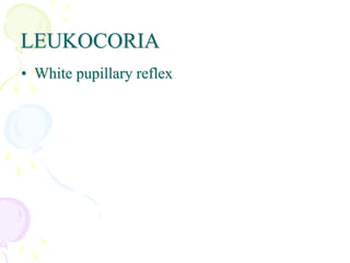
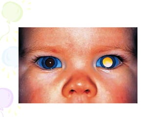
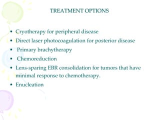
LEUKOCORIA
• White pupillary reflex
RETINOBLASTOMA
• the most common primary intraocular tumor of childhood.
• The incidence of retinoblastoma has been estimated to be 1 in 15,000
to 1 in 34,000 live births
• Both sexes are affected equally
• The average age at diagnosis is 18/24 months. Although cases have
been reported in adults, retinoblastoma is rare after age 4 years.
• Derived histogenically from primitive retinal cells that are capable
of differentiating into neuronal and glial elements.
• a hereditary form :
– Only 7 -10% of RB patients have positive family history.
– In affected families, the tumor frequently affects both eyes.
-- the average age at diagnosis is younger than non-heritable form
–ranging from newborn to 12 months
_ predisposed to a variety of cancers throughout life
-- 85% of heritable RB patients develop multiple , bilateral eye
tumors
NB. Bilaterality is a clinical marker for potentially transmissible
disease.
• About one third of all retinoblastomas are bilateral the
additional bilateral cases are caused by new germ-line
mutations.
• The great majority of retinoblastomas (90-93%)arise
sporadically --in patients with a negative family history.
• Sporadic tumors caused by somatic mutations are always
unilateral.
CLINICAL PRESENTATION
Depends on the size, location, growth pattern, and stage of the lesion at
diagnosis
- Leukocoria – 90%
caused by tumor in the vitreous cavity (endophytic
tumors) or total retinal detachment (exophytic tumors).
- Strabismus – about 1/3 of cases
- In underdeveloped countries, patients may present in the late stages
of the disease with a tumor that has extended extraocularly, causing
exophthalmos and an orbital mass.
• Other presenting Signs and Symptoms of Retinoblastoma
Uveitis
Hyphema
Glaucoma/rubeosis irides
Proptosis
Endophthalmitis/panophthalmitis
Orbital cellulitis
Visual obscuration
• The tumor does not cause pain or discomfort as it enlarges and
virtually never causes a red or inflamed eye early in the course of
the disease.
NATURAL HISTORY
• Retinoblastoma is a highly malignant neoplasm that
grows relentlessly and almost always has a fatal
outcome if untreated.
TREATMENT OPTIONS
• Cryotherapy for peripheral disease
• Direct laser photocoagulation for posterior disease
• Primary brachytherapy
• Chemoreduction
• Lens-sparing EBR consolidation for tumors that have
minimal response to chemotherapy.
• Enucleation
PERSISTENT HYPERPLASTIC PRIMARY
VITREOUS
• A congenital anomaly that is present at birth
• Characterized by the persistence of variable components of the
primary vitreous
• Usually unilateral
• Histopathologically, a plaque of fibrovascular tissue that resembles
primary vitreous adheres to the posterior lens capsule, and the
hyaloid artery is often patent.
• Rx :- Lensectomy, anterior vitrectomy
RETINOPATHY OF PREMATURITY
• bilateral disease and rarely present at birth
• In the 1950s accounted for almost half of causes of blindness in
children in European countries and USA; currently accounts for 10%
blindness.
• Classically, ROP develops in premature infants who have received
supplemental oxygen therapy.
• Infants weighing less than 1500 g at birth and those born at a
gestational age of less than 32 weeks are at risk for developing ROP.
Currently In industrialized countries due to improved monitoring
only neonates with ELBW [ <1000gm]are affected.
• Length of time in oxygen and mechanical ventilation are other risk
factors
Pathogenesis of ROP
• In the developing human fetus, the process of retinal blood vessel
development begins during the fourth month of gestation.
• Blood vessels grow outward from the optic disc and reach the
oraserrata nasally at 8 months and temporally around term.
Vascular proliferation and secondary vitreous fibrosis are thought to result
from the effect of oxygen on the immature, incompletely vascularized retina.
Treatment
• PHOTOCOAGULATION AND CRYOTHERAPY
• VITRECTOMY
OCULAR TOXOCARIASIS
• The most common inflammatory disease that simulates
retinoblastoma
• Ocular toxocariasis is an infestation by second-stage larvae of
the canine ascarid ,Toxocara canis, and an ocular
manifestation of visceral larval migrans.
• Leukocoria in toxocariasis results from the accumulation of
inflammatory cells or debris in the vitreous
• Ocular toxocariasis almost always occurs unilaterally in
children ages 6 to 11 years.
COATS' DISEASE
• Congenital retinal telangiectasia
• A developmental retinal vascular anomaly consisting of leaking
telangiectatic and aneurysmal retinal vessels with associated lipid
exudation
• Most frequently occurs in one eye of otherwise healthy, juvenile
boys.
0001 leukocoria.ppt
0001 leukocoria.ppt

0001 leukocoria.ppt

  • 2.
  • 3.
    RETINOBLASTOMA • the mostcommon primary intraocular tumor of childhood. • The incidence of retinoblastoma has been estimated to be 1 in 15,000 to 1 in 34,000 live births • Both sexes are affected equally • The average age at diagnosis is 18/24 months. Although cases have been reported in adults, retinoblastoma is rare after age 4 years. • Derived histogenically from primitive retinal cells that are capable of differentiating into neuronal and glial elements.
  • 4.
    • a hereditaryform : – Only 7 -10% of RB patients have positive family history. – In affected families, the tumor frequently affects both eyes. -- the average age at diagnosis is younger than non-heritable form –ranging from newborn to 12 months _ predisposed to a variety of cancers throughout life -- 85% of heritable RB patients develop multiple , bilateral eye tumors NB. Bilaterality is a clinical marker for potentially transmissible disease.
  • 5.
    • About onethird of all retinoblastomas are bilateral the additional bilateral cases are caused by new germ-line mutations. • The great majority of retinoblastomas (90-93%)arise sporadically --in patients with a negative family history. • Sporadic tumors caused by somatic mutations are always unilateral.
  • 7.
    CLINICAL PRESENTATION Depends onthe size, location, growth pattern, and stage of the lesion at diagnosis - Leukocoria – 90% caused by tumor in the vitreous cavity (endophytic tumors) or total retinal detachment (exophytic tumors). - Strabismus – about 1/3 of cases - In underdeveloped countries, patients may present in the late stages of the disease with a tumor that has extended extraocularly, causing exophthalmos and an orbital mass.
  • 8.
    • Other presentingSigns and Symptoms of Retinoblastoma Uveitis Hyphema Glaucoma/rubeosis irides Proptosis Endophthalmitis/panophthalmitis Orbital cellulitis Visual obscuration • The tumor does not cause pain or discomfort as it enlarges and virtually never causes a red or inflamed eye early in the course of the disease.
  • 11.
    NATURAL HISTORY • Retinoblastomais a highly malignant neoplasm that grows relentlessly and almost always has a fatal outcome if untreated.
  • 12.
    TREATMENT OPTIONS • Cryotherapyfor peripheral disease • Direct laser photocoagulation for posterior disease • Primary brachytherapy • Chemoreduction • Lens-sparing EBR consolidation for tumors that have minimal response to chemotherapy. • Enucleation
  • 13.
    PERSISTENT HYPERPLASTIC PRIMARY VITREOUS •A congenital anomaly that is present at birth • Characterized by the persistence of variable components of the primary vitreous • Usually unilateral • Histopathologically, a plaque of fibrovascular tissue that resembles primary vitreous adheres to the posterior lens capsule, and the hyaloid artery is often patent. • Rx :- Lensectomy, anterior vitrectomy
  • 14.
    RETINOPATHY OF PREMATURITY •bilateral disease and rarely present at birth • In the 1950s accounted for almost half of causes of blindness in children in European countries and USA; currently accounts for 10% blindness. • Classically, ROP develops in premature infants who have received supplemental oxygen therapy. • Infants weighing less than 1500 g at birth and those born at a gestational age of less than 32 weeks are at risk for developing ROP. Currently In industrialized countries due to improved monitoring only neonates with ELBW [ <1000gm]are affected. • Length of time in oxygen and mechanical ventilation are other risk factors
  • 15.
    Pathogenesis of ROP •In the developing human fetus, the process of retinal blood vessel development begins during the fourth month of gestation. • Blood vessels grow outward from the optic disc and reach the oraserrata nasally at 8 months and temporally around term.
  • 16.
    Vascular proliferation andsecondary vitreous fibrosis are thought to result from the effect of oxygen on the immature, incompletely vascularized retina.
  • 17.
    Treatment • PHOTOCOAGULATION ANDCRYOTHERAPY • VITRECTOMY
  • 18.
    OCULAR TOXOCARIASIS • Themost common inflammatory disease that simulates retinoblastoma • Ocular toxocariasis is an infestation by second-stage larvae of the canine ascarid ,Toxocara canis, and an ocular manifestation of visceral larval migrans. • Leukocoria in toxocariasis results from the accumulation of inflammatory cells or debris in the vitreous • Ocular toxocariasis almost always occurs unilaterally in children ages 6 to 11 years.
  • 19.
    COATS' DISEASE • Congenitalretinal telangiectasia • A developmental retinal vascular anomaly consisting of leaking telangiectatic and aneurysmal retinal vessels with associated lipid exudation • Most frequently occurs in one eye of otherwise healthy, juvenile boys.