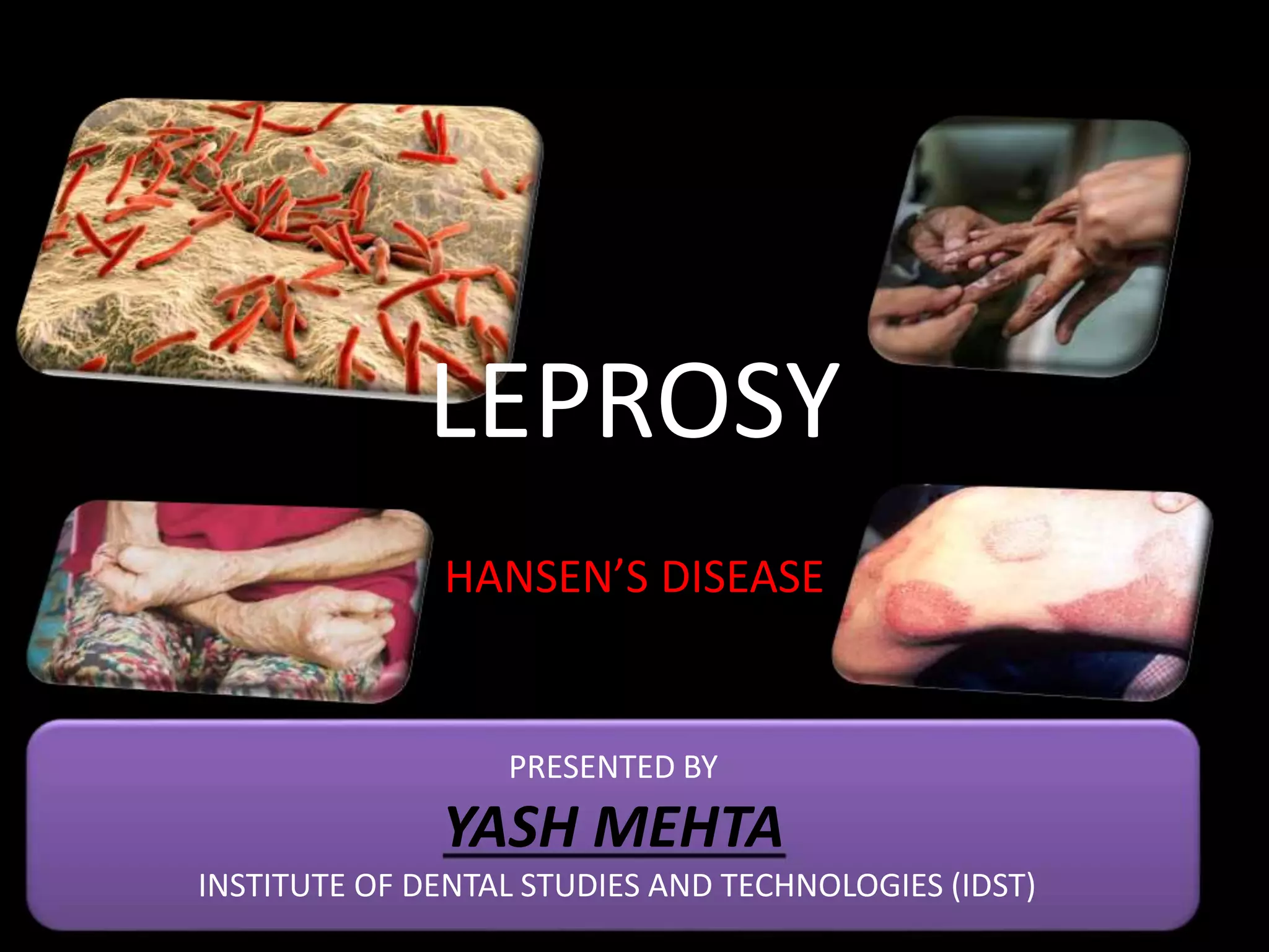
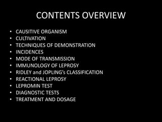
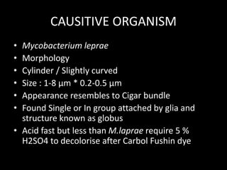
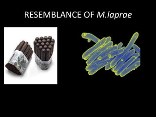
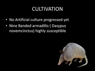
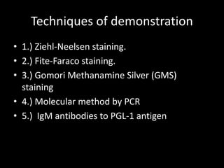
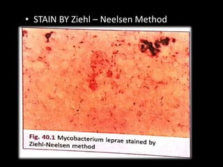
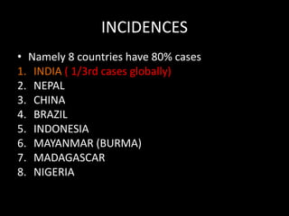
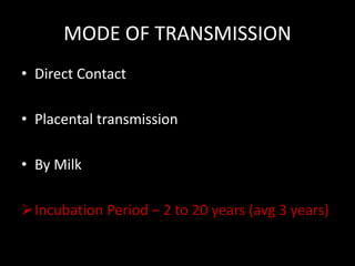
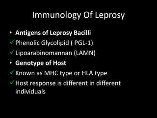
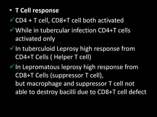
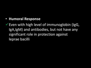
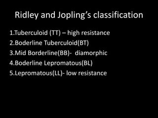
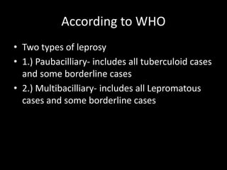
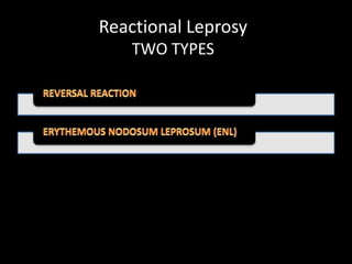
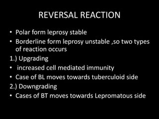
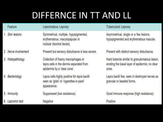
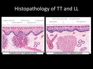
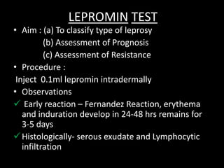
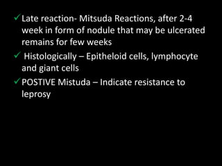
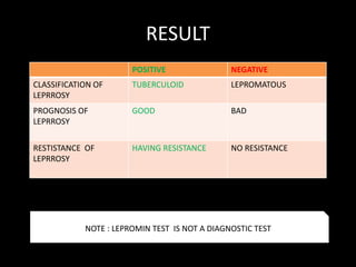
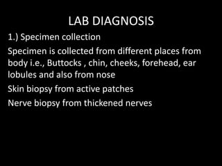
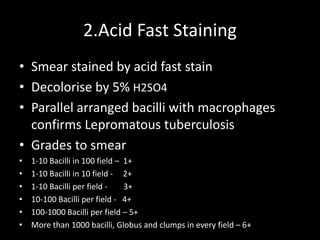
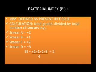
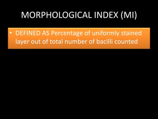
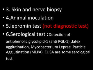
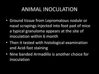
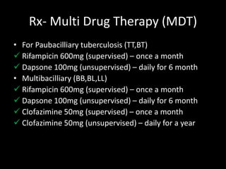
LEPROSY
HANSEN’S DISEASE
PRESENTED BY
YASH MEHTA
INSTITUTE OF DENTAL STUDIES AND TECHNOLOGIES (IDST)
CONTENTS OVERVIEW
• CAUSITIVE ORGANISM
• CULTIVATION
• TECHNIQUES OF DEMONSTRATION
• INCIDENCES
• MODE OF TRANSMISSION
• IMMUNOLOGY OF LEPROSY
• RIDLEY and JOPLING’s CLASSIFICATION
• REACTIONAL LEPROSY
• LEPROMIN TEST
• DIAGNOSTIC TESTS
• TREATMENT AND DOSAGE
CAUSITIVE ORGANISM
• Mycobacterium leprae
• Morphology
• Cylinder / Slightly curved
• Size : 1-8 µm * 0.2-0.5 µm
• Appearance resembles to Cigar bundle
• Found Single or In group attached by glia and
structure known as globus
• Acid fast but less than M.laprae require 5 %
H2SO4 to decolorise after Carbol Fushin dye
RESEMBLANCE OF M.laprae
CULTIVATION
• No Artificial culture progressed yet
• Nine Banded armadillo ( Dasypus
novemcinctus) highly susceptible
Techniques of demonstration
• 1.) Ziehl-Neelsen staining.
• 2.) Fite-Faraco staining.
• 3.) Gomori Methanamine Silver (GMS)
staining
• 4.) Molecular method by PCR
• 5.) IgM antibodies to PGL-1 antigen
• STAIN BY Ziehl – Neelsen Method
INCIDENCES
• Namely 8 countries have 80% cases
1. INDIA ( 1/3rd cases globally)
2. NEPAL
3. CHINA
4. BRAZIL
5. INDONESIA
6. MAYANMAR (BURMA)
7. MADAGASCAR
8. NIGERIA
MODE OF TRANSMISSION
• Direct Contact
• Placental transmission
• By Milk
Incubation Period – 2 to 20 years (avg 3 years)
Immunology Of Leprosy
• Antigens of Leprosy Bacilli
Phenolic Glycolipid ( PGL-1)
Lipoarabinomannan (LAMN)
• Genotype of Host
Known as MHC type or HLA type
Host response is different in different
individuals
• T Cell response
CD4 + T cell, CD8+T cell both activated
While in tubercular infection CD4+T cells
activated only
In tuberculoid Leprosy high response from
CD4+T Cells ( Helper T cell)
In Lepromatous leprosy high response from
CD8+T Cells (suppressor T cell),
but macrophage and suppressor T cell not
able to destroy bacilli due to CD8+T cell defect
• Humoral Response
Even with high level of immunoglobin (IgG,
IgA,IgM) and antibodies, but not have any
significant role in protection against
leprae bacilli
Ridley and Jopling’s classification
1.Tuberculoid (TT) – high resistance
2.Boderline Tuberculoid(BT)
3.Mid Borderline(BB)- diamorphic
4.Boderline Lepromatous(BL)
5.Lepromatous(LL)- low resistance
According to WHO
• Two types of leprosy
• 1.) Paubacilliary- includes all tuberculoid cases
and some borderline cases
• 2.) Multibacilliary- includes all Lepromatous
cases and some borderline cases
Reactional Leprosy
TWO TYPES
REVERSAL REACTION
• Polar form leprosy stable
• Borderline form leprosy unstable ,so two types
of reaction occurs
1.) Upgrading
• increased cell mediated immunity
• Case of BL moves towards tuberculoid side
2.) Downgrading
• Cases of BT moves towards Lepromatous side
DIFFERNCE IN TT AND LL
Histopathology of TT and LL
TUBERCULOID TUBERCULOSIS LEPROMATOUS TUBERCULOSIS
LEPROMIN TEST
• Aim : (a) To classify type of leprosy
(b) Assessment of Prognosis
(c) Assessment of Resistance
• Procedure :
Inject 0.1ml lepromin intradermally
• Observations
 Early reaction – Fernandez Reaction, erythema
and induration develop in 24-48 hrs remains for
3-5 days
Histologically- serous exudate and Lymphocytic
infiltration
Late reaction- Mitsuda Reactions, after 2-4
week in form of nodule that may be ulcerated
remains for few weeks
 Histologically – Epitheloid cells, lymphocyte
and giant cells
POSTIVE Mistuda – Indicate resistance to
leprosy
RESULT
POSITIVE NEGATIVE
CLASSIFICATION OF
LEPRROSY
TUBERCULOID LEPROMATOUS
PROGNOSIS OF
LEPRROSY
GOOD BAD
RESTISTANCE OF
LEPRROSY
HAVING RESISTANCE NO RESISTANCE
NOTE : LEPROMIN TEST IS NOT A DIAGNOSTIC TEST
LAB DIAGNOSIS
1.) Specimen collection
Specimen is collected from different places from
body i.e., Buttocks , chin, cheeks, forehead, ear
lobules and also from nose
Skin biopsy from active patches
Nerve biopsy from thickened nerves
2.Acid Fast Staining
• Smear stained by acid fast stain
• Decolorise by 5% H2SO4
• Parallel arranged bacilli with macrophages
confirms Lepromatous tuberculosis
• Grades to smear
• 1-10 Bacilli in 100 field – 1+
• 1-10 Bacilli in 10 field - 2+
• 1-10 Bacilli per field - 3+
• 10-100 Bacilli per field - 4+
• 100-1000 Bacilli per field – 5+
• More than 1000 bacilli, Globus and clumps in every field – 6+
BACTERIAL INDEX (BI) :
• MAY DEFINED AS PRESENT IN TISSUE
CALCULATION: total grades divided by total
number of smears e.g.,
Smear A = +2
Smear B = +1
Smear C = +2
Smear D = +3
BI = +2+1+2+3 = 2.
4
MORPHOLOGICAL INDEX (MI)
• DEFINED AS Percentage of uniformly stained
layer out of total number of bacilli counted
• 3. Skin and nerve biopsy
• 4.Animal inoculation
• 5.lepromin test (not diagnostic test)
• 6.Serological test :Detection of
antiphenolic glycolipid-1 (anti PGL-1) ,latex
agglutination, Mycobacterium Leprae Particle
Agglutination (MLPA), ELISA are some serological
test
ANIMAL INOCULATION
• Ground tissue from Lepromatous nodule or
nasal scrapings injected into foot pad of mice
a typical granuloma appears at the site of
inoculation within 6 month
• Then it tested with histological examination
and Acid-fast staining
• Nine banded Armadillo is another choice for
inoculation
Rx- Multi Drug Therapy (MDT)
• For Paubacilliary tuberculosis (TT,BT)
 Rifampicin 600mg (supervised) – once a month
 Dapsone 100mg (unsupervised) – daily for 6 month
• Multibacilliary (BB,BL,LL)
 Rifampicin 600mg (supervised) – once a month
 Dapsone 100mg (unsupervised) – daily for 6 month
 Clofazimine 50mg (supervised) – once a month
 Clofazimine 50mg (unsupervised) – daily for a year
REFRENCES
HARSH MOHAN TEXTBOOK OF PATHOLOGY- 6TH
EDITION
ROBBIN’S AND CONTRAN PATHOLOGIC BASES OF
DISEASE- 9TH EDITION
DR.CP BAVEJA MICROBIOLOGY FOR DENTAL
STUDENTS – 6TH EDITION
PRESCOTT’S PRINCIPLE OF MICROBIOLOGY
SOME RELATED IMAGES FROM GOOGLE IMAGES

Leprosy, its pathogenesis and microbiology

  • 1.
    LEPROSY HANSEN’S DISEASE PRESENTED BY YASHMEHTA INSTITUTE OF DENTAL STUDIES AND TECHNOLOGIES (IDST)
  • 2.
    CONTENTS OVERVIEW • CAUSITIVEORGANISM • CULTIVATION • TECHNIQUES OF DEMONSTRATION • INCIDENCES • MODE OF TRANSMISSION • IMMUNOLOGY OF LEPROSY • RIDLEY and JOPLING’s CLASSIFICATION • REACTIONAL LEPROSY • LEPROMIN TEST • DIAGNOSTIC TESTS • TREATMENT AND DOSAGE
  • 3.
    CAUSITIVE ORGANISM • Mycobacteriumleprae • Morphology • Cylinder / Slightly curved • Size : 1-8 µm * 0.2-0.5 µm • Appearance resembles to Cigar bundle • Found Single or In group attached by glia and structure known as globus • Acid fast but less than M.laprae require 5 % H2SO4 to decolorise after Carbol Fushin dye
  • 4.
  • 5.
    CULTIVATION • No Artificialculture progressed yet • Nine Banded armadillo ( Dasypus novemcinctus) highly susceptible
  • 6.
    Techniques of demonstration •1.) Ziehl-Neelsen staining. • 2.) Fite-Faraco staining. • 3.) Gomori Methanamine Silver (GMS) staining • 4.) Molecular method by PCR • 5.) IgM antibodies to PGL-1 antigen
  • 7.
    • STAIN BYZiehl – Neelsen Method
  • 8.
    INCIDENCES • Namely 8countries have 80% cases 1. INDIA ( 1/3rd cases globally) 2. NEPAL 3. CHINA 4. BRAZIL 5. INDONESIA 6. MAYANMAR (BURMA) 7. MADAGASCAR 8. NIGERIA
  • 9.
    MODE OF TRANSMISSION •Direct Contact • Placental transmission • By Milk Incubation Period – 2 to 20 years (avg 3 years)
  • 10.
    Immunology Of Leprosy •Antigens of Leprosy Bacilli Phenolic Glycolipid ( PGL-1) Lipoarabinomannan (LAMN) • Genotype of Host Known as MHC type or HLA type Host response is different in different individuals
  • 11.
    • T Cellresponse CD4 + T cell, CD8+T cell both activated While in tubercular infection CD4+T cells activated only In tuberculoid Leprosy high response from CD4+T Cells ( Helper T cell) In Lepromatous leprosy high response from CD8+T Cells (suppressor T cell), but macrophage and suppressor T cell not able to destroy bacilli due to CD8+T cell defect
  • 12.
    • Humoral Response Evenwith high level of immunoglobin (IgG, IgA,IgM) and antibodies, but not have any significant role in protection against leprae bacilli
  • 13.
    Ridley and Jopling’sclassification 1.Tuberculoid (TT) – high resistance 2.Boderline Tuberculoid(BT) 3.Mid Borderline(BB)- diamorphic 4.Boderline Lepromatous(BL) 5.Lepromatous(LL)- low resistance
  • 14.
    According to WHO •Two types of leprosy • 1.) Paubacilliary- includes all tuberculoid cases and some borderline cases • 2.) Multibacilliary- includes all Lepromatous cases and some borderline cases
  • 15.
  • 16.
    REVERSAL REACTION • Polarform leprosy stable • Borderline form leprosy unstable ,so two types of reaction occurs 1.) Upgrading • increased cell mediated immunity • Case of BL moves towards tuberculoid side 2.) Downgrading • Cases of BT moves towards Lepromatous side
  • 17.
  • 18.
    Histopathology of TTand LL TUBERCULOID TUBERCULOSIS LEPROMATOUS TUBERCULOSIS
  • 19.
    LEPROMIN TEST • Aim: (a) To classify type of leprosy (b) Assessment of Prognosis (c) Assessment of Resistance • Procedure : Inject 0.1ml lepromin intradermally • Observations  Early reaction – Fernandez Reaction, erythema and induration develop in 24-48 hrs remains for 3-5 days Histologically- serous exudate and Lymphocytic infiltration
  • 20.
    Late reaction- MitsudaReactions, after 2-4 week in form of nodule that may be ulcerated remains for few weeks  Histologically – Epitheloid cells, lymphocyte and giant cells POSTIVE Mistuda – Indicate resistance to leprosy
  • 21.
    RESULT POSITIVE NEGATIVE CLASSIFICATION OF LEPRROSY TUBERCULOIDLEPROMATOUS PROGNOSIS OF LEPRROSY GOOD BAD RESTISTANCE OF LEPRROSY HAVING RESISTANCE NO RESISTANCE NOTE : LEPROMIN TEST IS NOT A DIAGNOSTIC TEST
  • 22.
    LAB DIAGNOSIS 1.) Specimencollection Specimen is collected from different places from body i.e., Buttocks , chin, cheeks, forehead, ear lobules and also from nose Skin biopsy from active patches Nerve biopsy from thickened nerves
  • 23.
    2.Acid Fast Staining •Smear stained by acid fast stain • Decolorise by 5% H2SO4 • Parallel arranged bacilli with macrophages confirms Lepromatous tuberculosis • Grades to smear • 1-10 Bacilli in 100 field – 1+ • 1-10 Bacilli in 10 field - 2+ • 1-10 Bacilli per field - 3+ • 10-100 Bacilli per field - 4+ • 100-1000 Bacilli per field – 5+ • More than 1000 bacilli, Globus and clumps in every field – 6+
  • 24.
    BACTERIAL INDEX (BI): • MAY DEFINED AS PRESENT IN TISSUE CALCULATION: total grades divided by total number of smears e.g., Smear A = +2 Smear B = +1 Smear C = +2 Smear D = +3 BI = +2+1+2+3 = 2. 4
  • 25.
    MORPHOLOGICAL INDEX (MI) •DEFINED AS Percentage of uniformly stained layer out of total number of bacilli counted
  • 26.
    • 3. Skinand nerve biopsy • 4.Animal inoculation • 5.lepromin test (not diagnostic test) • 6.Serological test :Detection of antiphenolic glycolipid-1 (anti PGL-1) ,latex agglutination, Mycobacterium Leprae Particle Agglutination (MLPA), ELISA are some serological test
  • 27.
    ANIMAL INOCULATION • Groundtissue from Lepromatous nodule or nasal scrapings injected into foot pad of mice a typical granuloma appears at the site of inoculation within 6 month • Then it tested with histological examination and Acid-fast staining • Nine banded Armadillo is another choice for inoculation
  • 28.
    Rx- Multi DrugTherapy (MDT) • For Paubacilliary tuberculosis (TT,BT)  Rifampicin 600mg (supervised) – once a month  Dapsone 100mg (unsupervised) – daily for 6 month • Multibacilliary (BB,BL,LL)  Rifampicin 600mg (supervised) – once a month  Dapsone 100mg (unsupervised) – daily for 6 month  Clofazimine 50mg (supervised) – once a month  Clofazimine 50mg (unsupervised) – daily for a year
  • 30.
    REFRENCES HARSH MOHAN TEXTBOOKOF PATHOLOGY- 6TH EDITION ROBBIN’S AND CONTRAN PATHOLOGIC BASES OF DISEASE- 9TH EDITION DR.CP BAVEJA MICROBIOLOGY FOR DENTAL STUDENTS – 6TH EDITION PRESCOTT’S PRINCIPLE OF MICROBIOLOGY SOME RELATED IMAGES FROM GOOGLE IMAGES