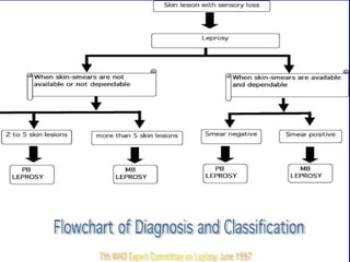
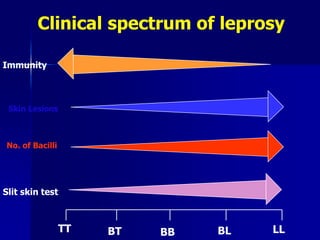
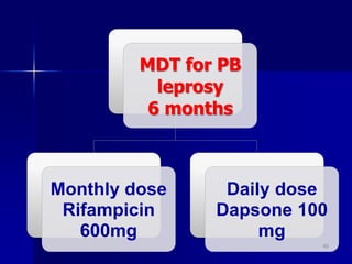
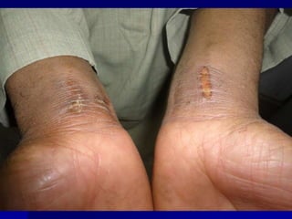
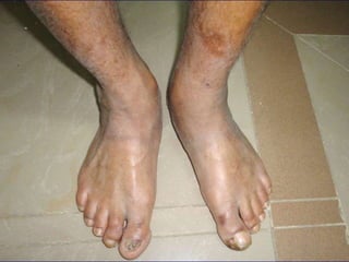
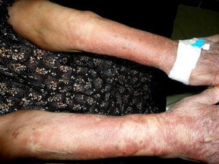
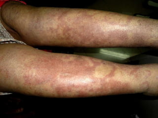
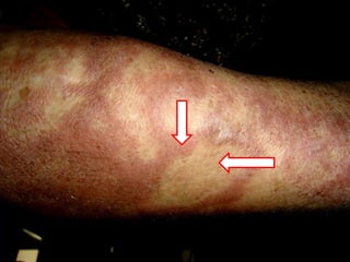
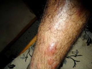
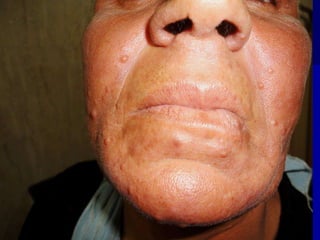
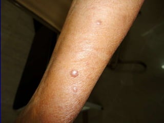
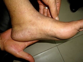
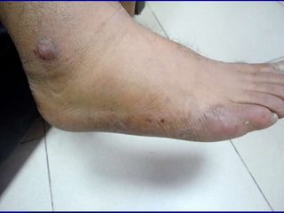
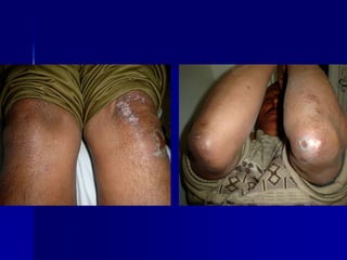
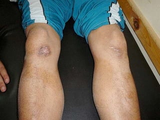
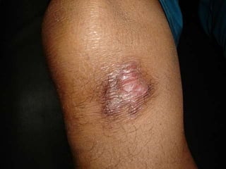
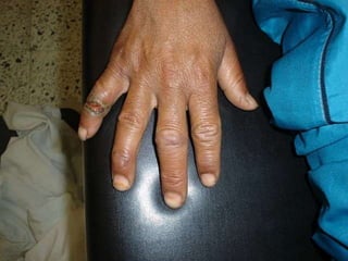
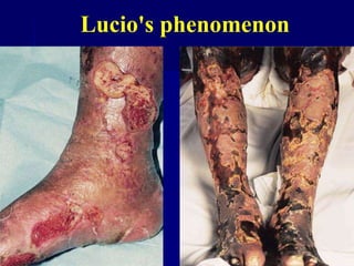
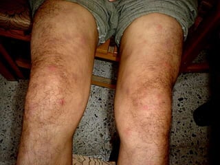
By
M.D.
It is a chronic infectious disease
characterized by lesions of the peripheral
nerve, skin, and mucus membrane of the
nasal mucosa and internal organs. World's
oldest recorded disease
3
Definition
 More prevalent in Africa, South east Asia
and Latin America.
 Commonest in India.
 Incidence now: 1/10000
 Males twice as females
 Children are more prone
 Two peaks: 10-15 and 35-45
Epidemiology
Acid fast bacillus
Staining: Z-N stain
Growth :Culture and Inoculation
Incubation period
Multiplication: very slow (Ms-ys) 5
Ateiology
Mode of infection
Although human-to-human transmission is
the primary source of infection, three other
species can carry and (rarely) transfer M.
leprae to humans: chimpanzees, mangabey
monkeys, and nine-banded armadillos.
1-Droplet infection
2-Contact through the skin (rare).
3-Arthropod-born infection (rare).
4-Through placenta and milk.
Mode of infection
1-Residence in an endemic area.
2-Poverty (malnutrition).
3-Contact with affected armadillo.
4-Genetic :DR2 &3 in TT, DQW in LL
5-Low immunity.
Predisposing factors
Ridley & Jopling Classification
Based on Host Immunity
TT BL LL
BT BB BL
9
Classification
WHO Classification
Based on Bacterial Load
Paucibacillary
1-5 skin lesions
Multibacillary
>6 skin lesions
PositiveNegative
LEPROSY
Paucibacillary
(PB)
Multibacillary
(MB)
Indeterminate Leprosy (IL)
Tuberculoid Leprosy (TL)
Borderline Tuberculoid (BT)
Borderline Borderline (BB)
Borderline Lepromatous(BL)
Lepromatous Leprosy (LL)
11
In Tuberculoid leprosy
1-T-Cells : CD4 predominate
2- Humoral immunity: low antibody titer
3-Lepromin test: +ve
In Lepromatous leprosy
1-T-Cells : CD8 predominate
2- Humoral immunity: high antibody titer
3-Lepromin test: -ve
Immunology
Skin lesions
Peripheral nerves
Internal organs
Bacteriology
Histopathology
Lepromin test
Course and prognosis
Clinical features
1-Type of leprosy
2-Mechanism
3-Clinical features
4-Histopathology
5-Lepromin test
6-Treatment
Reactions
BLBBBT
ManySomeFew(<5)Lesion no.
RoughlyLessWellLesions borders
SlightModerateMarkedSensory
impairment
Roughly
symmetrical
AsymmetricalAsymmetricalDistribution of
skin lesions
Less
asymmetrical
AsymmetricalAsymmetricalPeripheral nerves
MultibacillaryMultibacillaryPaucibacillaryType of leprosy
4+2+ / 3+- / 1+Slit skin smear
Diagnosis of Leprosy
Clinical Examination
Slit Skin Smear
Skin Biopsy
Pinch the site tight.
Incise.
Scrape & collect material
Smear on a slide.
Air dry & fix.
Stain (Z-N method)
17
Slit Skin Smear
0 – no bacilli in 100 fields
1+: 1-10 bacilli in 100 fields
2+: 1-10 bacilli in 10 fields
3+: 1-10 bacilli in 1 field
4+: 10-100 bacilli in 1 field
5+: 100-1000 in 1 field
6+: >1000 bacilli field (globi).
Bacteriological Index
BI is calculated by adding up the
index from site examined and
dividing by the total number
TT:-ve
+BT:0-2
LL & BL:5+ or 6+
The percentage of living bacilli (solid staining
bacilli) to the total number of bacilli
(fragmented and granular) in the smear.
Morphological index
Tuberculoid Leprosy
Lepromatous leprosy
Lepromatous leprosy
TT BT BB BL LL
Skin Lesions
No. of Bacilli
Slit skin test
Immunity
Clinical spectrum of leprosy
Other tests:
Histamine test: for the diagnosis of
indeterminate leprosy
Immunological tests
Test for detecting CMI
Test for detecting antibobies
Lepromin skin test :
To differentiate the two different
forms of leprosy apart, but it is not
used to diagnose the disease
Because: false negative and false
positive
Test for detecting CMI
Procedure to Lepromin Skin Test
A tiny sample (0.1) of leprosy antigen is injected
under the skin, usually in the forearm.
The site of the injection is marked, and is examined
for reaction, first after 3 days(early reaction-
Fernandez reaction:-redness and induration) and
then again after 21 days(late reaction-Mitsuda
reaction:-nodule>5mm).
Lepromin Skin Test
Test for detecting antibobies
1-Fluorescent leprosy antibody
absorption test(FLA-ABS test):-Screening
92.3% Sensitive & 100% specific
2-Monoclonal antibodies
3-ELISA
4-Radioimmunoassay: cell wall AG -7
TREATMENT
Basics about therapy
-MDT will cure you completely
-MDT is free of cost
-MDT is available in all health centres
-MDT should be taken as advised
(regular, full course)
1-Skin discoloration due to clofazimine
2-Urine discoloration due to rifampicin
3-In case of fever, pain in the nerves,
4-muscle weakness, joint pains they must
return immediately to the health centre
5-Appearance of new skin patches
Basics about therapy
MDT for PB
leprosy
6 months
Monthly dose
Rifampicin
600mg
Daily dose
Dapsone 100
mg
40
MDT for MB
leprosy
12 months
Monthly dose
Rifampicin 600mg
Clofazimine 300mg
Daily dose
Dapson 100mg
Clofazimine 50 mg
41
ADVERVE EFFECT OF ANTI-LEPROTIC DRUGS
DRUGS MINOR MAJOR
RIFAMPICIN RED URINE JAUNDICE
GIT UPSET HEPATITIS
FLU LIKE SYNDROME SHOCK
DAPSONE GIT UPSET DAPSONE SYNDROME
DRUG RASH AGRANULOCYTOSIS
ANAEMIA HEMOLYTIC ANAEMIA
CLOFAZIMINE GIT UPSET ACUTE PAIN ABDOMEN
DISCOLOURATION OF SKIN
ICHTHYOSIS
42
LEPRA REACTION
ADVERSE EFFECT OF ANTI-LEPROTIC DRUGS
DISABILITIES & DEFORMITIES
PSYCHO-SOCIAL PROBLEMS
43
Complications
DISABILITIES
Causes
1-Late diagnosis and late treatment with MDT
2-Advanced disease (MB leprosy)
3-Leprosy reactions which involve nerves
4-Lack of information on how to protect
insensitive parts
5-Only about 10-15% of leprosy affected person
develop significant deformities and disabilities.
1:
1-Leonine facies
2-Nasal deformity
3-Loss of outer third of eyebrow
45
1) Specific deformities
2) Paralytic deformities:
- result from damage to motor nerve.
-seen most often in the hand(claw
finger), less often in the feet &
occasionally in the
face(lagopthalomos,facial palsy
46
3)Anesthetic deformity :
--Occur as a consequence of neglected injuries
in part rendered insensitive because of
damage to sensory nerve.
- Found most often on the feet and
hand(ulceration, scar contracture,
shortening of digits & skeletal
disorganization of foot)
47
WHO Grade 0 Grade 1 Grade 2
EYES Normal
vision,lid
gap,blinking.
Corneal reflex
weak
Reduced
vision,lagophtha
lmos.
HANDS Normal
sensation &
m.power.
Loss of feeling in
the palm
Visible
damage:wounds,
claw hand,loss
of tissue etc.
FEET Normal
sensation &
m.power.
Loss of feeling in
the sole
Visible
damage:wound,f
oot drop,loss of
tissue.
48
Peripheral nerves
Sensory Motor Autonomic
Hypoaestesia /
anaestesia
Muscle paralysis Lack of sweating &
sebum
Ulcers Ulnar nerve Claw hand
Radial nerve Wrist drop
Lt. popliteal Foot drop
Post. tibial Claw toes
Facial lagophthalmous
Dry skin
Cracked skin
Ulcers
49
50
Clinical applications
Indeterminate Leprosy
TT: Erythematous plaque
Borderline Leprosy
Borderline Leprosy
Neural Leprosy
Neural Leprosy
Neural Leprosy
Type 1-reaction
Type 2 reaction
Lucio's phenomenon
THANK YOU

Leprosy lecture

  • 2.
  • 3.
    It is achronic infectious disease characterized by lesions of the peripheral nerve, skin, and mucus membrane of the nasal mucosa and internal organs. World's oldest recorded disease 3 Definition
  • 4.
     More prevalentin Africa, South east Asia and Latin America.  Commonest in India.  Incidence now: 1/10000  Males twice as females  Children are more prone  Two peaks: 10-15 and 35-45 Epidemiology
  • 5.
    Acid fast bacillus Staining:Z-N stain Growth :Culture and Inoculation Incubation period Multiplication: very slow (Ms-ys) 5 Ateiology
  • 6.
    Mode of infection Althoughhuman-to-human transmission is the primary source of infection, three other species can carry and (rarely) transfer M. leprae to humans: chimpanzees, mangabey monkeys, and nine-banded armadillos.
  • 7.
    1-Droplet infection 2-Contact throughthe skin (rare). 3-Arthropod-born infection (rare). 4-Through placenta and milk. Mode of infection
  • 8.
    1-Residence in anendemic area. 2-Poverty (malnutrition). 3-Contact with affected armadillo. 4-Genetic :DR2 &3 in TT, DQW in LL 5-Low immunity. Predisposing factors
  • 9.
    Ridley & JoplingClassification Based on Host Immunity TT BL LL BT BB BL 9 Classification
  • 10.
    WHO Classification Based onBacterial Load Paucibacillary 1-5 skin lesions Multibacillary >6 skin lesions PositiveNegative
  • 11.
    LEPROSY Paucibacillary (PB) Multibacillary (MB) Indeterminate Leprosy (IL) TuberculoidLeprosy (TL) Borderline Tuberculoid (BT) Borderline Borderline (BB) Borderline Lepromatous(BL) Lepromatous Leprosy (LL) 11
  • 12.
    In Tuberculoid leprosy 1-T-Cells: CD4 predominate 2- Humoral immunity: low antibody titer 3-Lepromin test: +ve In Lepromatous leprosy 1-T-Cells : CD8 predominate 2- Humoral immunity: high antibody titer 3-Lepromin test: -ve Immunology
  • 13.
    Skin lesions Peripheral nerves Internalorgans Bacteriology Histopathology Lepromin test Course and prognosis Clinical features
  • 14.
    1-Type of leprosy 2-Mechanism 3-Clinicalfeatures 4-Histopathology 5-Lepromin test 6-Treatment Reactions
  • 15.
    BLBBBT ManySomeFew(<5)Lesion no. RoughlyLessWellLesions borders SlightModerateMarkedSensory impairment Roughly symmetrical AsymmetricalAsymmetricalDistributionof skin lesions Less asymmetrical AsymmetricalAsymmetricalPeripheral nerves MultibacillaryMultibacillaryPaucibacillaryType of leprosy 4+2+ / 3+- / 1+Slit skin smear
  • 16.
    Diagnosis of Leprosy ClinicalExamination Slit Skin Smear Skin Biopsy
  • 17.
    Pinch the sitetight. Incise. Scrape & collect material Smear on a slide. Air dry & fix. Stain (Z-N method) 17 Slit Skin Smear
  • 21.
    0 – nobacilli in 100 fields 1+: 1-10 bacilli in 100 fields 2+: 1-10 bacilli in 10 fields 3+: 1-10 bacilli in 1 field 4+: 10-100 bacilli in 1 field 5+: 100-1000 in 1 field 6+: >1000 bacilli field (globi). Bacteriological Index
  • 22.
    BI is calculatedby adding up the index from site examined and dividing by the total number TT:-ve +BT:0-2 LL & BL:5+ or 6+
  • 23.
    The percentage ofliving bacilli (solid staining bacilli) to the total number of bacilli (fragmented and granular) in the smear. Morphological index
  • 25.
  • 26.
  • 27.
  • 29.
    TT BT BBBL LL Skin Lesions No. of Bacilli Slit skin test Immunity Clinical spectrum of leprosy
  • 30.
    Other tests: Histamine test:for the diagnosis of indeterminate leprosy Immunological tests Test for detecting CMI Test for detecting antibobies
  • 31.
    Lepromin skin test: To differentiate the two different forms of leprosy apart, but it is not used to diagnose the disease Because: false negative and false positive Test for detecting CMI
  • 32.
    Procedure to LeprominSkin Test A tiny sample (0.1) of leprosy antigen is injected under the skin, usually in the forearm. The site of the injection is marked, and is examined for reaction, first after 3 days(early reaction- Fernandez reaction:-redness and induration) and then again after 21 days(late reaction-Mitsuda reaction:-nodule>5mm). Lepromin Skin Test
  • 33.
    Test for detectingantibobies 1-Fluorescent leprosy antibody absorption test(FLA-ABS test):-Screening 92.3% Sensitive & 100% specific 2-Monoclonal antibodies 3-ELISA 4-Radioimmunoassay: cell wall AG -7
  • 34.
  • 35.
    Basics about therapy -MDTwill cure you completely -MDT is free of cost -MDT is available in all health centres -MDT should be taken as advised (regular, full course)
  • 36.
    1-Skin discoloration dueto clofazimine 2-Urine discoloration due to rifampicin 3-In case of fever, pain in the nerves, 4-muscle weakness, joint pains they must return immediately to the health centre 5-Appearance of new skin patches Basics about therapy
  • 40.
    MDT for PB leprosy 6months Monthly dose Rifampicin 600mg Daily dose Dapsone 100 mg 40
  • 41.
    MDT for MB leprosy 12months Monthly dose Rifampicin 600mg Clofazimine 300mg Daily dose Dapson 100mg Clofazimine 50 mg 41
  • 42.
    ADVERVE EFFECT OFANTI-LEPROTIC DRUGS DRUGS MINOR MAJOR RIFAMPICIN RED URINE JAUNDICE GIT UPSET HEPATITIS FLU LIKE SYNDROME SHOCK DAPSONE GIT UPSET DAPSONE SYNDROME DRUG RASH AGRANULOCYTOSIS ANAEMIA HEMOLYTIC ANAEMIA CLOFAZIMINE GIT UPSET ACUTE PAIN ABDOMEN DISCOLOURATION OF SKIN ICHTHYOSIS 42
  • 43.
    LEPRA REACTION ADVERSE EFFECTOF ANTI-LEPROTIC DRUGS DISABILITIES & DEFORMITIES PSYCHO-SOCIAL PROBLEMS 43 Complications
  • 44.
    DISABILITIES Causes 1-Late diagnosis andlate treatment with MDT 2-Advanced disease (MB leprosy) 3-Leprosy reactions which involve nerves 4-Lack of information on how to protect insensitive parts 5-Only about 10-15% of leprosy affected person develop significant deformities and disabilities.
  • 45.
    1: 1-Leonine facies 2-Nasal deformity 3-Lossof outer third of eyebrow 45 1) Specific deformities
  • 46.
    2) Paralytic deformities: -result from damage to motor nerve. -seen most often in the hand(claw finger), less often in the feet & occasionally in the face(lagopthalomos,facial palsy 46
  • 47.
    3)Anesthetic deformity : --Occuras a consequence of neglected injuries in part rendered insensitive because of damage to sensory nerve. - Found most often on the feet and hand(ulceration, scar contracture, shortening of digits & skeletal disorganization of foot) 47
  • 48.
    WHO Grade 0Grade 1 Grade 2 EYES Normal vision,lid gap,blinking. Corneal reflex weak Reduced vision,lagophtha lmos. HANDS Normal sensation & m.power. Loss of feeling in the palm Visible damage:wounds, claw hand,loss of tissue etc. FEET Normal sensation & m.power. Loss of feeling in the sole Visible damage:wound,f oot drop,loss of tissue. 48
  • 49.
    Peripheral nerves Sensory MotorAutonomic Hypoaestesia / anaestesia Muscle paralysis Lack of sweating & sebum Ulcers Ulnar nerve Claw hand Radial nerve Wrist drop Lt. popliteal Foot drop Post. tibial Claw toes Facial lagophthalmous Dry skin Cracked skin Ulcers 49
  • 50.
  • 51.
  • 53.
  • 67.
  • 68.
  • 69.
  • 129.
  • 130.
  • 131.
  • 133.
  • 134.
  • 135.
  • 144.