Downloaded 161 times
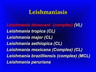
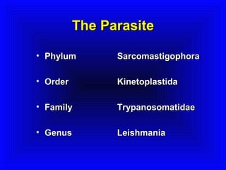
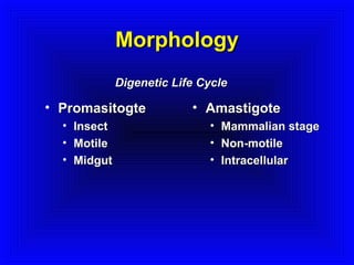
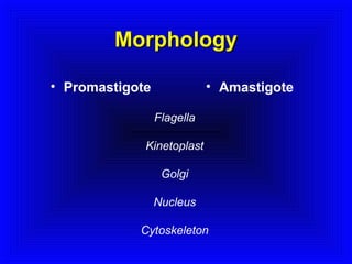
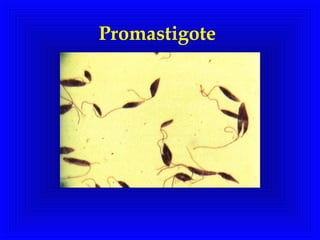
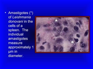
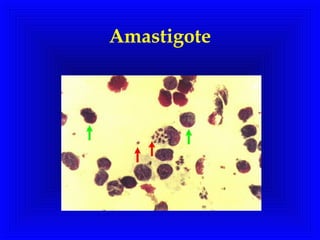
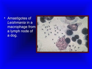
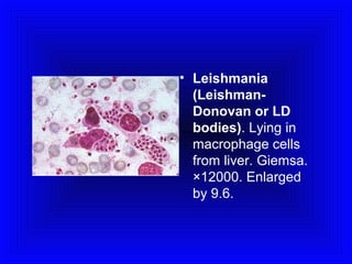
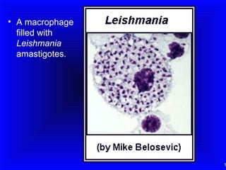
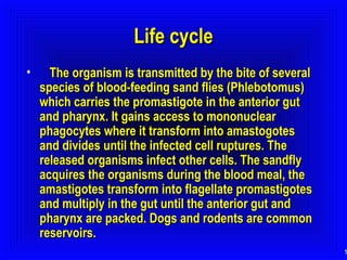
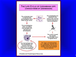
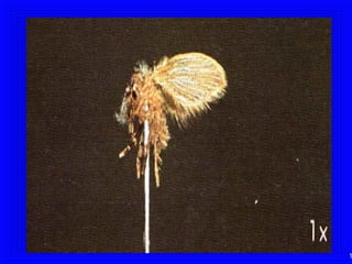
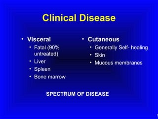
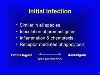
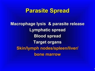
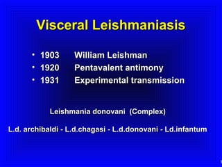
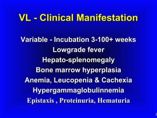

This document summarizes information about Leishmaniasis, a parasitic disease caused by protozoa of the genus Leishmania. It is transmitted by the bite of infected sand flies. The document outlines the life cycle between the promastigote stage in the sand fly and amastigote stage in mammals. It describes the clinical manifestations of visceral leishmaniasis and cutaneous leishmaniasis. Diagnosis involves clinical signs and microscopy or culture of parasites. Treatment currently involves pentavalent antimony compounds or pentamidine, with efforts ongoing to develop new drugs and delivery methods. Control relies on vector control, reservoir control, treatment, and working towards an effective vaccine.


