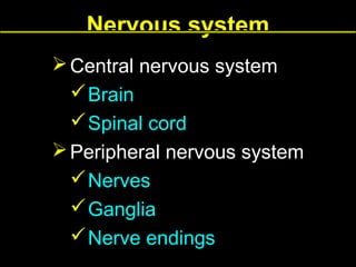
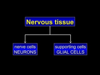
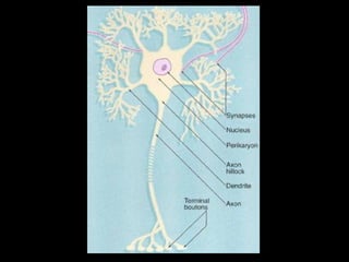
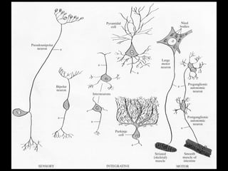
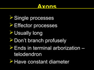
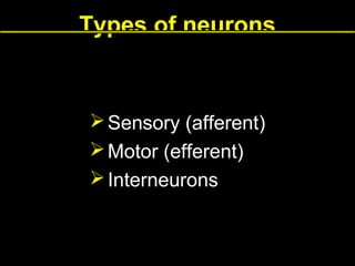
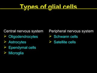
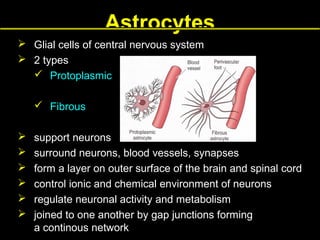
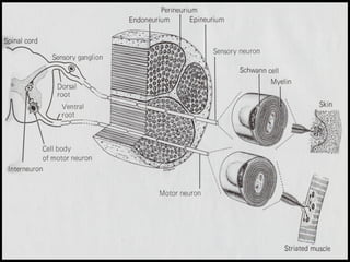
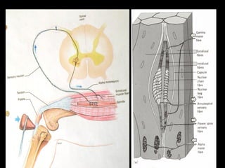
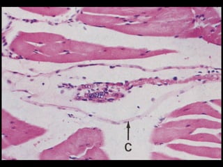
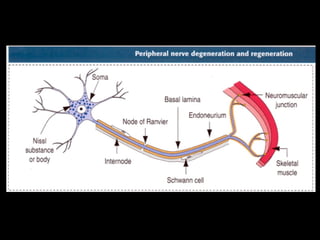
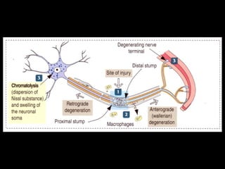
Nervous system
 Central nervous system
 Brain
 Spinal cord
 Peripheral nervous system
 Nerves
 Ganglia
 Nerve endings
Nervous tissue

nerve cells
NEURONS

supporting cells
GLIAL CELLS
Dendrites
 Multiple processes
 Receptor processes
 Usually short
 Branch profusely forming
a dendritic tree
 Become thinner as they subdivide
into branches
Axons
 Single processes
 Effector processes
 Usually long
 Don’t branch profusely
 Ends in terminal arborization –
telodendron
 Have constant diameter
Types of neurons

 Sensory (afferent)
 Motor (efferent)
 Interneurons
Types of neurons
 Bipolar neurons
 Retina
 Olfactory mucosa
 Cochlear and
vestibular ganglia
 Pseudounipolar
neurons
 Sensory neurons
 Multipolar neurons
 Motor neurons
 Interneurons
Nuclei of glial cells

dendrite

Axon hillock
Nissl bodies
rough endoplasmic reticulum
cisternaes and free ribosomes
Types of synapses

Axosomatic
Axodendritic
Axoaxonic
Types of synapses

 Excitatory
 Inhibitory
Types of glial cells

Central nervous system
 Oligodendrocytes
 Astrocytes
 Ependymal cells
 Microglia

Peripheral nervous system
 Schwann cells
 Satellite cells
Astrocytes
 Glial cells of central nervous system
 2 types
 Protoplasmic
 Fibrous







support neurons
surround neurons, blood vessels, synapses
form a layer on outer surface of the brain and spinal cord
control ionic and chemical environment of neurons
regulate neuronal activity and metabolism
joined to one another by gap junctions forming
a continous network
Astrocytes

Blood vessel
Microglial cells – Microglia
 glial cells of central nervous
system
 phagocytic cells – belong to
mononuclear phagocytic
system
 macrophages of CNS
 derive from precursor cells in
bone marrow
 involved in inflammation
processes in CNS
 phagocyte dead neurons and
an excess of neurons during
embryogenesis
Ependymal cells

 glial cells of CNS
 cuboidal cells
 line the ventricles of the brain
and the central canal of the spinal cord
Oligodendrocytes
 glial cells of CNS
 have only a few small processes
 myelin – forming cells in CNS
Cells forming the myelin sheath

PNS
Schwann cells

CNS
Oligodendrocytes
Schwann cell
Internode

Node of Ranvier
Peripheral nervous system
Myelinated fibers

Unmyelinated fibers

One Shwann cell→ one axon

One Schwann cell→ several axons
Axon

Shwann cell

Shwann cell
Oligodendrocyte

Axons
Central nervous system
Myelinated fibers

Unmyelinated fibers

Oligodendrocytes

Bare nerve fibers
– not imbedded in glial cells
Peripheral nervous system
 nerves
 sensory nerves
 motor nerves
 mixed nerves
 ganglia
 nerve endings
Epineurium

Perineurium
Endoneurium
Nerve
 Endoneurium – loose connective tissue
 Perineurium – a few layers of flattened
epithelium – like cells
 Epineurium – dense irregular connective
tissue containing blood vessels
Nucleolus

Satelite cells

Nucleus of nerve cell
Sensory ganglia
 lie in dorsal roots of spinal cord, outside of CNS
 contain cell bodies of sensory neurons –
pseudounipolar
 2 regions
 central region – nerve fibers
 peripheral region – cell bodies
 cell bodies are covered by satellite cells
 satellite cells – a kind of glial cells of PNS
Nervous system
 Somatic nervous system – voluntary
functions – control skeletal muscles
 Autonomic nervous system –
involuntary functions – control smooth
muscle, secretion of some glands, modulation of
cardiac rhythm
Autonomic nervous system
 Sympathetic system – presynaptic
neurons are located in thoracic and lumbar
segments of spinal cord

 Parasympathetic system – presynaptic
neurons have their nuclei in medulla, midbrain,
sacral portion of spinal cord
Nerve endings

 afferent – sensory receptors
 efferent
Sensory receptors

 exteroceptors
 proprioceptors
 interoceptors
Sensory receptors
 Nonencapsulated
 Free nerve endings
 Merkel endings
 Encapsulated
 Meissner corpuscle
 Pacinian corpuscle
 Ruffini corpuscle
Free nerve endings
 Found in skin and
corneal epithelium
 Terminate in
epidermis
 Devoid of Schwann
cells and myelin
 Respond to touch,
heat, cold, pain
Merkel ending
 Nerve endings are
attached to Merkel
cells
 Merkel cells –
modified epidermal
cells located in skin
 Mechanoreceptors –
sensitive to touch
Pacinian corpuscles
 Encapsulated receptors
 Ovoid structure resembling
a hemisected onion
 Nerve ending is
surrounded
by concentric lamellae of
flattened cells
 Found in hypodermis and
deed fascia tissues
 Respond to vibrations and
deep pressure
Meissner’s corpuscles
Encapsulated receptors
Contain Schwann cells that
form irregular, tortuous
lamellae
 Nerve fibers pass between
lamellae
 Found in the papillary layer
of hairless skin (lips,
fingers, hands, foots)
 Respond to touch


Proprioceptors

 Muscle spindles
 Golgi tendon organs
Degeneration and regeneration of
peripheral nerve - steps
1.
2.


3.
4.
5.
6.
7.
8.

Schwann cells proliferate and bridge the scar.
The axon degenerate
Anterograde degeneration – (refers to distal segment) – comprise the
whole distal segment including axon and myelin sheath
Retrograde degeneration – (refers to proximal segment) –extends for a
short distance
Debris is phagocyted by macrophages.
The cell body undergoes chromatolysis.
The muscle fiber shows a denervation atrophy.
Shwann cell prolipherate within a connective tissue sleeve.
The axon grows and penetrate the Schwann cell columns.
Schwann cells produce myelin sheath on the regenerated axon.

Lecture8 nervous system

  • 1.
    Nervous system  Centralnervous system  Brain  Spinal cord  Peripheral nervous system  Nerves  Ganglia  Nerve endings
  • 2.
  • 5.
    Dendrites  Multiple processes Receptor processes  Usually short  Branch profusely forming a dendritic tree  Become thinner as they subdivide into branches
  • 6.
    Axons  Single processes Effector processes  Usually long  Don’t branch profusely  Ends in terminal arborization – telodendron  Have constant diameter
  • 7.
    Types of neurons Sensory (afferent)  Motor (efferent)  Interneurons
  • 8.
    Types of neurons Bipolar neurons  Retina  Olfactory mucosa  Cochlear and vestibular ganglia  Pseudounipolar neurons  Sensory neurons  Multipolar neurons  Motor neurons  Interneurons
  • 9.
    Nuclei of glialcells dendrite Axon hillock
  • 10.
    Nissl bodies rough endoplasmicreticulum cisternaes and free ribosomes
  • 11.
  • 13.
    Types of synapses Excitatory  Inhibitory
  • 14.
    Types of glialcells Central nervous system  Oligodendrocytes  Astrocytes  Ependymal cells  Microglia Peripheral nervous system  Schwann cells  Satellite cells
  • 15.
    Astrocytes  Glial cellsof central nervous system  2 types  Protoplasmic  Fibrous       support neurons surround neurons, blood vessels, synapses form a layer on outer surface of the brain and spinal cord control ionic and chemical environment of neurons regulate neuronal activity and metabolism joined to one another by gap junctions forming a continous network
  • 18.
  • 19.
    Microglial cells –Microglia  glial cells of central nervous system  phagocytic cells – belong to mononuclear phagocytic system  macrophages of CNS  derive from precursor cells in bone marrow  involved in inflammation processes in CNS  phagocyte dead neurons and an excess of neurons during embryogenesis
  • 20.
    Ependymal cells  glialcells of CNS  cuboidal cells  line the ventricles of the brain and the central canal of the spinal cord
  • 21.
    Oligodendrocytes  glial cellsof CNS  have only a few small processes  myelin – forming cells in CNS
  • 23.
    Cells forming themyelin sheath PNS Schwann cells CNS Oligodendrocytes
  • 24.
  • 27.
    Peripheral nervous system Myelinatedfibers Unmyelinated fibers One Shwann cell→ one axon One Schwann cell→ several axons Axon Shwann cell Shwann cell
  • 28.
  • 29.
    Central nervous system Myelinatedfibers Unmyelinated fibers Oligodendrocytes Bare nerve fibers – not imbedded in glial cells
  • 30.
    Peripheral nervous system nerves  sensory nerves  motor nerves  mixed nerves  ganglia  nerve endings
  • 31.
  • 32.
    Nerve  Endoneurium –loose connective tissue  Perineurium – a few layers of flattened epithelium – like cells  Epineurium – dense irregular connective tissue containing blood vessels
  • 41.
  • 42.
    Sensory ganglia  liein dorsal roots of spinal cord, outside of CNS  contain cell bodies of sensory neurons – pseudounipolar  2 regions  central region – nerve fibers  peripheral region – cell bodies  cell bodies are covered by satellite cells  satellite cells – a kind of glial cells of PNS
  • 43.
    Nervous system  Somaticnervous system – voluntary functions – control skeletal muscles  Autonomic nervous system – involuntary functions – control smooth muscle, secretion of some glands, modulation of cardiac rhythm
  • 45.
    Autonomic nervous system Sympathetic system – presynaptic neurons are located in thoracic and lumbar segments of spinal cord  Parasympathetic system – presynaptic neurons have their nuclei in medulla, midbrain, sacral portion of spinal cord
  • 46.
    Nerve endings  afferent– sensory receptors  efferent
  • 47.
    Sensory receptors  exteroceptors proprioceptors  interoceptors
  • 48.
    Sensory receptors  Nonencapsulated Free nerve endings  Merkel endings  Encapsulated  Meissner corpuscle  Pacinian corpuscle  Ruffini corpuscle
  • 49.
    Free nerve endings Found in skin and corneal epithelium  Terminate in epidermis  Devoid of Schwann cells and myelin  Respond to touch, heat, cold, pain
  • 50.
    Merkel ending  Nerveendings are attached to Merkel cells  Merkel cells – modified epidermal cells located in skin  Mechanoreceptors – sensitive to touch
  • 51.
    Pacinian corpuscles  Encapsulatedreceptors  Ovoid structure resembling a hemisected onion  Nerve ending is surrounded by concentric lamellae of flattened cells  Found in hypodermis and deed fascia tissues  Respond to vibrations and deep pressure
  • 53.
    Meissner’s corpuscles Encapsulated receptors ContainSchwann cells that form irregular, tortuous lamellae  Nerve fibers pass between lamellae  Found in the papillary layer of hairless skin (lips, fingers, hands, foots)  Respond to touch  
  • 56.
  • 63.
    Degeneration and regenerationof peripheral nerve - steps 1. 2.   3. 4. 5. 6. 7. 8. Schwann cells proliferate and bridge the scar. The axon degenerate Anterograde degeneration – (refers to distal segment) – comprise the whole distal segment including axon and myelin sheath Retrograde degeneration – (refers to proximal segment) –extends for a short distance Debris is phagocyted by macrophages. The cell body undergoes chromatolysis. The muscle fiber shows a denervation atrophy. Shwann cell prolipherate within a connective tissue sleeve. The axon grows and penetrate the Schwann cell columns. Schwann cells produce myelin sheath on the regenerated axon.