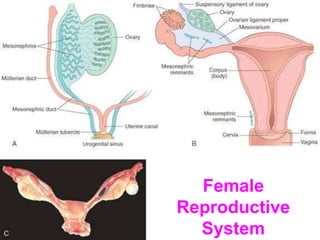
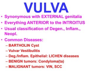
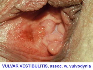
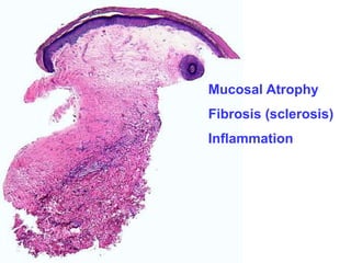
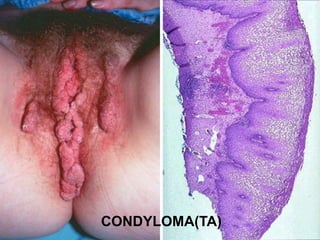
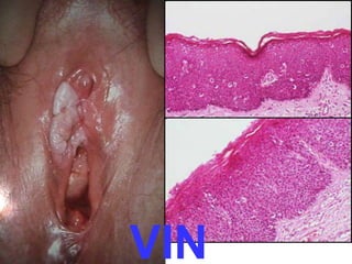
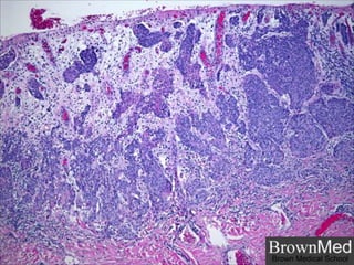
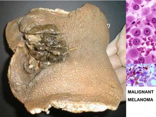
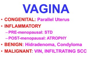
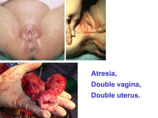
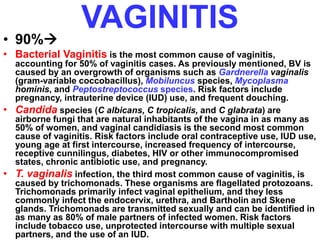
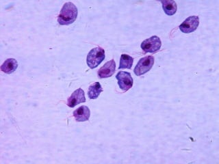
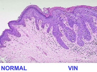
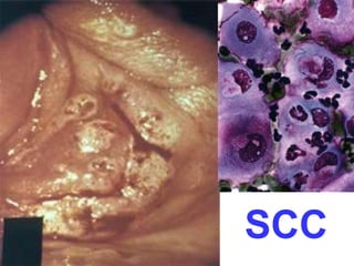
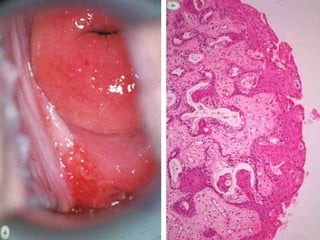
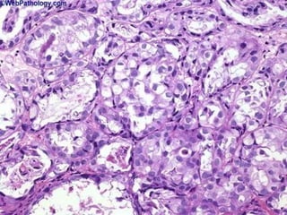
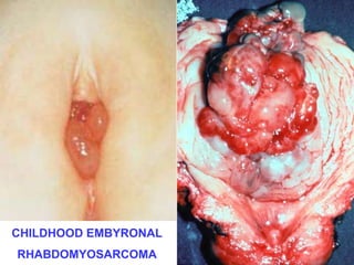
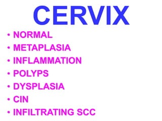
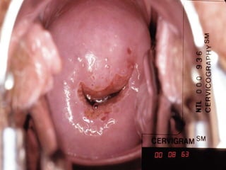
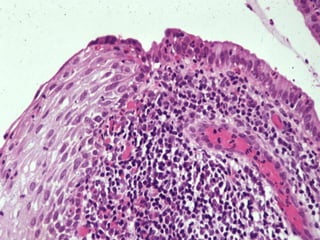
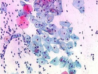
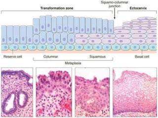
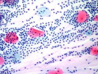
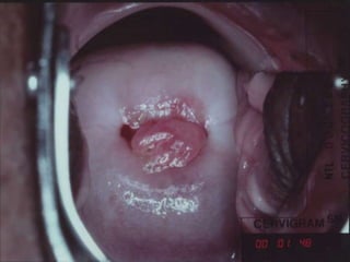
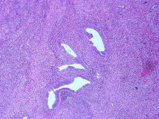
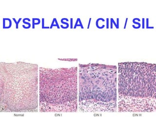
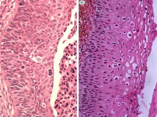
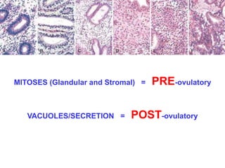
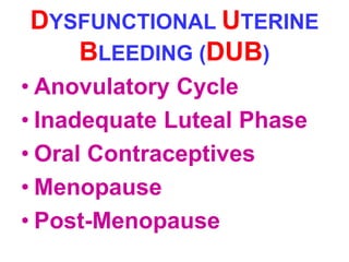
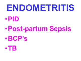
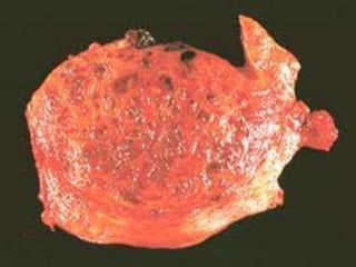
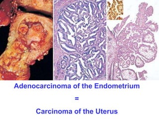
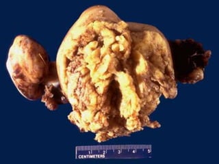
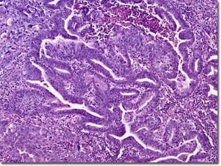
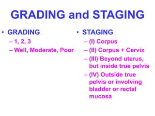
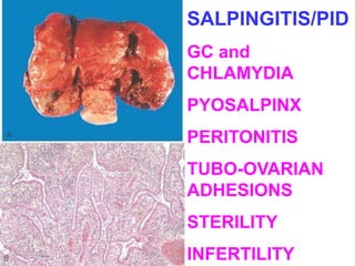
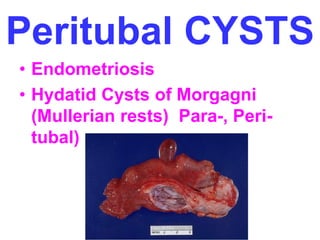
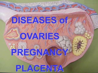
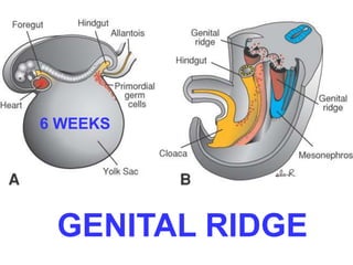
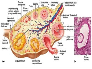
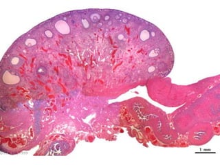
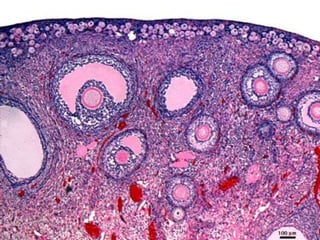
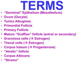
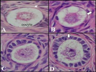
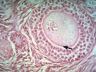
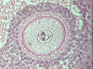
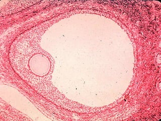
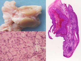
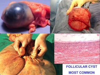
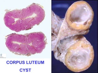
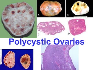
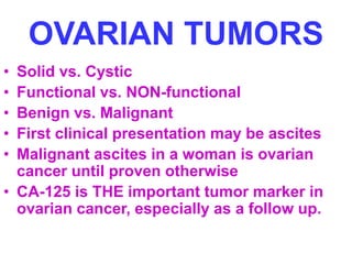
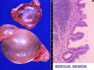
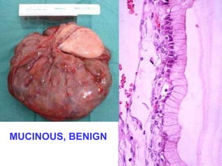
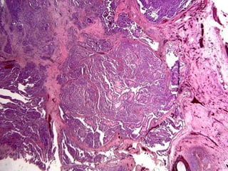
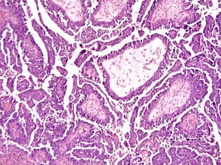
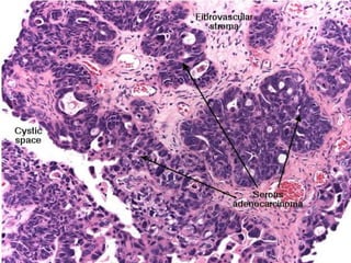
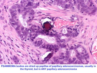
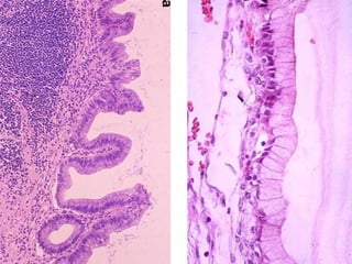
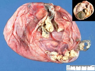
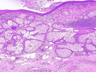
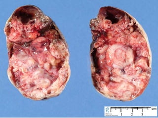
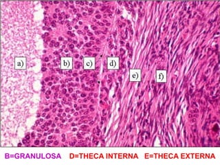
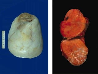
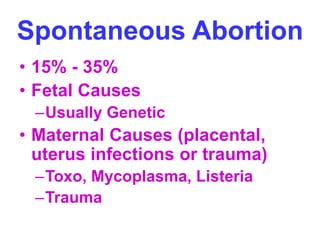
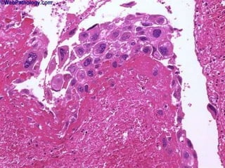
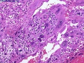
Female 
Reproductive 
System
TOPICS 
–Vulva 
–Vagina 
– Cervix, uterus 
–Body, uterus 
–Tubes 
– Ovaries 
– Placenta
VULVA 
• Synonymous with EXTERNAL genitalia 
• Everything ANTERIOR to the INTROITUS 
• Usual classification of Degen., Inflam., 
Neopl. 
• Common Diseases: 
– BARTHOLIN Cyst 
– Vulvar Vestibulitis 
– Deg./Inflam. Epithelial: LICHEN diseases 
– BENIGN tumors: Condyloma(ta) 
– MALIGNANT tumors: VIN, SCC
Result from 
Inflammation/Obstruction 
of the Bartholin glands 
(i.e., greater vestibular 
glands) 
Often result in abscesses 
Surgical removal is 
curative when local 
procedures are 
inadequate or often 
recurrent 
NEVER become 
malignant
VULVAR VESTIBULITIS, assoc. w. vulvodynia
“LICHEN” DISORDERS 
LICHEN Sclerosis (atrophic skin) 
LICHEN Simplex Chronicus 
(hypertrophic skin) 
Common features of 
FIBROSIS and INFLAMMATION
Mucosal Atrophy 
Fibrosis (sclerosis) 
Inflammation
LICHEN SIMPLEX 
CHRONICUS
The types of lichen lesions 
which show HYPER-plastic 
mucosal changes are often 
regarded as being potentially 
malignant
CONDYLOMA(TA)
VIN, SCC 
• Like condylomas, HIGHLY 
linked to HPV 
• VIN=changes leading to SCC-in- 
situ, look like “plaques” 
• BEYOND VIN = 
INFILTRATION
VIN
MALIGNANT 
MELANOMA
VAGINA 
• CONGENITAL: Parallel Uterus 
• INFLAMMATORY 
–PRE-menopausal: STD 
–POST-menopausal: ATROPHY 
• BENIGN: Hidradenoma, Condyloma 
• MALIGNANT: VIN, INFILTRATING SCC
CONGENITAL 
• Imperforate hymen 
(hematocolpos) 
• Atresia 
• Absence (agenesis) 
• Septate 
• Double (didelphys)
Atresia, 
Double vagina, 
Double uterus.
VAGINITIS 
• 90% 
• Bacterial Vaginitis is the most common cause of vaginitis, 
accounting for 50% of vaginitis cases. As previously mentioned, BV is 
caused by an overgrowth of organisms such as Gardnerella vaginalis 
(gram-variable coccobacillus), Mobiluncus species, Mycoplasma 
hominis, and Peptostreptococcus species. Risk factors include 
pregnancy, intrauterine device (IUD) use, and frequent douching. 
• Candida species (C albicans, C tropicalis, and C glabrata) are 
airborne fungi that are natural inhabitants of the vagina in as many as 
50% of women, and vaginal candidiasis is the second most common 
cause of vaginitis. Risk factors include oral contraceptive use, IUD use, 
young age at first intercourse, increased frequency of intercourse, 
receptive cunnilingus, diabetes, HIV or other immunocompromised 
states, chronic antibiotic use, and pregnancy. 
• T. vaginalis infection, the third most common cause of vaginitis, is 
caused by trichomonads. These organisms are flagellated protozoans. 
Trichomonads primarily infect vaginal epithelium, and they less 
commonly infect the endocervix, urethra, and Bartholin and Skene 
glands. Trichomonads are transmitted sexually and can be identified in 
as many as 80% of male partners of infected women. Risk factors 
include tobacco use, unprotected intercourse with multiple sexual 
partners, and the use of an IUD.
VAGINAL NEOPLASIA 
• VIN 
• INFILTRATING SCC 
• ADENOSIS (D.E.S.)  
•ADENOCARCINOMA 
(Di-Ethyl-Stilbestrol)
VIN
NORMAL VIN
SCC
CHILDHOOD EMBYRONAL 
RHABDOMYOSARCOMA
CERVIX 
• NORMAL 
• METAPLASIA 
• INFLAMMATION 
• POLYPS 
• DYSPLASIA 
• CIN 
• INFILTRATING SCC
DYSPLASIA / CIN / SIL
INFILTRATION
How have we “CURED” Cervical Carcinoma?
ENDOMETRIUM 
• FUNCTIONAL HISTOLOGY 
• D.U.B. (Dysfunctional Uterine Bleeding) 
• INFLAMMATION 
• ADENOMYOSIS/ENDOMETRIOSIS 
• POLYPS/HYPERPLASIA 
• ADENOCARCINOMA and/or STROMAL 
• LEIOMYOMYOMAS, -SARCOMAS 
• MITOSES differentiate benign from malignant
MITOSES (Glandular and Stromal) = PRE-ovulatory 
VACUOLES/SECRETION = POST-ovulatory
DYSFUNCTIONAL UTERINE 
BLEEDING (DUB) 
• Anovulatory Cycle 
• Inadequate Luteal Phase 
• Oral Contraceptives 
• Menopause 
• Post-Menopause
ENDOMETRITIS 
•PID 
•Post-partum Sepsis 
• BCP’s 
•TB
ADENOMYOSIS 
• Defined as normal endometrial glands 
deep within the myometrium
ENDOMETRIOSIS 
Defined as normal endometrial glands OUTSIDE 
the confines of the myometrium 
Reverse menstruation vs. Embryologic “rest” 
theories 
EXTREMELY common cause of cyclical 
abdominal/pelvic pain 
Broad Ligament, Ovary (“chocolate cysts”), 
Peritoneum, Bowel, Umbilicus
“CHOCOLATE” 
CYST
Adenocarcinoma of the Endometrium 
= 
Carcinoma of the Uterus
ADENOCARCINOMA 
of the ENDOMETRIUM 
• Papillary, Polypoid 
• Clear Cell 
• Adeno-Squamous 
• Mucinous 
• Serous 
• Preceded by hyperplasia (EIN), dysplasia 
• Estrogenic, DES effects 
• Ass. w.: obesity, diabetes, hypertension, infertility 
• Stromal “sarcomatous” conditions can co-exist, 
i.e., “adenosarcoma”
GRADING and STAGING 
• GRADING 
– 1, 2, 3 
– Well, Moderate, Poor 
• STAGING 
– (I) Corpus 
– (II) Corpus + Cervix 
– (III) Beyond uterus, 
but inside true pelvis 
– (IV) Outside true 
pelvis or involving 
bladder or rectal 
mucosa
Fallopian Tubes 
•Inflammation 
•Cysts 
•Neoplasms
SALPINGITIS/PID 
GC and 
CHLAMYDIA 
PYOSALPINX 
PERITONITIS 
TUBO-OVARIAN 
ADHESIONS 
STERILITY 
INFERTILITY
Peritubal CYSTS 
• Endometriosis 
• Hydatid Cysts of Morgagni 
(Mullerian rests) Para-, Peri-tubal)
TUBAL NEOPLASMS 
Adenocarcinomas 
Leiomyomas
DISEASES of 
OVARIES 
PREGNANCY 
PLACENTA
DISEASES of 
OVARIES 
• DEGENERATIVE? 
• INFLAMMATORY? 
• CYSTS 
• TUMORS 
– Müllerian (“Germinal”) 
– Germ Cell 
– Sex Cord/Stromal 
– Metastatic
DISEASES of 
PREGNANCY 
•EARLY Pregnancy 
•LATE Pregnancy
DISEASES of 
PLACENTA 
• “BENIGN” tumors (MOLES) 
• MALIGNANT tumors 
(CHORIOCARCINOMA)
6 WEEKS 
GENITAL RIDGE
Everything 
you can see 
or feel is 
lined by 
serosa (i.e., 
mesothelial 
cells, 
visceral and 
parietal
TERMS 
• “Germinal” Epithelium (Mesothelium) 
• Ovum (Oocyte) 
• Tunica Albuginea 
• Primordial Follicle 
• Primary Follicle 
• Mature “Graffian” follicle (antral or secondary) 
• Granulosa cells ( Estrogen) 
• Thecal cells ( Estrogen) 
• Corpus luteum ( Progesterone) 
• “Atretic” follicle 
• Corpus Albicans 
• “Stroma”
B=GRANULOSA D=THECA INTERNA E=THECA EXTERNA
ESTROGEN 
• Controlled by FSH and LH 
• Develop, Lactate Breast Lobules 
• Proliferate Endometrial Glands 
• “Cardioprotective” 
• “Bone Mass” protective
PROGESTERONE 
• Controlled by FSH and LH 
• SECRETE Endometrial Glands 
• IMPLANTATION of the blastocyst 
• Lactation
DISEASES of 
OVARIES 
•CYSTS: 
–Follicular 
–Luteal
FOLLICULAR CYST 
MOST COMMON
CORPUS LUTEUM 
CYST
POLY-Cystic Ovarian Disease 
(Stein-Leventhal syndrome) 
5% Prevalence 
Anovulation 
Oligomenorrhea 
Obesity 
Hirsutism
Polycystic Ovaries
OVARIAN TUMORS 
• MÜLLERIAN (MAJORITY) 
– Serous (Benign, Borderline, Malignant) 
– Mucinous (Benign, Borderline, Malignant) 
– Endometroid (Benign, Borderline, Malignant) 
– Adenosarcoma (Carcinoma AND Sarcoma) 
– Mesodermal Mixed (MULTIPHASIC Sarcoma) 
– Clear Cell 
– Brenner (almost always benign) 
– Transitional (almost always look like Brenner) 
• Germ Cell 
• SEX-CORD/STROMAL 
• METASTATIC
OVARIAN TUMORS 
• Solid vs. Cystic 
• Functional vs. NON-functional 
• Benign vs. Malignant 
• First clinical presentation may be ascites 
• Malignant ascites in a woman is ovarian 
cancer until proven otherwise 
• CA-125 is THE important tumor marker in 
ovarian cancer, especially as a follow up.
SEROUS, BENIGN
MUCINOUS, BENIGN
PSAMMOMA bodies are dried up papillae of papillary adenocarcinomas, usually in 
the thyroid, but in ANY papillary adenocarcinoma
OTHER MÜLLERIAN 
• ENDOMETRIOD, malignant 
– (looks like endometrium) 
• CLEAR CELL, malignant 
– (clear cells, reminiscent of renal clear cell ca.) 
• CYSTADENOFIBROMA, benign 
– (BENIGN “FIBROUS” COMPONENT) 
• BRENNER TUMOR, benign 
– (transitional cell nests) 
• CARCINOMA with SARCOMA 
– (adenosarcoma, mixed Müllerian)
“GERM CELL” Tumors 
• Teratomas (usually benign in ovary), i.e., 
“mature” cystic teratoma or dermoid cyst 
• “Immature” teratomas are regarded as 
malignant 
• Dysgerminoma (look exactly like the 
testicular seminoma), malignant 
• Endodermal Sinus (Yolk Sac), malignant, 
Just like testicular 
• Choriocarcinoma, malignant, just like 
testicular
Dysgerminoma:Female::Seminoma:Male
ENDODERMAL SINUS 
TUMOR, aka 
YOLK SAC TUMOR
CHORIOCARCINOMA, 
Just like testis or placenta
SEX-CORD/STROMAL 
TUMORS 
• Chiefly benign and NON-cystic, 
i.e., “solid”, often functional 
(hyper-estrogen-ism) 
• Granulosa-Theca 
• Fibroma-Theca 
• Sertoli-Leydig (Androblastoma)
CALL-EXNER 
BODIES
B=GRANULOSA D=THECA INTERNA E=THECA EXTERNA
DISEASES of 
PREGNANCY 
•EARLY Pregnancy 
•LATE Pregnancy
EARLY PREGNANCY 
• SPONTANEOUS ABORTION 
• ECTOPIC PREGNANCY
Spontaneous Abortion 
• 15% - 35% 
• Fetal Causes 
–Usually Genetic 
• Maternal Causes (placental, 
uterus infections or trauma) 
–Toxo, Mycoplasma, Listeria 
–Trauma
Ectopic Pregnancy 
• Chiefly TUBAL, but ovarian or 
abdominal rare 
•1% OF NORMAL WOMEN 
•35%-50% OF WOMEN with 
previous SALPINGITIS/PID 
• + HCG, Abdominal pain, 1st 
trimester, ultrasound
LATE PREGNANCY 
• PLACENTAL ANOMALIES 
• TWIN PLACENTAS 
• PLACENTAL INFLAMMATIONS 
• TOXEMIA (ECLAMPSIA/PRE-ECLAMPSIA) 
• INTRAUTERINE GROWTH 
RETARDATION
PLACENTAL ANOMALIES 
• Accessory Lobes 
• Bipartite Placenta 
• Circumvallate Placenta 
• Placenta Accreta, chorion going 
DIRECTLY to the myometrium
CIRCUMVALLATE
PLACENTA ACCRETA 
NO DECIDUA BETWEEN VILLI AND MYOMETRIUM
MRI of Placenta PREVIA, or LOW-LYING placenta, usually 
anatomically normal, but just lies LOWER than it should.
MONOCHORIONIC = MONOZYGOTIC
TOXEMIA of PREGNANCY 
(PRE-eclampsia) 
• Hypertension 
• Proteinuria 
• Edema 
• Related to Placental Ischemia 
• Risk for DIC, convulsions (eclampsia)
Intrauterine Growth Retardation 
• Fetal causes: Genetic, malformations 
• Maternal Causes, vascular diseases, 
toxemia, infections, placental 
diseases 
• Placenta size (350-700g) ~ Fetal size 
(7.5lb)
Placental Infections 
• Villitis vs. chorionamnionitis vs. funisitis 
• ASCENDING vs. hematogenous 
• ASCENDING are usually bacterial, and 
chorionamnionitis 
• Hematogenous are often TORCH, and 
villitis
Placental Neoplasms, 
i.e. gestational trophoblastic disease 
• Benign: MOLES (Hydatidiform moles) 
• Malignant: CHORIOCARCINOMA 
• BOTH are associated with increased or 
persistent levels of the placental 
hormone HCG
Hydatidiform Mole 
• 1/1000 in USA 
• 1% in Indonesia 
• Also called NON-invasive mole in 
its most common benign variant, 
but can also be “invasive” 
• Complete (2% chorioCA incidence) 
or partial (0% incidence) 
• Grapelike clusters, i.e., swollen villi
The MAIN thing 
differentiating benign 
from malignant from 
worrisome trophoblastic 
neoplasms is 
INVASIVENESS 
of the trophoblast
23 female
23 female

23 female

  • 1.
  • 2.
    TOPICS –Vulva –Vagina – Cervix, uterus –Body, uterus –Tubes – Ovaries – Placenta
  • 3.
    VULVA • Synonymouswith EXTERNAL genitalia • Everything ANTERIOR to the INTROITUS • Usual classification of Degen., Inflam., Neopl. • Common Diseases: – BARTHOLIN Cyst – Vulvar Vestibulitis – Deg./Inflam. Epithelial: LICHEN diseases – BENIGN tumors: Condyloma(ta) – MALIGNANT tumors: VIN, SCC
  • 4.
    Result from Inflammation/Obstruction of the Bartholin glands (i.e., greater vestibular glands) Often result in abscesses Surgical removal is curative when local procedures are inadequate or often recurrent NEVER become malignant
  • 5.
  • 6.
    “LICHEN” DISORDERS LICHENSclerosis (atrophic skin) LICHEN Simplex Chronicus (hypertrophic skin) Common features of FIBROSIS and INFLAMMATION
  • 8.
    Mucosal Atrophy Fibrosis(sclerosis) Inflammation
  • 9.
  • 10.
    The types oflichen lesions which show HYPER-plastic mucosal changes are often regarded as being potentially malignant
  • 11.
  • 12.
    VIN, SCC •Like condylomas, HIGHLY linked to HPV • VIN=changes leading to SCC-in- situ, look like “plaques” • BEYOND VIN = INFILTRATION
  • 13.
  • 16.
  • 17.
    VAGINA • CONGENITAL:Parallel Uterus • INFLAMMATORY –PRE-menopausal: STD –POST-menopausal: ATROPHY • BENIGN: Hidradenoma, Condyloma • MALIGNANT: VIN, INFILTRATING SCC
  • 18.
    CONGENITAL • Imperforatehymen (hematocolpos) • Atresia • Absence (agenesis) • Septate • Double (didelphys)
  • 19.
    Atresia, Double vagina, Double uterus.
  • 20.
    VAGINITIS • 90% • Bacterial Vaginitis is the most common cause of vaginitis, accounting for 50% of vaginitis cases. As previously mentioned, BV is caused by an overgrowth of organisms such as Gardnerella vaginalis (gram-variable coccobacillus), Mobiluncus species, Mycoplasma hominis, and Peptostreptococcus species. Risk factors include pregnancy, intrauterine device (IUD) use, and frequent douching. • Candida species (C albicans, C tropicalis, and C glabrata) are airborne fungi that are natural inhabitants of the vagina in as many as 50% of women, and vaginal candidiasis is the second most common cause of vaginitis. Risk factors include oral contraceptive use, IUD use, young age at first intercourse, increased frequency of intercourse, receptive cunnilingus, diabetes, HIV or other immunocompromised states, chronic antibiotic use, and pregnancy. • T. vaginalis infection, the third most common cause of vaginitis, is caused by trichomonads. These organisms are flagellated protozoans. Trichomonads primarily infect vaginal epithelium, and they less commonly infect the endocervix, urethra, and Bartholin and Skene glands. Trichomonads are transmitted sexually and can be identified in as many as 80% of male partners of infected women. Risk factors include tobacco use, unprotected intercourse with multiple sexual partners, and the use of an IUD.
  • 22.
    VAGINAL NEOPLASIA •VIN • INFILTRATING SCC • ADENOSIS (D.E.S.)  •ADENOCARCINOMA (Di-Ethyl-Stilbestrol)
  • 23.
  • 24.
  • 25.
  • 28.
  • 29.
    CERVIX • NORMAL • METAPLASIA • INFLAMMATION • POLYPS • DYSPLASIA • CIN • INFILTRATING SCC
  • 37.
  • 39.
  • 40.
    How have we“CURED” Cervical Carcinoma?
  • 41.
    ENDOMETRIUM • FUNCTIONALHISTOLOGY • D.U.B. (Dysfunctional Uterine Bleeding) • INFLAMMATION • ADENOMYOSIS/ENDOMETRIOSIS • POLYPS/HYPERPLASIA • ADENOCARCINOMA and/or STROMAL • LEIOMYOMYOMAS, -SARCOMAS • MITOSES differentiate benign from malignant
  • 43.
    MITOSES (Glandular andStromal) = PRE-ovulatory VACUOLES/SECRETION = POST-ovulatory
  • 44.
    DYSFUNCTIONAL UTERINE BLEEDING(DUB) • Anovulatory Cycle • Inadequate Luteal Phase • Oral Contraceptives • Menopause • Post-Menopause
  • 45.
    ENDOMETRITIS •PID •Post-partumSepsis • BCP’s •TB
  • 46.
    ADENOMYOSIS • Definedas normal endometrial glands deep within the myometrium
  • 48.
    ENDOMETRIOSIS Defined asnormal endometrial glands OUTSIDE the confines of the myometrium Reverse menstruation vs. Embryologic “rest” theories EXTREMELY common cause of cyclical abdominal/pelvic pain Broad Ligament, Ovary (“chocolate cysts”), Peritoneum, Bowel, Umbilicus
  • 50.
  • 56.
    Adenocarcinoma of theEndometrium = Carcinoma of the Uterus
  • 59.
    ADENOCARCINOMA of theENDOMETRIUM • Papillary, Polypoid • Clear Cell • Adeno-Squamous • Mucinous • Serous • Preceded by hyperplasia (EIN), dysplasia • Estrogenic, DES effects • Ass. w.: obesity, diabetes, hypertension, infertility • Stromal “sarcomatous” conditions can co-exist, i.e., “adenosarcoma”
  • 60.
    GRADING and STAGING • GRADING – 1, 2, 3 – Well, Moderate, Poor • STAGING – (I) Corpus – (II) Corpus + Cervix – (III) Beyond uterus, but inside true pelvis – (IV) Outside true pelvis or involving bladder or rectal mucosa
  • 61.
    Fallopian Tubes •Inflammation •Cysts •Neoplasms
  • 62.
    SALPINGITIS/PID GC and CHLAMYDIA PYOSALPINX PERITONITIS TUBO-OVARIAN ADHESIONS STERILITY INFERTILITY
  • 63.
    Peritubal CYSTS •Endometriosis • Hydatid Cysts of Morgagni (Mullerian rests) Para-, Peri-tubal)
  • 64.
  • 65.
    DISEASES of OVARIES PREGNANCY PLACENTA
  • 66.
    DISEASES of OVARIES • DEGENERATIVE? • INFLAMMATORY? • CYSTS • TUMORS – Müllerian (“Germinal”) – Germ Cell – Sex Cord/Stromal – Metastatic
  • 67.
    DISEASES of PREGNANCY •EARLY Pregnancy •LATE Pregnancy
  • 68.
    DISEASES of PLACENTA • “BENIGN” tumors (MOLES) • MALIGNANT tumors (CHORIOCARCINOMA)
  • 69.
  • 72.
    Everything you cansee or feel is lined by serosa (i.e., mesothelial cells, visceral and parietal
  • 75.
    TERMS • “Germinal”Epithelium (Mesothelium) • Ovum (Oocyte) • Tunica Albuginea • Primordial Follicle • Primary Follicle • Mature “Graffian” follicle (antral or secondary) • Granulosa cells ( Estrogen) • Thecal cells ( Estrogen) • Corpus luteum ( Progesterone) • “Atretic” follicle • Corpus Albicans • “Stroma”
  • 80.
  • 83.
    ESTROGEN • Controlledby FSH and LH • Develop, Lactate Breast Lobules • Proliferate Endometrial Glands • “Cardioprotective” • “Bone Mass” protective
  • 84.
    PROGESTERONE • Controlledby FSH and LH • SECRETE Endometrial Glands • IMPLANTATION of the blastocyst • Lactation
  • 85.
    DISEASES of OVARIES •CYSTS: –Follicular –Luteal
  • 86.
  • 87.
  • 88.
    POLY-Cystic Ovarian Disease (Stein-Leventhal syndrome) 5% Prevalence Anovulation Oligomenorrhea Obesity Hirsutism
  • 89.
  • 90.
    OVARIAN TUMORS •MÜLLERIAN (MAJORITY) – Serous (Benign, Borderline, Malignant) – Mucinous (Benign, Borderline, Malignant) – Endometroid (Benign, Borderline, Malignant) – Adenosarcoma (Carcinoma AND Sarcoma) – Mesodermal Mixed (MULTIPHASIC Sarcoma) – Clear Cell – Brenner (almost always benign) – Transitional (almost always look like Brenner) • Germ Cell • SEX-CORD/STROMAL • METASTATIC
  • 91.
    OVARIAN TUMORS •Solid vs. Cystic • Functional vs. NON-functional • Benign vs. Malignant • First clinical presentation may be ascites • Malignant ascites in a woman is ovarian cancer until proven otherwise • CA-125 is THE important tumor marker in ovarian cancer, especially as a follow up.
  • 92.
  • 93.
  • 97.
    PSAMMOMA bodies aredried up papillae of papillary adenocarcinomas, usually in the thyroid, but in ANY papillary adenocarcinoma
  • 99.
    OTHER MÜLLERIAN •ENDOMETRIOD, malignant – (looks like endometrium) • CLEAR CELL, malignant – (clear cells, reminiscent of renal clear cell ca.) • CYSTADENOFIBROMA, benign – (BENIGN “FIBROUS” COMPONENT) • BRENNER TUMOR, benign – (transitional cell nests) • CARCINOMA with SARCOMA – (adenosarcoma, mixed Müllerian)
  • 100.
    “GERM CELL” Tumors • Teratomas (usually benign in ovary), i.e., “mature” cystic teratoma or dermoid cyst • “Immature” teratomas are regarded as malignant • Dysgerminoma (look exactly like the testicular seminoma), malignant • Endodermal Sinus (Yolk Sac), malignant, Just like testicular • Choriocarcinoma, malignant, just like testicular
  • 105.
  • 106.
    ENDODERMAL SINUS TUMOR,aka YOLK SAC TUMOR
  • 107.
    CHORIOCARCINOMA, Just liketestis or placenta
  • 108.
    SEX-CORD/STROMAL TUMORS •Chiefly benign and NON-cystic, i.e., “solid”, often functional (hyper-estrogen-ism) • Granulosa-Theca • Fibroma-Theca • Sertoli-Leydig (Androblastoma)
  • 109.
  • 110.
  • 112.
    DISEASES of PREGNANCY •EARLY Pregnancy •LATE Pregnancy
  • 113.
    EARLY PREGNANCY •SPONTANEOUS ABORTION • ECTOPIC PREGNANCY
  • 114.
    Spontaneous Abortion •15% - 35% • Fetal Causes –Usually Genetic • Maternal Causes (placental, uterus infections or trauma) –Toxo, Mycoplasma, Listeria –Trauma
  • 115.
    Ectopic Pregnancy •Chiefly TUBAL, but ovarian or abdominal rare •1% OF NORMAL WOMEN •35%-50% OF WOMEN with previous SALPINGITIS/PID • + HCG, Abdominal pain, 1st trimester, ultrasound
  • 117.
    LATE PREGNANCY •PLACENTAL ANOMALIES • TWIN PLACENTAS • PLACENTAL INFLAMMATIONS • TOXEMIA (ECLAMPSIA/PRE-ECLAMPSIA) • INTRAUTERINE GROWTH RETARDATION
  • 118.
    PLACENTAL ANOMALIES •Accessory Lobes • Bipartite Placenta • Circumvallate Placenta • Placenta Accreta, chorion going DIRECTLY to the myometrium
  • 122.
  • 123.
    PLACENTA ACCRETA NODECIDUA BETWEEN VILLI AND MYOMETRIUM
  • 124.
    MRI of PlacentaPREVIA, or LOW-LYING placenta, usually anatomically normal, but just lies LOWER than it should.
  • 125.
  • 126.
    TOXEMIA of PREGNANCY (PRE-eclampsia) • Hypertension • Proteinuria • Edema • Related to Placental Ischemia • Risk for DIC, convulsions (eclampsia)
  • 127.
    Intrauterine Growth Retardation • Fetal causes: Genetic, malformations • Maternal Causes, vascular diseases, toxemia, infections, placental diseases • Placenta size (350-700g) ~ Fetal size (7.5lb)
  • 128.
    Placental Infections •Villitis vs. chorionamnionitis vs. funisitis • ASCENDING vs. hematogenous • ASCENDING are usually bacterial, and chorionamnionitis • Hematogenous are often TORCH, and villitis
  • 129.
    Placental Neoplasms, i.e.gestational trophoblastic disease • Benign: MOLES (Hydatidiform moles) • Malignant: CHORIOCARCINOMA • BOTH are associated with increased or persistent levels of the placental hormone HCG
  • 131.
    Hydatidiform Mole •1/1000 in USA • 1% in Indonesia • Also called NON-invasive mole in its most common benign variant, but can also be “invasive” • Complete (2% chorioCA incidence) or partial (0% incidence) • Grapelike clusters, i.e., swollen villi
  • 133.
    The MAIN thing differentiating benign from malignant from worrisome trophoblastic neoplasms is INVASIVENESS of the trophoblast