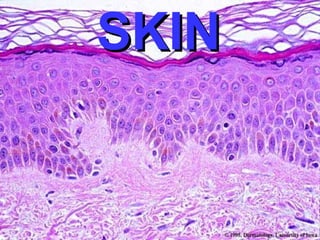
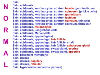
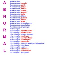
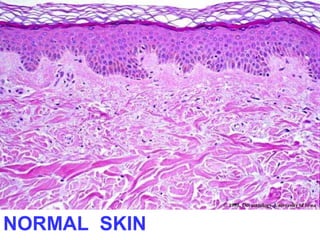
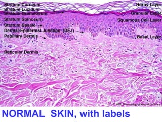
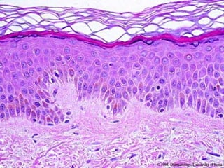
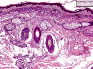
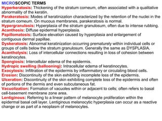
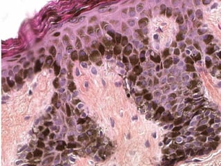
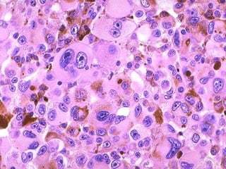
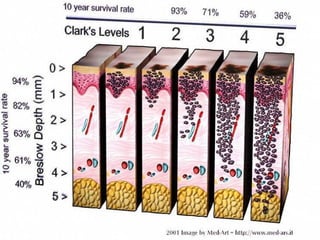
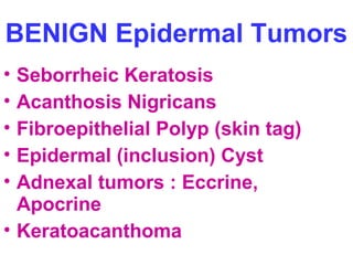
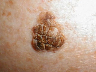
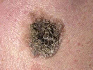
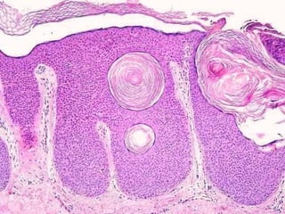
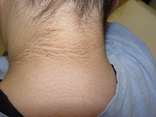
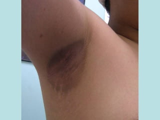
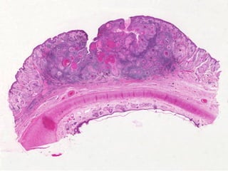
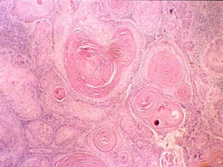
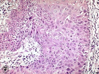
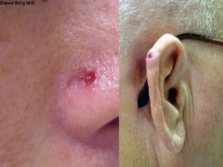
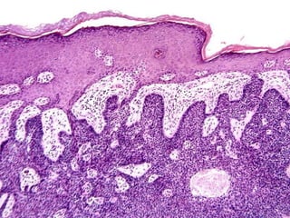
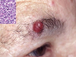
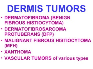
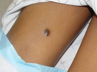
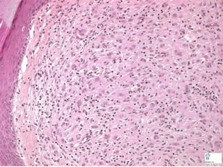
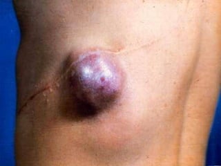
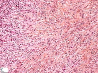
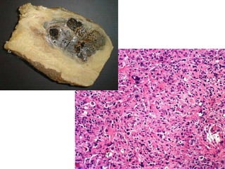
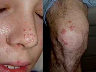
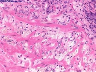
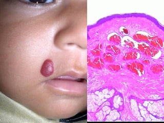
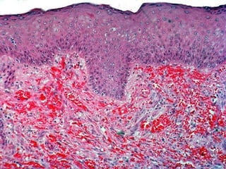
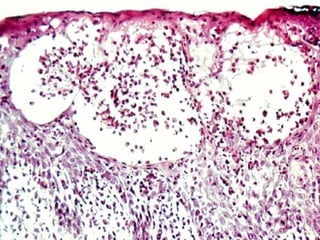
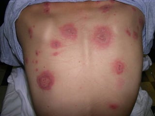
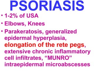
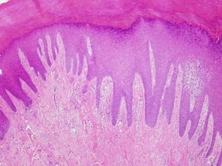
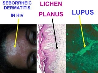
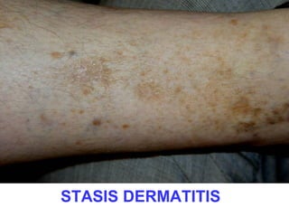
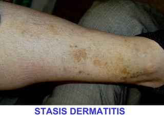
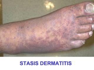
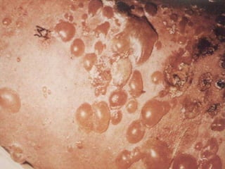
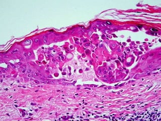
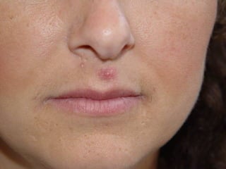
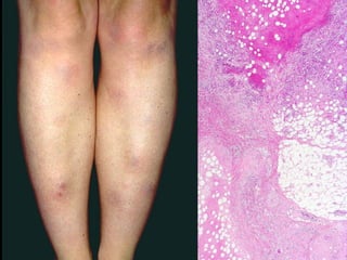
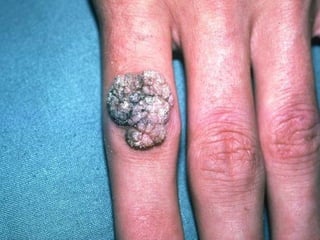
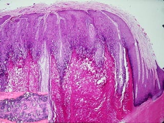
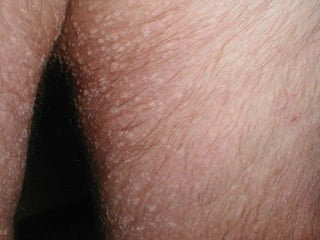
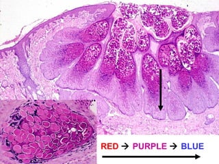
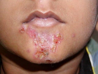
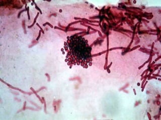
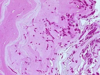
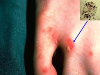
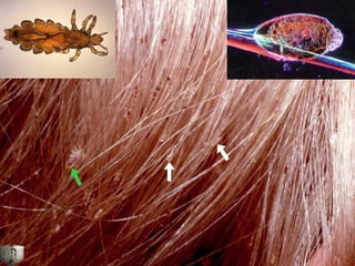
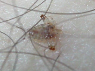
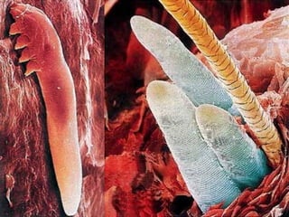
SSKKIINN
Skin 
Skin, epidermis 
Skin, epidermis, keratinocytes, stratum basale (germinativum) 
Skin, epidermis, keratinocytes, stratum spinosum (prickle cells) 
Skin, epidermis, keratinocytes, stratum granulosum 
Skin, epidermis, keratinocytes, stratum lucidum 
Skin, epidermis, keratinocytes, stratum corneum, thin skin 
Skin, epidermis, keratinocytes, stratum corneum, thick skin 
Skin, epidermis, melanocytes 
Skin, epidermis, Langerhans cells 
Skin, epidermis, Merkel cells 
Skin, epidermis, appendage(s) 
Skin, epidermis, appendage, hair follicle 
Skin, epidermis, appendage, hair follicle, shaft 
Skin, epidermis, appendage, hair follicle, sebaceous gland 
Skin, epidermis, appendage, sweat gland, eccrine 
Skin, epidermis, appendage, sweat gland, apocrine 
Skin, basement membrane 
Skin, dermis 
Skin, dermis, papillary 
Skin, dermis, reticular 
Skin, hypodermis (sub-cutis, pannus) 
N 
O 
R 
M 
A 
L
Macroscopic 
Macroscopic, macule 
Macroscopic, patch 
Macroscopic, papule 
Macroscopic, nodule 
Macroscopic, plaque 
Macroscopic, vesicle 
Macroscopic, bulla 
Macroscopic, blister 
Macroscopic, pustule 
Macroscopic, wheal 
Macroscopic, scale 
Macroscopic, lichenification 
Macroscopic, excoriation 
Macroscopic, onycholysis 
microscopic 
microscopic, hyperkeratosis 
microscopic, parakeratosis 
microscopic, hypergranulosis 
microscopic, acanthosis 
microscopic, papillomatosis 
microscopic, acantholysis 
microscopic, spongiosis 
microscopic, hydropic swelling (ballooning) 
microscopic, exocytosis 
microscopic, erosion 
microscopic, ulceration 
microscopic, vacuolization 
microscopic, lentiginous 
A 
B 
N 
O 
R 
M 
A 
L
Pigmentation disorders 
Pigmentation disorders, vitiligo 
Pigmentation disorders, freckle (ephelis) 
Pigmentation disorders, melasma 
Pigmentation disorders, lentigo 
Pigmentation disorders, nevus 
Pigmentation disorders, nevus, melanocytic 
Pigmentation disorders, nevus, dysplastic 
Pigmentation disorders, malignant melanoma 
Epidermal neoplasms 
Epidermal neoplasms, benign 
Epidermal neoplasms, benign, seborrheic keratosis 
Epidermal neoplasms, benign, acanthosis nigricans 
Epidermal neoplasms, benign, fibroepithelial polyp (skin tag) 
Epidermal neoplasms, benign, epithelial inclusion cyst (wen) 
Epidermal neoplasms, benign, appendage tumors 
Epidermal neoplasms, benign, keratoacanthoma 
Epidermal neoplasms, malignant, actinic keratosis 
Epidermal neoplasms, malignant, squamous cell carcinoma (SCC) 
Epidermal neoplasms, malignant, basal cell carcinoma (BCC) 
Epidermal neoplasms, malignant, Merkel cell tumor 
Dermal neoplasms 
Dermal neoplasms, fibrous histiocytoma (dermatofibroma) 
Dermal neoplasms, dermatofibrosarcoma protuberans 
Dermal neoplasms, xanthomas 
Dermal neoplasms, vascular tumors 
Tumors of cellular “immigrants”, Langerhans cells 
Tumors of cellular “immigrants”, t- cell lymphomas (Mycosis Fungoides) 
Tumors of cellular “immigrants”, mast cells 
A 
B 
N 
O 
R 
M 
A 
L
Epidermis, maturation disorder, ichthyosis 
Epidermis/Dermis, inflammation, acute 
Epidermis/Dermis, inflammation, acute, urticaria 
Epidermis/Dermis, inflammation, acute, eczema 
Epidermis/Dermis, inflammation, acute, erythema multiforme 
Epidermis/Dermis, inflammation, chronic 
Epidermis/Dermis, inflammation, chronic, psoriasis 
Epidermis/Dermis, inflammation, chronic, seborrheic dermatitis 
Epidermis/Dermis, inflammation, chronic, lichen planus 
Epidermis/Dermis, inflammation, chronic, lupus erythematosus 
Epidermis/Dermis, infection/infestation 
Epidermis/Dermis, infection/infestation, (verrucae) 
Epidermis/Dermis, infection/infestation, molluscum contagiosum 
Epidermis/Dermis, infection/infestation, impetigo 
Epidermis/Dermis, infection/infestation, fungus 
Epidermis/Dermis, infection/infestation, arthropods 
Epidermis/Dermis, infection/infestation, arthropods, bites 
Epidermis/Dermis, infection/infestation, arthropods, stings 
Epidermis/Dermis, infection/infestation, arthropods, infestations 
Epidermis/Dermis, bullae (blisters) 
Epidermis/Dermis, bullae, pemphigus 
Epidermis/Dermis, bullae, bullous pemphigoid 
Epidermis/Dermis, bullae, dermatitis herpetiformis 
Epidermis/Dermis, bullae, epidermolysis bullosa 
Epidermis/Dermis, bullae, porphyria 
Epidermis/Dermis, adnexae (appendages), acne vulgaris 
Hypodermis (pannus), inflammation (panniculitis) 
Hypodermis (pannus), inflammation, erythema nodosum 
Hypodermis (pannus), inflammation, erythema induratum 
A 
B 
N 
O 
R 
M 
A 
L
NO RMAL S KIN
NO RMAL S KIN, with labels
MACRO-scopic (clinical) 
TERMS • macule 
• patch 
• papule 
• nodule 
• plaque 
• vesicle 
• bulla 
• blister 
• pustule 
• wheal 
• scale 
• lichenification 
• excoriation 
• onycholysis
MACROSCOPIC TERMS 
Macule: Circumscribed lesion of up to 5 mm in diameter characterized by 
flatness and usually discolored (often red) 
Patch: Circumscribed lesion of more than 5 mm in diameter characterized by 
flatness and usually discolored (often red) 
Papule: Elevated dome-shaped or flat-topped lesion 5 mm or less across. 
Nodule: Elevated lesion with spherical contour greater than 5 mm across. 
Plaque: Elevated flat-topped lesion, usually greater than 5 mm across (may be 
caused by coalescent papules). 
Vesicle: Fluid-filled raised lesion 5 mm or less across. 
Bulla: Fluid-filled raised lesion greater than 5 mm across. 
Blister: Common term used for vesicle or bulla. 
Pustule: Discrete, pus-filled, raised lesion. 
Wheal: Itchy, transient, elevated lesion with variable blanching and erythema 
formed as the result of dermal edema. 
Scale: Dry, horny, platelike excrescence; usually the result of imperfect 
cornification (i.e., keratinization). 
Lichenification: Thickened and rough skin characterized by prominent skin 
markings; usually the result of repeated rubbing in susceptible persons. 
Excoriation: Traumatic lesion characterized by breakage of the epidermis, 
causing a raw linear area (i.e., a deep scratch) 
Onycholysis: Separation of nail plate from nail bed.
micro-scopic 
(histologic) TERMS 
• hyperkeratosis 
• parakeratosis 
• hypergranulosis 
• acanthosis 
• papillomatosis 
• acantholysis 
• spongiosis 
• hydropic swelling (ballooning) 
• exocytosis 
• erosion 
• ulceration 
• vacuolization 
• lentiginous
MICROSCOPIC TERMS 
Hyperkeratosis: Thickening of the stratum corneum, often associated with a qualitative 
abnormality of the keratin. 
Parakeratosis: Modes of keratinization characterized by the retention of the nuclei in the 
stratum corneum. On mucous membranes, parakeratosis is normal. 
Hypergranulosis: Hyperplasia of the stratum granulosum, often due to intense rubbing. 
Acanthosis: Diffuse epidermal hyperplasia. 
Papillomatosis: Surface elevation caused by hyperplasia and enlargement of 
contiguous dermal papillae. 
Dyskeratosis: Abnormal keratinization occurring prematurely within individual cells or 
groups of cells below the stratum granulosum. Generally the same as DYSPLASIA. 
Acantholysis: Loss of intercellular connections resulting in loss of cohesion between 
keratinocytes. 
Spongiosis: Intercellular edema of the epidermis. 
Hydropic swelling (ballooning): Intracellular edema of keratinocytes. 
Exocytosis: Infiltration of the epidermis by inflammatory or circulating blood cells. 
Erosion: Discontinuity of the skin exhibiting incomplete loss of the epidermis. 
Ulceration: Discontinuity of the skin exhibiting complete loss of the epidermis and often 
of portions of the dermis and even subcutaneous fat. 
Vacuolization: Formation of vacuoles within or adjacent to cells; often refers to basal 
cell-basement membrane zone area. 
Lentiginous: Referring to a linear pattern of melanocyte proliferation within the 
epidermal basal cell layer. Lentiginous melanocytic hyperplasia can occur as a reactive 
change or as part of a neoplasm of melanocytes.
SKIN PATHOLOGY 
• DEGENERATION 
•INFLAMMATION, i.e., 
DERMATOSES 
•NEOPLASMS: 
Epidermis, Dermis, 
Benign, Malignant
SKIN PATHOLOGY 
• Pigmentation 
• Dermatoses, acute 
• Epidermal tumors, 
• Dermatoses, chronic 
benign 
• Blisters (Bullae) 
• Epidermal tumors 
premalignant 
• Appendage (adnexal) 
• Epidermal tumors, 
disorders 
malignant 
• Panniculitis 
• Dermal tumors 
• Infection/Infestation 
• “Immigrant” tumors 
• Maturation 
disorders
PIGMENTATION DISORDERS 
• VITILIGO 
• FRECKLE (EPHELIS) 
• MELASMA 
• LENTIGO 
• NEVUS 
• “DYSPLASTIC” NEVUS 
• MALIGNANT MELANOMA
NEVI 
• Many, many adjectives and classifications. 
• The MAIN things to differentiate from 
melanomas 
• Junctional (more pigmented, 
more closely associated with 
melanoma) 
• Intradermal 
• Compound (both)
MALIGNANT MELANOMA 
• Incidence rising, VERY much 
• Related to SUN like ALL skin cancers are 
• The only primary skin cancer that can kill 
you (except for the RARE Merkel cell tumor) 
• QUICKLY METASTASIZES 
• Has both VERTICAL and HORIZONTAL 
growth phase but prognosis is 100% related 
to the VERTICAL, (BRESLOW staging, TNM 
too) 
• DIFFICULT to differentiate from NEVUS 
clinically and often microscopically
BENIGN Epidermal Tumors 
• Seborrheic Keratosis 
• Acanthosis Nigricans 
• Fibroepithelial Polyp (skin tag) 
• Epidermal (inclusion) Cyst 
• Adnexal tumors : Eccrine, 
Apocrine 
• Keratoacanthoma
ADNEXAL TUMORS 
• HAIR FOLLICLES 
• SEBACEOUS GLANDS 
• SWEAT GLANDS 
–ECCRINE 
–APOCRINE
PREMALIGNANT/MALIGNANT 
• ACTINIC (Solar) KERATOSIS, i.e. precursor 
to SCC 
• SQUAMOUS CELL CARCINOMA, 
squamous “pearls”, intercellular bridges 
• BASAL CELL CARCINOMA, by far, MOST 
COMMON, BLUE palisading nests 
• MERKEL CELL CARCINOMA (TUMOR), 
VERY MALIGNANT AND LETHAL, LOOK 
LIKE SMALL CELL CA. OF LUNG
GENERAL COMMENTS 
• BOTH SCC and BCC related to SUN (i.e., 
radiation) exposure. 
• SCC also related to As, carcinogens, chaw, 
betel nut, HPV, familial, etc. 
• BOTH SCC and BCC can do local damage 
but very rarely metastasize or kill. 
• MERKEL CELL tumors metastasize early 
and extensively, like melanomas.
DERMIS TUMORS 
• DERMATOFIBROMA (BENIGN 
FIBROUS HISTIOCYTOMA) 
• DERMATOFIBROSARCOMA 
PROTUBERANS (DFP) 
• MALIGNANT FIBROUS HISTIOCYTOMA 
(MFH) 
• XANTHOMA 
• VASCULAR TUMORS of various types
Cellular “Immigrants” 
• Langerhans cells 
(Histiocytosis) 
• Mycosis Fungoides (T-Cell 
cutaneous lymphoma) 
• Mastocytosis (mast cell 
tumors)
DERMATOSES 
•ACUTE 
–URTICARIA 
(i.e., “HIVES”) 
–ECZEMA 
–ERYTHEMA 
MULTIFORME 
•CHRONIC 
–PSORIASIS 
–SEBORRHEIC 
DERMATITIS 
–LICHEN PLANUS 
–LUPUS 
ERTHYMATOSUS
URTICARIA 
• DERMAL EDEMA 
• DILATATION of VASCULAR 
SPACES 
• EARLY PERIVASCULAR 
CUFFING OF 
INFLAMMATORY CELLS
ECZEMA 
(aka, acute eczematous dermatitis) 
• A myriad of ACUTE 
inflammatory disorders, with 
allergic, drug related, sun 
related etiologies 
• The common histologic feature 
is SPONGIOSIS
PSORIASIS • 1-2% of USA 
• Elbows, Knees 
• Parakeratosis, generalized 
epidermal hyperplasia, 
elongation of the rete pegs, 
extensive chronic inflammatory 
cell infiltrates, “MUNRO” 
intraepidermal microabscesses
SEBORRHEIC 
DERMATITIS 
IN HIV 
LICHEN 
PLANUS LUPUS
STASIS DERMATITIS
STASIS DERMATITIS
STASIS DERMATITIS
STASIS DERMATITIS
STASIS DERMATITIS
BULLOUS DISEASES 
•PEMPHIGUS(VULGARIS) 
• BULLOUS PEMPHIGOID 
• DERMATITIS HERPETIFORMIS 
• EPIDERMOLYSIS BULLOSA 
• PORPHYRIA 
• “ACANTHOLYSIS” is the common 
unifying finding, as is basement 
membrane immunoglobulins
ACNE VULGARIS 
• Bread and Butter of dermatology 
practice 
• Sebaceous duct blockage with 
secondary inflammation is main 
feature 
• bacterial lipases of 
Propionibacterium acnes break 
down sebaceous oils, and the 
resulting fatty acids acts as 
irritants
PANNICULITIS 
•ERYTHEMA NODOSUM, 
(red nodules on legs) 
• ERYTHEMA INDURATUM
INFECTION/INFESTATION 
• VERRUCAE, viral (HPV) 
• MULLUSCUM CONTAGIOSUM, viral 
• IMPETIGO, bacterial, staph strep 
• FUNGI 
• ARTHROPODS
RED  PURPLE  BLUE
TINEAS… 
• …Capitis (Scalp ringworm) 
• …Barbae 
• …Corporis (Ringworm) 
• …Cruris (Jock itch) 
• …Pedis (Athlete’s foot) 
• …Onychomycosis (nail)
TINEAS 
• Trichophyton species 
• Microsporum species 
• Epidermophyton species
ARTHROPODS 
•Bites 
•Stings 
•INFESTATIONS
ARTHROPODS 
• Scabies 
• Pediculosis 
•Demodex 
•Ticks, Mites
26 skin
26 skin
26 skin
26 skin
26 skin
26 skin

26 skin

  • 1.
  • 2.
    Skin Skin, epidermis Skin, epidermis, keratinocytes, stratum basale (germinativum) Skin, epidermis, keratinocytes, stratum spinosum (prickle cells) Skin, epidermis, keratinocytes, stratum granulosum Skin, epidermis, keratinocytes, stratum lucidum Skin, epidermis, keratinocytes, stratum corneum, thin skin Skin, epidermis, keratinocytes, stratum corneum, thick skin Skin, epidermis, melanocytes Skin, epidermis, Langerhans cells Skin, epidermis, Merkel cells Skin, epidermis, appendage(s) Skin, epidermis, appendage, hair follicle Skin, epidermis, appendage, hair follicle, shaft Skin, epidermis, appendage, hair follicle, sebaceous gland Skin, epidermis, appendage, sweat gland, eccrine Skin, epidermis, appendage, sweat gland, apocrine Skin, basement membrane Skin, dermis Skin, dermis, papillary Skin, dermis, reticular Skin, hypodermis (sub-cutis, pannus) N O R M A L
  • 3.
    Macroscopic Macroscopic, macule Macroscopic, patch Macroscopic, papule Macroscopic, nodule Macroscopic, plaque Macroscopic, vesicle Macroscopic, bulla Macroscopic, blister Macroscopic, pustule Macroscopic, wheal Macroscopic, scale Macroscopic, lichenification Macroscopic, excoriation Macroscopic, onycholysis microscopic microscopic, hyperkeratosis microscopic, parakeratosis microscopic, hypergranulosis microscopic, acanthosis microscopic, papillomatosis microscopic, acantholysis microscopic, spongiosis microscopic, hydropic swelling (ballooning) microscopic, exocytosis microscopic, erosion microscopic, ulceration microscopic, vacuolization microscopic, lentiginous A B N O R M A L
  • 4.
    Pigmentation disorders Pigmentationdisorders, vitiligo Pigmentation disorders, freckle (ephelis) Pigmentation disorders, melasma Pigmentation disorders, lentigo Pigmentation disorders, nevus Pigmentation disorders, nevus, melanocytic Pigmentation disorders, nevus, dysplastic Pigmentation disorders, malignant melanoma Epidermal neoplasms Epidermal neoplasms, benign Epidermal neoplasms, benign, seborrheic keratosis Epidermal neoplasms, benign, acanthosis nigricans Epidermal neoplasms, benign, fibroepithelial polyp (skin tag) Epidermal neoplasms, benign, epithelial inclusion cyst (wen) Epidermal neoplasms, benign, appendage tumors Epidermal neoplasms, benign, keratoacanthoma Epidermal neoplasms, malignant, actinic keratosis Epidermal neoplasms, malignant, squamous cell carcinoma (SCC) Epidermal neoplasms, malignant, basal cell carcinoma (BCC) Epidermal neoplasms, malignant, Merkel cell tumor Dermal neoplasms Dermal neoplasms, fibrous histiocytoma (dermatofibroma) Dermal neoplasms, dermatofibrosarcoma protuberans Dermal neoplasms, xanthomas Dermal neoplasms, vascular tumors Tumors of cellular “immigrants”, Langerhans cells Tumors of cellular “immigrants”, t- cell lymphomas (Mycosis Fungoides) Tumors of cellular “immigrants”, mast cells A B N O R M A L
  • 5.
    Epidermis, maturation disorder,ichthyosis Epidermis/Dermis, inflammation, acute Epidermis/Dermis, inflammation, acute, urticaria Epidermis/Dermis, inflammation, acute, eczema Epidermis/Dermis, inflammation, acute, erythema multiforme Epidermis/Dermis, inflammation, chronic Epidermis/Dermis, inflammation, chronic, psoriasis Epidermis/Dermis, inflammation, chronic, seborrheic dermatitis Epidermis/Dermis, inflammation, chronic, lichen planus Epidermis/Dermis, inflammation, chronic, lupus erythematosus Epidermis/Dermis, infection/infestation Epidermis/Dermis, infection/infestation, (verrucae) Epidermis/Dermis, infection/infestation, molluscum contagiosum Epidermis/Dermis, infection/infestation, impetigo Epidermis/Dermis, infection/infestation, fungus Epidermis/Dermis, infection/infestation, arthropods Epidermis/Dermis, infection/infestation, arthropods, bites Epidermis/Dermis, infection/infestation, arthropods, stings Epidermis/Dermis, infection/infestation, arthropods, infestations Epidermis/Dermis, bullae (blisters) Epidermis/Dermis, bullae, pemphigus Epidermis/Dermis, bullae, bullous pemphigoid Epidermis/Dermis, bullae, dermatitis herpetiformis Epidermis/Dermis, bullae, epidermolysis bullosa Epidermis/Dermis, bullae, porphyria Epidermis/Dermis, adnexae (appendages), acne vulgaris Hypodermis (pannus), inflammation (panniculitis) Hypodermis (pannus), inflammation, erythema nodosum Hypodermis (pannus), inflammation, erythema induratum A B N O R M A L
  • 6.
  • 7.
    NO RMAL SKIN, with labels
  • 10.
    MACRO-scopic (clinical) TERMS• macule • patch • papule • nodule • plaque • vesicle • bulla • blister • pustule • wheal • scale • lichenification • excoriation • onycholysis
  • 11.
    MACROSCOPIC TERMS Macule:Circumscribed lesion of up to 5 mm in diameter characterized by flatness and usually discolored (often red) Patch: Circumscribed lesion of more than 5 mm in diameter characterized by flatness and usually discolored (often red) Papule: Elevated dome-shaped or flat-topped lesion 5 mm or less across. Nodule: Elevated lesion with spherical contour greater than 5 mm across. Plaque: Elevated flat-topped lesion, usually greater than 5 mm across (may be caused by coalescent papules). Vesicle: Fluid-filled raised lesion 5 mm or less across. Bulla: Fluid-filled raised lesion greater than 5 mm across. Blister: Common term used for vesicle or bulla. Pustule: Discrete, pus-filled, raised lesion. Wheal: Itchy, transient, elevated lesion with variable blanching and erythema formed as the result of dermal edema. Scale: Dry, horny, platelike excrescence; usually the result of imperfect cornification (i.e., keratinization). Lichenification: Thickened and rough skin characterized by prominent skin markings; usually the result of repeated rubbing in susceptible persons. Excoriation: Traumatic lesion characterized by breakage of the epidermis, causing a raw linear area (i.e., a deep scratch) Onycholysis: Separation of nail plate from nail bed.
  • 12.
    micro-scopic (histologic) TERMS • hyperkeratosis • parakeratosis • hypergranulosis • acanthosis • papillomatosis • acantholysis • spongiosis • hydropic swelling (ballooning) • exocytosis • erosion • ulceration • vacuolization • lentiginous
  • 13.
    MICROSCOPIC TERMS Hyperkeratosis:Thickening of the stratum corneum, often associated with a qualitative abnormality of the keratin. Parakeratosis: Modes of keratinization characterized by the retention of the nuclei in the stratum corneum. On mucous membranes, parakeratosis is normal. Hypergranulosis: Hyperplasia of the stratum granulosum, often due to intense rubbing. Acanthosis: Diffuse epidermal hyperplasia. Papillomatosis: Surface elevation caused by hyperplasia and enlargement of contiguous dermal papillae. Dyskeratosis: Abnormal keratinization occurring prematurely within individual cells or groups of cells below the stratum granulosum. Generally the same as DYSPLASIA. Acantholysis: Loss of intercellular connections resulting in loss of cohesion between keratinocytes. Spongiosis: Intercellular edema of the epidermis. Hydropic swelling (ballooning): Intracellular edema of keratinocytes. Exocytosis: Infiltration of the epidermis by inflammatory or circulating blood cells. Erosion: Discontinuity of the skin exhibiting incomplete loss of the epidermis. Ulceration: Discontinuity of the skin exhibiting complete loss of the epidermis and often of portions of the dermis and even subcutaneous fat. Vacuolization: Formation of vacuoles within or adjacent to cells; often refers to basal cell-basement membrane zone area. Lentiginous: Referring to a linear pattern of melanocyte proliferation within the epidermal basal cell layer. Lentiginous melanocytic hyperplasia can occur as a reactive change or as part of a neoplasm of melanocytes.
  • 14.
    SKIN PATHOLOGY •DEGENERATION •INFLAMMATION, i.e., DERMATOSES •NEOPLASMS: Epidermis, Dermis, Benign, Malignant
  • 15.
    SKIN PATHOLOGY •Pigmentation • Dermatoses, acute • Epidermal tumors, • Dermatoses, chronic benign • Blisters (Bullae) • Epidermal tumors premalignant • Appendage (adnexal) • Epidermal tumors, disorders malignant • Panniculitis • Dermal tumors • Infection/Infestation • “Immigrant” tumors • Maturation disorders
  • 16.
    PIGMENTATION DISORDERS •VITILIGO • FRECKLE (EPHELIS) • MELASMA • LENTIGO • NEVUS • “DYSPLASTIC” NEVUS • MALIGNANT MELANOMA
  • 22.
    NEVI • Many,many adjectives and classifications. • The MAIN things to differentiate from melanomas • Junctional (more pigmented, more closely associated with melanoma) • Intradermal • Compound (both)
  • 27.
    MALIGNANT MELANOMA •Incidence rising, VERY much • Related to SUN like ALL skin cancers are • The only primary skin cancer that can kill you (except for the RARE Merkel cell tumor) • QUICKLY METASTASIZES • Has both VERTICAL and HORIZONTAL growth phase but prognosis is 100% related to the VERTICAL, (BRESLOW staging, TNM too) • DIFFICULT to differentiate from NEVUS clinically and often microscopically
  • 31.
    BENIGN Epidermal Tumors • Seborrheic Keratosis • Acanthosis Nigricans • Fibroepithelial Polyp (skin tag) • Epidermal (inclusion) Cyst • Adnexal tumors : Eccrine, Apocrine • Keratoacanthoma
  • 41.
    ADNEXAL TUMORS •HAIR FOLLICLES • SEBACEOUS GLANDS • SWEAT GLANDS –ECCRINE –APOCRINE
  • 45.
    PREMALIGNANT/MALIGNANT • ACTINIC(Solar) KERATOSIS, i.e. precursor to SCC • SQUAMOUS CELL CARCINOMA, squamous “pearls”, intercellular bridges • BASAL CELL CARCINOMA, by far, MOST COMMON, BLUE palisading nests • MERKEL CELL CARCINOMA (TUMOR), VERY MALIGNANT AND LETHAL, LOOK LIKE SMALL CELL CA. OF LUNG
  • 46.
    GENERAL COMMENTS •BOTH SCC and BCC related to SUN (i.e., radiation) exposure. • SCC also related to As, carcinogens, chaw, betel nut, HPV, familial, etc. • BOTH SCC and BCC can do local damage but very rarely metastasize or kill. • MERKEL CELL tumors metastasize early and extensively, like melanomas.
  • 55.
    DERMIS TUMORS •DERMATOFIBROMA (BENIGN FIBROUS HISTIOCYTOMA) • DERMATOFIBROSARCOMA PROTUBERANS (DFP) • MALIGNANT FIBROUS HISTIOCYTOMA (MFH) • XANTHOMA • VASCULAR TUMORS of various types
  • 65.
    Cellular “Immigrants” •Langerhans cells (Histiocytosis) • Mycosis Fungoides (T-Cell cutaneous lymphoma) • Mastocytosis (mast cell tumors)
  • 67.
    DERMATOSES •ACUTE –URTICARIA (i.e., “HIVES”) –ECZEMA –ERYTHEMA MULTIFORME •CHRONIC –PSORIASIS –SEBORRHEIC DERMATITIS –LICHEN PLANUS –LUPUS ERTHYMATOSUS
  • 68.
    URTICARIA • DERMALEDEMA • DILATATION of VASCULAR SPACES • EARLY PERIVASCULAR CUFFING OF INFLAMMATORY CELLS
  • 70.
    ECZEMA (aka, acuteeczematous dermatitis) • A myriad of ACUTE inflammatory disorders, with allergic, drug related, sun related etiologies • The common histologic feature is SPONGIOSIS
  • 75.
    PSORIASIS • 1-2%of USA • Elbows, Knees • Parakeratosis, generalized epidermal hyperplasia, elongation of the rete pegs, extensive chronic inflammatory cell infiltrates, “MUNRO” intraepidermal microabscesses
  • 77.
    SEBORRHEIC DERMATITIS INHIV LICHEN PLANUS LUPUS
  • 78.
  • 79.
  • 80.
  • 81.
  • 82.
  • 83.
    BULLOUS DISEASES •PEMPHIGUS(VULGARIS) • BULLOUS PEMPHIGOID • DERMATITIS HERPETIFORMIS • EPIDERMOLYSIS BULLOSA • PORPHYRIA • “ACANTHOLYSIS” is the common unifying finding, as is basement membrane immunoglobulins
  • 87.
    ACNE VULGARIS •Bread and Butter of dermatology practice • Sebaceous duct blockage with secondary inflammation is main feature • bacterial lipases of Propionibacterium acnes break down sebaceous oils, and the resulting fatty acids acts as irritants
  • 89.
    PANNICULITIS •ERYTHEMA NODOSUM, (red nodules on legs) • ERYTHEMA INDURATUM
  • 91.
    INFECTION/INFESTATION • VERRUCAE,viral (HPV) • MULLUSCUM CONTAGIOSUM, viral • IMPETIGO, bacterial, staph strep • FUNGI • ARTHROPODS
  • 95.
  • 97.
    TINEAS… • …Capitis(Scalp ringworm) • …Barbae • …Corporis (Ringworm) • …Cruris (Jock itch) • …Pedis (Athlete’s foot) • …Onychomycosis (nail)
  • 98.
    TINEAS • Trichophytonspecies • Microsporum species • Epidermophyton species
  • 107.
  • 108.
    ARTHROPODS • Scabies • Pediculosis •Demodex •Ticks, Mites

Editor's Notes

  • #3 Normal histology review.
  • #4 Basic microscopic and macroscopic descriptive terms, the essentials of describing skin lesions properly
  • #5 Classifications and major examples of the classifications.
  • #6 Classifications and major examples of the classifications.
  • #7 Histology review (no labels)‏
  • #8 Histology review (labels)‏
  • #9 Voila!
  • #10 Appendages: hair follicles, sebaceous glands, sweat glands (eccrine, apocrine). Is this thick skin or thin skin?
  • #18 Vitiligo
  • #19 Freckles (ephelis)‏
  • #20 Melasma
  • #21 Lentigo
  • #22 Lentigo
  • #24 Intradermal nevus
  • #25 Intradermal nevus
  • #26 Junctional nevus
  • #27 Junctional nevus. Why is this called “Junctional”?
  • #28 Malignant melanomas are malignant proliferations of melanocytes.
  • #29 What is the ABCDEE principle?
  • #30 What kind of malignant cell would have pigment in it?
  • #31 Clark vs. Breslow (better) classification
  • #33 Seborrheic keratosis
  • #34 Seborrheic keratosis, a bit more pigmented than the previous one
  • #35 Squamous “horn cysts” in seborrheic keratosis
  • #36 Acanthosis nigricans, often associated with diabetes mellitus
  • #37 Acanthosis nigricans, often associated with diabetes mellitus
  • #38 Fibroepithelial polyp, or “skin tag”
  • #39 Fibroepithelial polyp, or “skin tag”
  • #40 Epidermal inclusion cyst, the overlying skin looks normal.
  • #41 Epidermal inclusion cyst
  • #43 Keratoacanthoma, the MAIN lesion to differentiate from squamous cell carcinoma
  • #44 Keratoacanthoma, the MAIN lesion to differentiate from squamous cell carcinoma
  • #45 Keratoacanthoma, the MAIN lesion to differentiate from squamous cell carcinoma
  • #48 Actinic keratosis
  • #49 Actinic keratosis vs. squamous cell carcinoma
  • #50 Squamous cell carcinoma, infiltrating. What is squamous cell carcinoma-in-situ usually called? Ans: Bowen’s disease
  • #51 Squamous cell carcinoma, infiltrating. Note the “pearls”. Does the presence of pearls make this well differentiated? Ans: Yes.
  • #52 Squamous dysplasia, perhaps actinic keratosis, or something leading into squamous cell carcinoma.
  • #53 By far, the commonest malignancy of skin, BCC, i.e., Basal Cell Carcinoma, typical appearance.
  • #54 By far, the commonest malignancy of skin, BCC, i.e., Basal Cell Carcinoma, typical appearance. Note the PERIPHERAL PALISADING!!!
  • #55 Merkel cell tumor, very highly malignant and usually fatal, looks EXACTLY like a small cell carcinoma of the lung? Ans: yes.
  • #57 Benign fibrous histiocytoma, or dermatofibroma
  • #58 Benign fibrous histiocytoma, or dermatofibroma
  • #59 Large fibrous histiocytoma, perhaps a dermatofibrosarcoma protuberans?
  • #61 Malignant fibrous histiocytoma.
  • #62 Xanthomas filled with cholesterol and lipids, to give the “foamy” appearance.
  • #63 Xanthoma filled with cholesterol and lipids, to give the “foamy” appearance.
  • #64 Hemangioma, often a congenital “birth mark”, which can regress significantly with aging.
  • #65 Kaposi’s sarcoma
  • #67 Icthyosis, usually genetic.
  • #68 Inflammatory skin diseases are called dermatoses.
  • #72 Eczema
  • #73 Eczema with spongiosis
  • #74 Pustules can be thought of as extreme “spongiosis”
  • #75 Pustules, ulcerated
  • #77 Classical psoriasis, parakeratosis, hyperplasia, rete peg elongation, chronic inflammation, microabscesses (of Munro)‏
  • #84 Know the various types of “bullous” diseases. What is a bulla? What is acantholysis?
  • #85 Pemphigus, ruptured, scabbed bullae
  • #86 Pemphigus, fresh bullae
  • #87 Acantholysis in the bullous family of diseases
  • #94 Papillomatous epidermal hyperplasia is the most consistent feature of verrucae (warts)‏
  • #95 Molluscum contagiosum.
  • #96 Molluscum contagiosum.
  • #97 Impetigo
  • #100 Ringworm of scalp
  • #101 Tinea barbae
  • #102 Ringworm
  • #103 Tinea cruris, or jock itch
  • #104 Athlete’s foot, or tinea pedis
  • #105 Onychomycosis
  • #106 PAS stain of hyphae
  • #107 PAS stain of hyphae
  • #110 Scabies in it’s most common location
  • #111 Body lice (pediculosis)‏
  • #112 Pubic louse (phthirus pubis)‏
  • #113 Demodex follicularis, a mite larva
  • #114 Tick
  • #115 Tick doing what a tick does best.