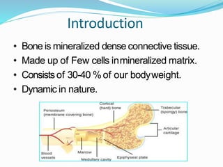
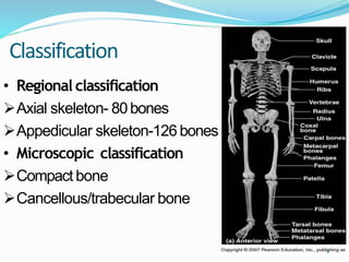
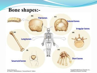
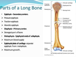
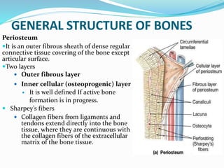
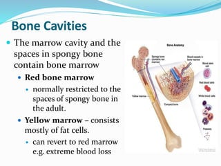
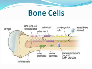
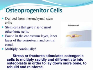
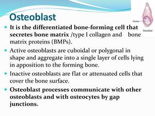
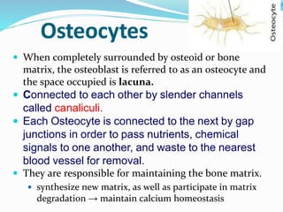
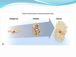
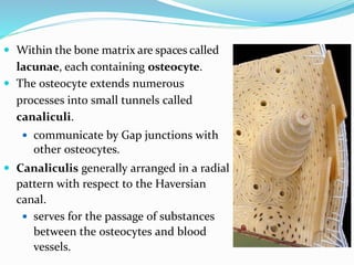
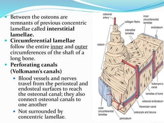
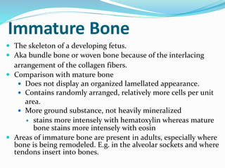
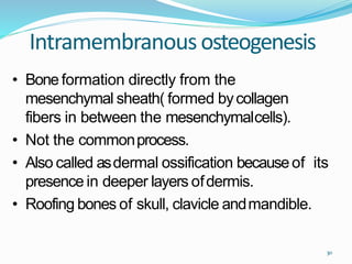
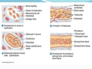
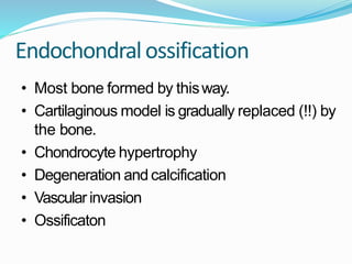
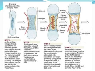
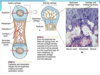
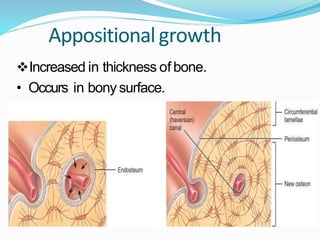
Dr. Anshu Sharma
Assistant Prof.
Dept. of Orthopaediscs, GMC&H.
Introduction
• Bone is mineralized dense connective tissue.
• Made up of Few cells inmineralized matrix.
• Consistsof 30-40 %of our bodyweight.
• Dynamic in nature.
Functions
• Framework of thebody.
• Attachment of muscle and tendon.
• Permit the movements in body parts.
• Protection of organs.
• Haematopoiesis
• Reservoir of minerals andfat.
• Regional classification
Axial skeleton- 80bones
Appedicular skeleton-126 bones
• Microscopic classification
Compact bone
Cancellous/trabecular bone
Classification
4
Longbones
Short bones
Flatbones
Sesamoidbones
Sutural bones
Irregular bones
Bone shapes:-
Parts of a Long Bone
• Epiphysis–Secondarycenters.
 Pressureepiphysis
 Tractionepiphysis
 Atavisticepiphysis
• Diaphysis–Primarycenter.
 Strongestpart ofbone
• Metaphysis- Epiphysialendsof adiaphysis.
 Maximumbloodsupply
• Epiphysialplateofcartilage-separate
epiphysis from metaphysis.
 Maximumgrowth.
Epiphysial/Growthplate
• Zoneof resting cells
• Zoneof proliferating cells
• Zoneof hypertrophic cells
• Zoneof calcifiedcells
Increase in length
Hyaline cartilage that separates the epiphysis from diaphysis.
Zone where bones elongate by a growth process
GENERAL STRUCTURE OF BONES
Periosteum
It is an outer fibrous sheath of dense regular
connective tissue covering of the bone except
articular surface.
Two layers
 Outer fibrous layer
 Inner cellular (osteoprogenic) layer
 It is well defined If active bone
formation is in progress.
 Sharpey’s fibers
 Collagen fibers from ligaments and
tendons extend directly into the bone
tissue, where they are continuous with
the collagen fibers of the extracellular
matrix of the bone tissue.
Bone Cavities
 The marrow cavity and the
spaces in spongy bone
contain bone marrow
 Red bone marrow
 normally restricted to the
spaces of spongy bone in
the adult.
 Yellow marrow – consists
mostly of fat cells.
 can revert to red marrow
e.g. extreme blood loss
Endosteum
 Is often only one cell thick
a layer of connective tissue
cells lining Bone cavities.
 Made up of osteoprogenitor
cells that can differentiate
into osteoblasts, and bone-
lining cells endosteal cells
layer.
BLOOD & NERVE SUPPLY
 Bone is supplied with blood by
 Periosteal arteries:-Enterthe Volkmann’scanals. Supplyouter 1/3rd of
cortex.
 Nutrient artery:-Suppliesmedullary cavity, inner 2/3rdof cortex.
 Metaphyseal arteries
 Nerves accompany the blood vessels that supply bones.
 The periosteum is rich in sensory nerves sensitive to tearing or
tension
 Bone tissue lacks lymphatic vessels; lymphatic drainage occurs only
from the periosteum.
Composition of Bone
Composition ofBone
• Cellular structures in groundsubstance.
• Ground substancemade up of:-
(A) Organic matrix(35%)
Collagen type 1
Glycosaminoglycans
Proteoglycans, BMP, osteonectin and
osteocalcin.
(B) Inorganic content (65%)
 largely made up of Ca++ , PO4--- and OH-.
• Theseions arranged parallel to the direction of
collagen fibers by forming acrystalline structure
called asHydroxyapatite .
Ca10(Po4)6(OH)2
Living bone contain 20%of water.
• Inorganic content give rigidity tothe bone
• Organic content give the elasticity tothe bone.
• Lackof inorganic content- Softbone.
• Lackof organic content- Brittle bone.
Bone Cells
Osteoprogenitor Cells
 Derived from mesenchymal stem
cells.
 Stem cells that give rise to most
other bone cells.
 Found in the endosteum layer, inner
layer of the periosteum and central
canal.
 Multiply continually!
Stress or fractures stimulates osteogenic
cells to multiply rapidly and differentiate into
osteoblasts in order to lay down more bone, to
rebuild and reinforce.
Osteoblast
 It is the differentiated bone-forming cell that
secretes bone matrix /type I collagen and bone
matrix proteins (BMPs).
 Active osteoblasts are cuboidal or polygonal in
shape and aggregate into a single layer of cells lying
in apposition to the forming bone.
 Inactive osteoblasts are flat or attenuated cells that
cover the bone surface.
 Osteoblast processes communicate with other
osteoblasts and with osteocytes by gap
junctions.
Osteocytes
 When completely surrounded by osteoid or bone
matrix, the osteoblast is referred to as an osteocyte and
the space occupied is lacuna.
 Connected to each other by slender channels
called canaliculi.
 Each Osteocyte is connected to the next by gap
junctions in order to pass nutrients, chemical
signals to one another, and waste to the nearest
blood vessel for removal.
 They are responsible for maintaining the bone matrix.
 synthesize new matrix, as well as participate in matrix
degradation → maintain calcium homeostasis
Bone-Lining Cells
 Bone-lining cells are derived from osteoblasts
and cover bone that is not remodeling.
 Layer of flat cells with attenuated cytoplasm
 on external bone surfaces = Periosteal cells.
 on internal bone surfaces = Endosteal cells.
 Function
 Maintenance and Nutritional support of the
osteocytes.
 Regulate the movement of calcium and phosphate
into and out of the bone.
Osteoclasts
 These are phagocytotic cells derived from fusion of hemopoietic
progenitor cells (stem cells) of neutrophilic granulocyte and monocyte
lineages→ multinucleated cells.
 These are bone-resorbing cells present on bone surfaces where bone is
being removed or remodeled.
 Shallowgrooveformed byosteoclasts called asHowship’slacunae(Resorption
bay) .
Mature Bone/lamellar bone
 Composed of cylindrical
units called osteons or
Haversian systems.
 Osteons consist of
concentric lamellae of bone
matrix surrounding a
central canal, the osteonal
(Haversian) canal, which
contains the vascular and
nerve supply of the osteon.
 Within the bone matrix are spaces called
lacunae, each containing osteocyte.
 The osteocyte extends numerous
processes into small tunnels called
canaliculi.
 communicate by Gap junctions with
other osteocytes.
 Canaliculis generally arranged in a radial
pattern with respect to the Haversian
canal.
 serves for the passage of substances
between the osteocytes and blood
vessels.
 Between the osteons are
remnants of previous concentric
lamellae called interstitial
lamellae.
 Circumferential lamellae
follow the entire inner and outer
circumferences of the shaft of a
long bone.
 Perforating canals
(Volkmann’s canals)
 Blood vessels and nerves
travel from the periosteal and
endosteal surfaces to reach
the osteonal canal; they also
connect osteonal canals to
one another
 Not surrounded by
concentric lamellae.
Immature Bone
 The skeleton of a developing fetus.
 Aka bundle bone or woven bone because of the interlacing
arrangement of the collagen fibers.
 Comparison with mature bone
 Does not display an organized lamellated appearance.
 Contains randomly arranged, relatively more cells per unit
area.
 More ground substance, not heavily mineralized
 stains more intensely with hematoxylin whereas mature
bone stains more intensely with eosin
 Areas of immature bone are present in adults, especially where
bone is being remodeled. E.g. in the alveolar sockets and where
tendons insert into bones.
Bone Formation
 Intramembranous ossification
 Flat bones of the skull and mandible are formed in
this way.
 Endochondral ossification
 Gradual replacement of cartilage by bone.
 Most bones of the body are formed in this way
including long bones.
30
Intramembranousosteogenesis
• Bone formation directly from the
mesenchymal sheath( formed bycollagen
fibers in between the mesenchymalcells).
• Not the commonprocess.
• Also called asdermal ossification becauseof its
presence in deeper layers ofdermis.
• Roofing bones of skull, clavicle andmandible.
Intramembranous ossification
• Mesenchymal cells condensation
• Secretion of organic matrix by the
mesenchymal cells which consists of collagen
fibers. Thusforming the mesenchymalsheath.
• Localized differentiation of osteoblasts within
sheath
Endochondralossification
• Most bone formed by thisway.
• Cartilaginous model is gradually replaced (!!) by
the bone.
• Chondrocyte hypertrophy
• Degeneration and calcification
• Vascular invasion
• Ossificaton
Appositionalgrowth
Increased in thickness of bone.
• Occurs in bony surface.
Thank you…!!!!!

Constitution of Bone

  • 1.
    Dr. Anshu Sharma AssistantProf. Dept. of Orthopaediscs, GMC&H.
  • 2.
    Introduction • Bone ismineralized dense connective tissue. • Made up of Few cells inmineralized matrix. • Consistsof 30-40 %of our bodyweight. • Dynamic in nature.
  • 3.
    Functions • Framework ofthebody. • Attachment of muscle and tendon. • Permit the movements in body parts. • Protection of organs. • Haematopoiesis • Reservoir of minerals andfat.
  • 4.
    • Regional classification Axialskeleton- 80bones Appedicular skeleton-126 bones • Microscopic classification Compact bone Cancellous/trabecular bone Classification 4
  • 5.
  • 6.
    Parts of aLong Bone • Epiphysis–Secondarycenters.  Pressureepiphysis  Tractionepiphysis  Atavisticepiphysis • Diaphysis–Primarycenter.  Strongestpart ofbone • Metaphysis- Epiphysialendsof adiaphysis.  Maximumbloodsupply • Epiphysialplateofcartilage-separate epiphysis from metaphysis.  Maximumgrowth.
  • 7.
    Epiphysial/Growthplate • Zoneof restingcells • Zoneof proliferating cells • Zoneof hypertrophic cells • Zoneof calcifiedcells Increase in length Hyaline cartilage that separates the epiphysis from diaphysis. Zone where bones elongate by a growth process
  • 8.
    GENERAL STRUCTURE OFBONES Periosteum It is an outer fibrous sheath of dense regular connective tissue covering of the bone except articular surface. Two layers  Outer fibrous layer  Inner cellular (osteoprogenic) layer  It is well defined If active bone formation is in progress.  Sharpey’s fibers  Collagen fibers from ligaments and tendons extend directly into the bone tissue, where they are continuous with the collagen fibers of the extracellular matrix of the bone tissue.
  • 9.
    Bone Cavities  Themarrow cavity and the spaces in spongy bone contain bone marrow  Red bone marrow  normally restricted to the spaces of spongy bone in the adult.  Yellow marrow – consists mostly of fat cells.  can revert to red marrow e.g. extreme blood loss
  • 10.
    Endosteum  Is oftenonly one cell thick a layer of connective tissue cells lining Bone cavities.  Made up of osteoprogenitor cells that can differentiate into osteoblasts, and bone- lining cells endosteal cells layer.
  • 11.
    BLOOD & NERVESUPPLY  Bone is supplied with blood by  Periosteal arteries:-Enterthe Volkmann’scanals. Supplyouter 1/3rd of cortex.  Nutrient artery:-Suppliesmedullary cavity, inner 2/3rdof cortex.  Metaphyseal arteries  Nerves accompany the blood vessels that supply bones.  The periosteum is rich in sensory nerves sensitive to tearing or tension  Bone tissue lacks lymphatic vessels; lymphatic drainage occurs only from the periosteum.
  • 13.
  • 14.
    Composition ofBone • Cellularstructures in groundsubstance. • Ground substancemade up of:- (A) Organic matrix(35%) Collagen type 1 Glycosaminoglycans Proteoglycans, BMP, osteonectin and osteocalcin.
  • 15.
    (B) Inorganic content(65%)  largely made up of Ca++ , PO4--- and OH-. • Theseions arranged parallel to the direction of collagen fibers by forming acrystalline structure called asHydroxyapatite . Ca10(Po4)6(OH)2 Living bone contain 20%of water.
  • 16.
    • Inorganic contentgive rigidity tothe bone • Organic content give the elasticity tothe bone. • Lackof inorganic content- Softbone. • Lackof organic content- Brittle bone.
  • 17.
  • 18.
    Osteoprogenitor Cells  Derivedfrom mesenchymal stem cells.  Stem cells that give rise to most other bone cells.  Found in the endosteum layer, inner layer of the periosteum and central canal.  Multiply continually! Stress or fractures stimulates osteogenic cells to multiply rapidly and differentiate into osteoblasts in order to lay down more bone, to rebuild and reinforce.
  • 19.
    Osteoblast  It isthe differentiated bone-forming cell that secretes bone matrix /type I collagen and bone matrix proteins (BMPs).  Active osteoblasts are cuboidal or polygonal in shape and aggregate into a single layer of cells lying in apposition to the forming bone.  Inactive osteoblasts are flat or attenuated cells that cover the bone surface.  Osteoblast processes communicate with other osteoblasts and with osteocytes by gap junctions.
  • 20.
    Osteocytes  When completelysurrounded by osteoid or bone matrix, the osteoblast is referred to as an osteocyte and the space occupied is lacuna.  Connected to each other by slender channels called canaliculi.  Each Osteocyte is connected to the next by gap junctions in order to pass nutrients, chemical signals to one another, and waste to the nearest blood vessel for removal.  They are responsible for maintaining the bone matrix.  synthesize new matrix, as well as participate in matrix degradation → maintain calcium homeostasis
  • 22.
    Bone-Lining Cells  Bone-liningcells are derived from osteoblasts and cover bone that is not remodeling.  Layer of flat cells with attenuated cytoplasm  on external bone surfaces = Periosteal cells.  on internal bone surfaces = Endosteal cells.  Function  Maintenance and Nutritional support of the osteocytes.  Regulate the movement of calcium and phosphate into and out of the bone.
  • 23.
    Osteoclasts  These arephagocytotic cells derived from fusion of hemopoietic progenitor cells (stem cells) of neutrophilic granulocyte and monocyte lineages→ multinucleated cells.  These are bone-resorbing cells present on bone surfaces where bone is being removed or remodeled.  Shallowgrooveformed byosteoclasts called asHowship’slacunae(Resorption bay) .
  • 25.
    Mature Bone/lamellar bone Composed of cylindrical units called osteons or Haversian systems.  Osteons consist of concentric lamellae of bone matrix surrounding a central canal, the osteonal (Haversian) canal, which contains the vascular and nerve supply of the osteon.
  • 26.
     Within thebone matrix are spaces called lacunae, each containing osteocyte.  The osteocyte extends numerous processes into small tunnels called canaliculi.  communicate by Gap junctions with other osteocytes.  Canaliculis generally arranged in a radial pattern with respect to the Haversian canal.  serves for the passage of substances between the osteocytes and blood vessels.
  • 27.
     Between theosteons are remnants of previous concentric lamellae called interstitial lamellae.  Circumferential lamellae follow the entire inner and outer circumferences of the shaft of a long bone.  Perforating canals (Volkmann’s canals)  Blood vessels and nerves travel from the periosteal and endosteal surfaces to reach the osteonal canal; they also connect osteonal canals to one another  Not surrounded by concentric lamellae.
  • 28.
    Immature Bone  Theskeleton of a developing fetus.  Aka bundle bone or woven bone because of the interlacing arrangement of the collagen fibers.  Comparison with mature bone  Does not display an organized lamellated appearance.  Contains randomly arranged, relatively more cells per unit area.  More ground substance, not heavily mineralized  stains more intensely with hematoxylin whereas mature bone stains more intensely with eosin  Areas of immature bone are present in adults, especially where bone is being remodeled. E.g. in the alveolar sockets and where tendons insert into bones.
  • 29.
    Bone Formation  Intramembranousossification  Flat bones of the skull and mandible are formed in this way.  Endochondral ossification  Gradual replacement of cartilage by bone.  Most bones of the body are formed in this way including long bones.
  • 30.
    30 Intramembranousosteogenesis • Bone formationdirectly from the mesenchymal sheath( formed bycollagen fibers in between the mesenchymalcells). • Not the commonprocess. • Also called asdermal ossification becauseof its presence in deeper layers ofdermis. • Roofing bones of skull, clavicle andmandible.
  • 31.
    Intramembranous ossification • Mesenchymalcells condensation • Secretion of organic matrix by the mesenchymal cells which consists of collagen fibers. Thusforming the mesenchymalsheath. • Localized differentiation of osteoblasts within sheath
  • 32.
    Endochondralossification • Most boneformed by thisway. • Cartilaginous model is gradually replaced (!!) by the bone. • Chondrocyte hypertrophy • Degeneration and calcification • Vascular invasion • Ossificaton
  • 35.
    Appositionalgrowth Increased in thicknessof bone. • Occurs in bony surface.
  • 36.