-Joint also called an articulation or arthrosis is a point of contact between two bones.
Arthrology is the science of studying joints.
Joint is a junction between two or more bones or cartilages.
It is a device to permit movement.
With the exception of the hyoid bone, every bone in the body is connected to or forms a joint.
There are 230 joints in the body.
-Joint Functions
Hold the skeletal bones together
Allow the skeleton some flexibility so gross movement can occur
Make bone growth possible
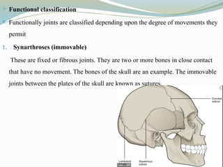
-Functional classification
Functionally joints are classified depending upon the degree of movements they permit
Synarthroses (immovable)
These are fixed or fibrous joints. They are two or more bones in close contact that have no movement. The bones of the skull are an example. The immovable joints between the plates of the skull are known as sutures.
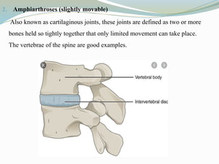
-Amphiarthroses (slightly movable)
Also known as cartilaginous joints, these joints are defined as two or more bones held so tightly together that only limited movement can take place. The vertebrae of the spine are good examples.
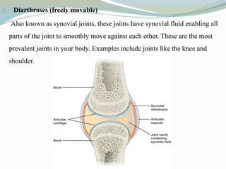
-Diarthroses (freely movable)
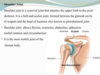
Also known as synovial joints, these joints have synovial fluid enabling all parts of the joint to smoothly move against each other. These are the most prevalent joints in your body. Examples include joints like the knee and shoulder.
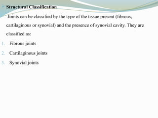
-Structural Classification
Joints can be classified by the type of the tissue present (fibrous, cartilaginous or synovial) and the presence of synovial cavity. They are classified as:
Fibrous joints
Cartilaginous joints
Synovial joints
-Fibrous Joints
Fibrous joints are connected by dense, tough connective tissue that is rich in collagen fibers.
These are fixed or immovable joints.
They lack synovial cavity.
There are three types of fibrous joints.
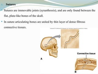
Sutures
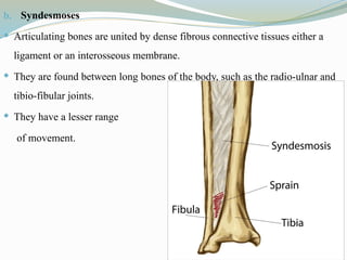
Syndesmoses
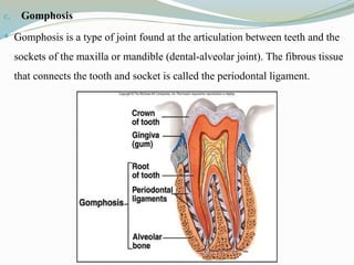
Gomphosis
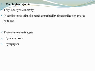
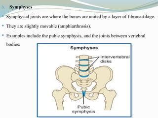
-Cartilaginous joints
They lack synovial cavity.
In cartilaginous joint, the bones are united by fibrocartilage or hyaline cartilage.
There are two main types
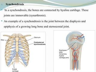
Synchondroses
Symphyses
-Synovial joints
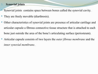
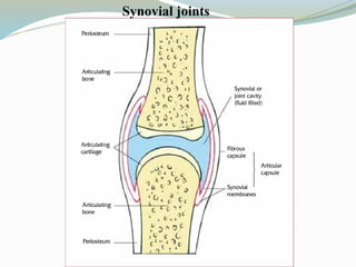
Synovial joints contains space between bones called the synovial cavity.
They are freely movable (diarthrosis).
Other characteristics of synovial joints are presence of articular cartilage and articular capsule a fibrous connective tissue structure that is attached to each bone just outside the area of the bone’s articulating surface (periosteum).
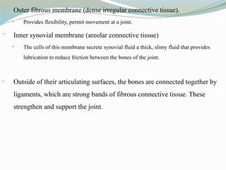
Articular capsule consists of two layers the outer fibrous membrane and the inner synovial membrane.
-Synovial fluid
Synovial fluid supplies oxygen and nutrients and removes carbon dioxide and metabolic wastes from the chondrocytes in the surrounding cartilage.
Synovial fluid lubricates the articulating joints and reduces friction between the bones.
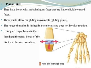
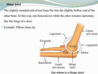
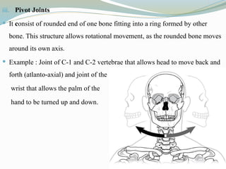
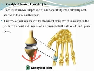
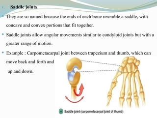
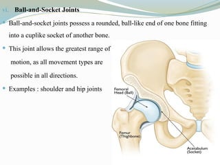
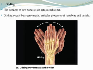
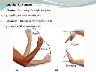
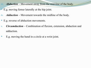
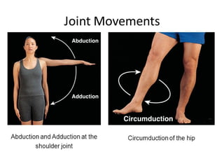
-Classification Of Synovial Joints
Classification of joints is based on the type and degree of movement permitted and depending on the shape of their articular surfaces.