Downloaded 169 times

![3/21/2010
Pinus leaf
Xeromorphic
GYMNOSPERM LEAF Low ratio of surface to volume
Epidermis heavily cuticularized/ thick-walled
Pinus leaf
Presence of hypodermis—thick wall; compact
yp ; p
(except with stoma)
Guard cells sunken (overtopped by subsidiary cells)
Vascular bundles surrounded by transfusion and
endodermis respectively
mesophyll not differentiated
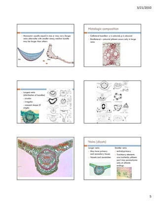
Other features
Resin ducts
Vascular bundles– x adaxial side; p abaxial side
Xylem is endarch
Transfusion tissue
2 kinds of cells: a] living parenchyma cells with non-
lignified walls and b] thin-walled but lignified tracheids Development of the leaf
with bordered pits
Parenchyma cells- deeply staining
Tracheids (near xylem)
Albuminous cells (near phloem) – dense cytoplasm and
prominent nuclei
Universally present in gymnos
Function: water storage or auxiliary conducting system
7](https://image.slidesharecdn.com/leaf-100321103606-phpapp01/85/Leaf-7-320.jpg)

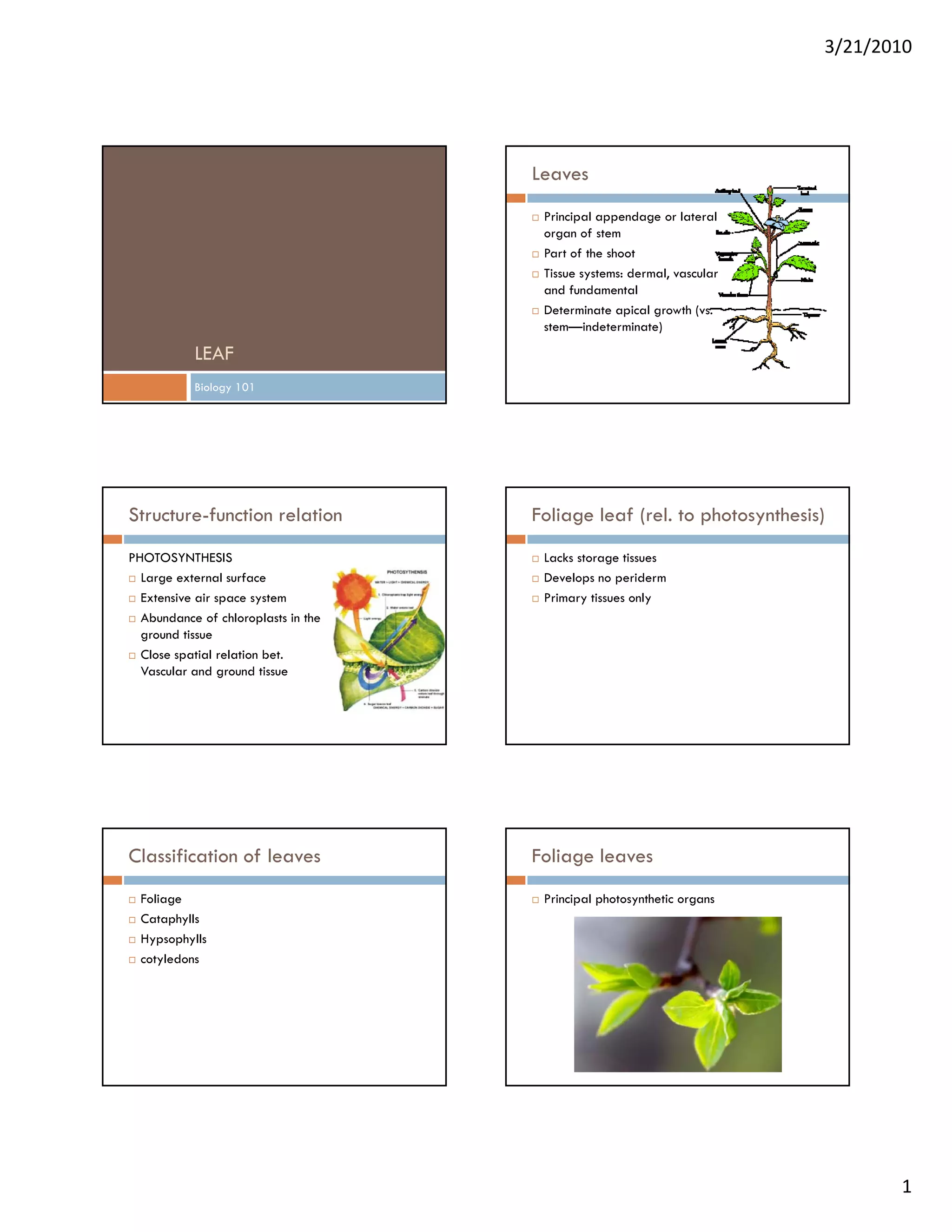
This document discusses leaf structure and function. It begins by defining leaves and their basic anatomy. It then covers leaf classification, morphology, histology, and development. The key structures discussed include the epidermis, mesophyll, vascular bundles, petiole, and abscission zone. Gymnosperm and angiosperm leaves are compared in terms of their tissues and support structures. Leaf development starts from the shoot apical meristem and progresses through initiation, outgrowth, and maturation of tissues.


![Plant growth and development [compatibility mode]](https://cdn.slidesharecdn.com/ss_thumbnails/plantgrowthanddevelopmentcompatibilitymode-130903083906--thumbnail.jpg?width=640&height=640&fit=bounds)


