
Recommended
More Related Content
What's hot
What's hot (20)
Similar to PATELLOFEMORAL PAIN (Harleen kaur Nagi).pptx
Similar to PATELLOFEMORAL PAIN (Harleen kaur Nagi).pptx (20)
Recently uploaded
Recently uploaded (20)
PATELLOFEMORAL PAIN (Harleen kaur Nagi).pptx
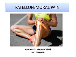
- 1. PATELLOFEMORAL PAIN DR HARLEEN KAUR NAGI (PT) MPT (SPORTS)
- 2. INTRODUCTION • Patellofemoral pain (PFP) is a common musculoskeletal related condition that is characterized by insidious onset of poorly defined pain, localized to the anterior retro patellar and/or peripatellar region of the knee. • An overuse injury in sports medicine. • Commonly known as “runner’s knee.
- 3. PREVALENCE AND INCIDENCE • Prevalence ranges from 3% to 85% for idiopathic anterior knee pain (AKP) or PFP and its associated diagnoses. • Patellofemoral pain occurs across the life span, from young children to older sedentary individuals. • More common in Military recruits and athletes • Female : Male :: 2 : 1
- 4. Anatomy and Biomechanics of Patellofemoral Complex • Interface between articular surface of the patella and trochlear groove. • Modified plane joint • 3 degrees of freedom
- 5. . • Anteriorly: Patellar tendon limits the excursion of patella from the tibia. . • The superficial and deep lateral retinaculum on the lateral side . • Medially: medial patellofemoral ligament. PASSIVE STRUCTURES/ DYNAMIC STABILIZERS
- 6. . • Quadriceps Muscle . • Resultant pull of the 4 muscles that constitute the Quadriceps & patellar tendon . • Clinicaly: Q angle ACTIVE STRUCTURES/STATIC STABILIZERS
- 7. Patellofemoral joint reaction forces depend upon the knee flexion angle and as the knee is flexed, the patellofemoral compressive load is increased.
- 8. Activity Patellofemoral compressive force Stance phase of walking (knee flexion is about 20°) 25 - 50% body weight Ascending stairs 2 – 3 times body weight Running 5 – 6 times body weight Flexion greater than 90° 8 times the body weight Squatting 20 times the body weight
- 9. ETIOLOGY Intrinsic factors: • Altered Biomechanics of leg • Altered biomechanics of foot • Anatomic Anomalies • Medial-Lateral patellar Mobility • Soft-tissue tightness • Muscle Imbalance
- 10. Altered Biomechanics of leg • Increase in Q angle = ↑ lateral patellofemoral contact pressure. • Excessive laterally tilted patella • Other malalignments: femoral ante version, genu valgum and external tibial torsion.
- 11. Subtalar joint pronation alters tibial rotation During terminal knee extension tibia remains internally rotated To compensate: internal rotation of femur- ↑ Q angle → internal rotation of tibia → ↑ Q angle Altered Biomechanics of the foot
- 12. Anatomic Anomalies • Dysplasia or hypoplasia of patella or trochlea. • Patella Alta
- 13. Gastrosoleus ↓dorsiflexio n ↑ subtalar pronation ↑ valgus force= ↑ Q angle Hamstrings Knee flexion at heel strike → increased quadriceps activity ↑ PFJ compression Iliotibial Band Increased lateral tracking and lateral tilt of the patella ↑ PFJ compression Soft Tissue Tightness
- 14. Restricts full excursion of patella in Trochlear groove Causes lateral tracking along with TFL Quadriceps
- 15. Muscle Imbalance Hip muscles weakness Abductors & External Rotators Excessive adduction & Internal Rotation ↑ Q angle Quadriceps weakness ↓ activity of VMO vastus lateralis activates before VMO Maltracking of the patella
- 16. Extrinsic Factors • Excessive duration or frequency of physical activities • Errors in training such as sudden increase in mileage. • Change of training surface. • Inappropriate foot wear such as high heels.
- 17. CLINICAL FEATURES Pain Swelling Stiffness Crepitus Pseudo- locking Giving away Popping sensation
- 18. PAIN HISTORY: Onset : Insidious or Gradual, can be precipitated by Trauma Area: peri-patellar, retro-patellar, ‘circle sign’ Type: Diffuse dull ache, sometimes sharp.
- 19. Aggravating Factors • Descending stairs > Ascending • Going uphill or walking on incline • Standing up from squatting
- 20. • Malalignment:genu-varum (bowleg) or genu- valgum (knock-knee). • Tibial Torsion: Medial →Genu varum Lateral→Genu valgum • Size, shape, position of the patella: grasshopper/ squinting/ patellar alta • Subtalar joint Pronation Observation: Posture- Standing Anterior View
- 21. Lateral view: • Patellar alta, camel sign • Genu recurvatum
- 22. Local Observation: • Wasting of quadriceps • Echymosis • Swelling Palpation: • Warmth, Edema, Tenderness • In PFPS: Lateral retinacular tenderness
- 23. EXAMINATION • Active & Passive ROM of Hip, knee and Ankle. • Patellar tracking while knee Flexion-Extension. • Abrupt lateral deviation of patella during terminal knee extension (J-sign).
- 24. Muscle Strength Testing Quadriceps Hip abductors Hip Internal Rotators Flexibility Testing ITB Rectus Femoris Hamstrings Hip Flexors Gastrocnemius
- 25. Single Leg Press Step Down Functional Performance Testing
- 26. Balance and Reach Test Bilateral Squatting
- 27. Special Tests 1.Patellar tilt test:
- 28. 2. Patellar Glide Test •Passive translation of the patella, measured as % of patellar width •25%: Normal, >50 : laxity of medial constraints
- 29. 3. Vastus Medialis Co-ordination Test: • Lack of co-ordinated full extension: Positive Test
- 30. 4. Patellar Apprehension Test: •Knee flexed to 30° •Push the patella as lateral as possible. •Positive Test: Pain / Apprehension
- 31. 5. Waldron’s Test •Phase I- Press the patella against femus while flexing the knee passively. •Phase II- slow, full squat while pressing the patella against femur. Presence of Pain and Crepitus
- 32. 6. Patellar Grind Test: • Knee is in slight flexion • Press the patella distally (with the hand on the superior border of the patella)
- 33. 7. Eccentric Step Test: • Patient stands on 15 cm (6 inches) stool • Steps down. First with uninvolved and then involved leg
- 34. MANAGEMENT • 1. RELATIVE REST!! • PFPS is an overuse/ overload syndrome • Runners: reduce mileage • Cyclists: lower gear, high pedal revolutions per minute. • Swimming: Breast stroke to be avoided.
- 35. 2.ICE: • Ice particularly after exercise 3. Electrotherapy: • TENS, ultrasonic therapy, Electrical stimulation 4. Gentle mobilization of patella
- 36. Strengthening exercises Start from non-weight bearing → weight bearing Particularly hip abductors and external rotators Stabilizes pelvis and controls hip internal rotation Pelvic and hip-stabilizing muscles: Transverse abdominus, Gluteus medius, and Gluteus minimus
- 37. Open v/s Closed Kinetic Chain Exercises: • Open kinetic chain (OKC) exercises have been reported to exacerbate symptoms in PFPS patients. • CKC place less stress on PFJ
- 38. TAPING To maintain the patella correctly within the femoral trochlea during full knee range of motion. McConnell Technique is most commonly used. McConnell’s Rehabilitation Program: Patellar taping + stretching of lateral tight structures + VMO strengthening Aim of taping: To medialize the patella, to improve patellar tracking
- 39. • Correcting Lateral Glide: • Correcting External Rotation:
- 40. The effect of taping should be assessed immediately using a pain provoking activity. Acute cases may initially need tape applied 24hrs a day until the pain reduces.
- 41. Knee braces and sleeves • The Palumbo dynamic patellar brace consists of a lateral pad that ’floats’ over the patella, maintaining effective position during knee motion. • Cho-Pat knee strap functions dynamically , improves patellar tracking and spreads pressure uniformly over the surface area.
- 42. Orthoses • Reducing excessive pronation in individuals with PFPS will result in reduced internal rotation of the lower limb. • Greater than 15 mm: - foot orthoses in runners.
- 43. MEDICAL MANAGEMENT • NSAID’s • Intra-articular hyaluronic acid (HA) injections: It forms viscous synovial fluid that lubricates joints, absorbs mechanical shock and protects the articular cartilage.
- 44. SURGICAL INTERVENTION • If symptoms persist despite completing 6 – 12 months of rehabilitation
- 45. Lateral Retinacular Release Proximal Realignment of extensor mechanism Repair or reconstruction of patellofemoral ligament Repair of patello- femoral articular cartilage lesion eg. Mosaic plasty Interposition trochleoplasty